Which part of the lower tract is involved in the micturition reflex?
The lower tract is involved in the micturition reflex. The bladder is a hollow organ that functions as reservoir for the storage and periodic elimination of urine. The bladder’s walls are made up of three layers of smooth muscle, known as the detrusor.
Where is the micturition center in the brain?
This reflex is integrated in the pontine micturition center, which is located in the rostral brainstem. Also, a sacral micturition center is located at S2–S4, through which the bladder can contract independently of cortical and pontine input.
What is bladder emptying and micturition reflex?
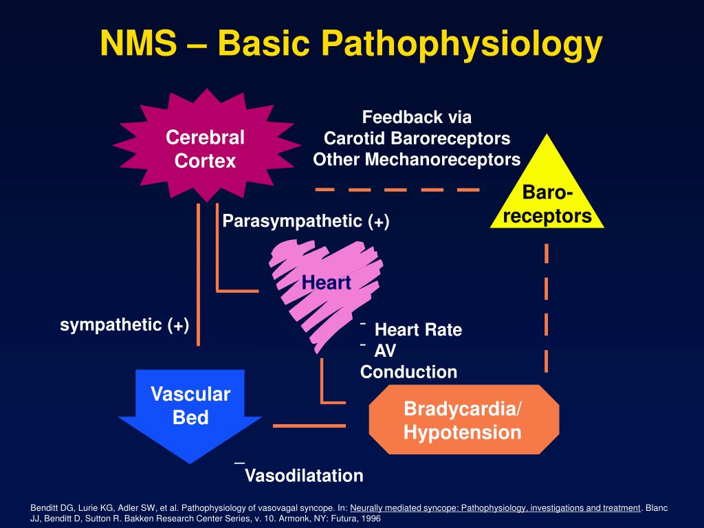
Bladder Emptying and the Micturition Reflex. The micturition or emptying phase displays a coordinated relaxation of the inner and outer urethral sphincters, under sympathetic and somatic regulation respectively, with strong contractions of the detrusor muscle due to parasympathetic impulses.
What is the volume of urine that initiates the micturition reflex?
Stimulus: Volume of urine that initiates micturition reflex is 300-400 ml 2. Receptor: Stretch receptors in bladder wall 3. Afferent: Pelvic parasympathetic

What is the micturition reflex?
The micturition reflex is peripherally mediated by components of the somatic and the autonomic nervous systems. The bladder receives its motor innervation through the parasympathetic pelvic nerves. The principal nuclei innervating the urinary bladder of the cat consist of motor neurons located in the gray matter of the intermediolateral cell column of the sacral spinal cord and motor neurons in the ventral gray matter of the sacral spinal cord in the region of Onuf's nucleus. In humans, sacral nerve blocks have revealed that the detrusor nucleus has a rostral–caudal extension going from the S3 to S4 segment. The precise intramedullary location of detrusor motor neurons and their histological characteristics in the sacral spinal cord have not been described.
Which nerve is the target of the micturition reflex?
Many of the sensory afferent nerve fibers contained in the sacral spinal nerves originate in the pudendal nerve, thereby making the pudendal nerve an ideal target for neuromodulating inhibition of the micturition reflex.
What are bladder reflexes?
These interneurons synapse with preganglionic efferent parasympathetic nerves to complete this reflex pathway. The bladder-bladder reflex is an excitatory one, which becomes activated by the sensing of a full bladder but is inhibited until it is socially appropriate to void. Interneurons activated by bladder afferent fibers also synapse with urethral parasympathetic efferent nerves to form a “bladder-urethral reflex” (Leng and Chancellor, 2005). This is an inhibitory reflex, which relaxes the smooth muscle of the proximal urethra and bladder outlet to open immediately before the onset of bladder contraction.
What is the term for a detrusor contraction without urethral relaxation?
Detrusor contraction without urethral relaxation is called reflex dyssynergia. 31.
Which neuron receives input from the cerebral cortex, cerebellum, basal ganglia, thal?
Micturition depends on a spinobulbospinal reflex that is relayed through the pontine micturition center, which receives input from the cerebral cortex, cerebellum, basal ganglia, thalamus, and hypothalamus, and is the final common pathway to the bladder motor neurons.
Which nerves are involved in reflex connections?
Reflex connections between the pelvic (parasympathetic) nerve afferents and the lumbar (sympathetic) motor neurons also have been demonstrated. 6,20,32 The effect seems similar to that observed in the pudendal (somatic) nerve—that is, as the pelvic motor neuron begins to fire to initiate a detrusor contraction, the hypogastric nerve becomes silent (see Figure 3-3, C and D ). When the pelvic nerve stops firing as the bladder is emptied, the hypogastric nerve discharges once more. Presumably, the effect of the hypogastric nerve is on bladder relaxation and urethral contraction.
Is the perineal to bladder reflex a sacral cord reflex?
Although during this early postnatal period there is a weak supraspinal bladder reflex present ( Kruse and De Groat, 1990 ), the perineal-to-blad der reflex is so prominent that thoracic cord transections of the spinal cord did not abolish it. Thus, the perineal-to-bladder reflex is a sacral cord reflex.
How do cortical areas prevent micturition?
They partially inhibit the reflex except when micturition is desired. They can prevent micturition by contraction of external urethral sphincter. When it is time to urinate, the cortical areas facilitate the sacral centre to initiate micturition reflex and inhibit the external urethral sphincter.
Which of the following is controlled by facilitatory and inhibitory higher centres?
1. The reflex is controlled by facilitatory and inhibitory higher centres.
What is the pontine micturition center?
Pontine Micturition Center. The pontine micturition center (PMC) in the brainstem is activated via afferent signals from the urinary bladder as it is filling. This center sends inhibitory impulses to the spinal reflex arcs to enable bladder voiding. In the absence of any other regulation, the afferents from the bladder and urethra to ...
What is the micturition phase?
The micturition or emptying phase displays a coordinated relaxation of the inner and outer urethral sphincters, under sympathetic and somatic regulation respectively, with strong contractions of the detrusor muscle due to parasympathetic impulses . The distension of the urinary bladder wall causes wall tension to rise very slightly.
What is the function of the afferents in the bladder?
In the absence of any other regulation, the afferents from the bladder and urethra to the midbrain and pons and the efferents to the spinal cord would act as an on-off switch, to cause either reflex voiding or storage depending only on the urine volume stored in the bladder. This means that during the filling or storage phase, the voiding reflex is off, but it is switched on to the highest level when the bladder is distended beyond a critical point, activating the tension receptors in the wall.
What is the detrusor in the bladder?
The detrusor is the smooth or involuntary muscle of the bladder wall. The urethral muscles consist of the external and internal sphincter. The internal sphincter and detrusor muscle are both under autonomic control. The external sphincter, however, is a voluntary muscle under the control of voluntary nerves.
What is the term for the relaxation of the striated sphincter?
Micturition is thus characterized by: relaxation of the striated sphincter (somatic innervation) relaxation of the smooth muscle sphincter and opening of the bladder neck (sympathetic innervation) detrusor contraction (parasympathetic innervation) The distension of the urinary bladder wall causes wall tension to rise very slightly.
What is the guarding reflex?
Urethral reflexes, called ‘the guarding reflex,’ also play a part in inhibiting involuntary bladder emptying during this process. The afferents are all conveyed through the pelvic nerves to initiate a spinal reflex.
What is the act of micturition?
The act of micturition is an autonomic reflex at the level of the spinal cord. This reflex also helps to complete micturition when the act is voluntarily initiated, or when it follows a period of inhibition by the brain, by relaxing the external sphincter. The control of this process is mediated via afferent signals from stretch ...
Where is the micturition reflex located?
The micturition reflex is a bladder-to-bladder contraction reflex for which the reflex center is located in the rostral pontine tegmentum (pontine micturition center: PMC). There are two afferent pathways from the bladder to the brain. One is the dorsal system and the other is the spinothalamic tract.
What is the micturition reflex?
The micturition reflex involves a coordinated and sustained contraction of the detrusor muscle (the detrusor reflex) along with simultaneous relaxation of the urethra. 20. Moreover, depending on the size of the bladder, the micturition reflex may also result in urine storage.
Introduction
Micturition, also known as urination, is the process of expelling urine from the bladder. The purpose of urination is to eliminate metabolic products and toxic wastes from the body that have been filtered from the blood by the kidneys.
Relevant Anatomy & Physiology
The urinary tract comprises of two mutually dependent components: the upper tract, which contains the kidneys and ureters, and the lower tract consisting of the bladder and urethra. The lower tract is involved in the micturition reflex.
Mechanism
Micturition is a complex and highly distributed process, involving pathways at multiple levels of the brain, spinal cord and PNS, in addition to being mediated by multiple neurotransmitters. At the most basic level, the micturition reflex is triggered when the bladder fills with urine.
Where is the pontine micturition center located?
The Pontine micturition center(PMC) is located in the medial dorsal pons, close to, or includes the lateral dorsal tegmental nucleus and locus coeruleus.[4] Upon stimulation, PMC exerts dual effects of producing detrusor muscle contraction and urethral sphincter relaxation with consequent micturition. External urethral sphincter relaxation is mediated indirectly by suppressing Onuf's nucleus located in the sacral spinal cord. A PET(positron emission tomography) scan is the imaging modality that detects how tissues and organs are functioning by measuring blood flow, the extent of oxygen and sugar use. Increased blood flow has been demonstrated by PET scan in right dorsomedial pons during voiding. In contrast, PET scan during withholding of urine showed higher blood flow in the right ventral pons. [5][6]There are also suggestions that specific nuclei that control micturition predominantly exist in the right pons.[4] The pontine micturition center (PMC) coordinates the mechanical process of micturition, thus the sphincter and detrusor muscle activities of the urinary bladder. Afferents from the bladder relay on the PMC before to higher brain centers. And the higher brain centers of micturition communicate with PMC through the periaqueductal gray matter to either provoke or suppress the voiding reflex. When the urinary bladder becomes distended, the stretch receptors of the bladder wall transmits an afferent signal to the pons, which subsequently notifies the brain. This afferent signal results in a perception of bladder fullness or desire to urinate. The higher brain determines the appropriateness of initiating voiding reflex. Under typical situations, the higher brain suppresses PMC by transmitting inhibitory signals via the periaqueductal gray matter. Upon the PMC inhibition, the urge to urinate disappears, which allows delaying voiding until finding a socially acceptable place and time. In an appropriate time and place, the brain withdraws the suppression of the PMC, which allows initiating a micturition reflex. Regardless, the individual may consciously contract the pelvic floor muscles to keep the external urethral sphincter closed or suppress the voiding urgency by distracting techniques.
Where is the PMC located?
The PMC, also known as Barrington's nucleus, is located in the brainstem and works with other regions of the brain to coordinate micturition. The primary role of the PMC when it is activated is to stimulate urination. It achieves this through activation of parasympathetic neurons, triggering the detrusor muscle to contract and indirectly inhibits the somatic nerves that maintain the external sphincter closed in contraction. [3]
What is the neural circuitry that controls urination?
It involves pathways at various levels of the brain, the spinal cord, and the peripheral nervous system and is under the mediation of multiple neurotransmitters.[1] To fully understand the role that the Pontine Micturition Center (PMC) plays in regulating urination requires a basic understanding of the structures and innervations involved.
What is the best way to access posterior pontine lesions?
Neurosurgical access to posterior pontine lesions bears higher chances of injury to PMC and accessed through the rhomboid fossa. Telovelar route or cerebellomedullary fissure approach is used to access the superior and posterior pontine lesions. The suprafacial triangle is an opening to the brainstem, which is located above the facial nerve, and bounded medially by medial longitudinal fasciculus(MLF) or the median sulcus, laterally by superior cerebellar peduncles and inferiorly by the facial nerve or facial colliculus. Although it is considered as a safe entry triangle, bipolar stimulation on the surface is recommended to localize the facial nerve course, which might have been distorted by the lesson. To preserve the MLF, entry to this zone should always be 2 mm from the midline. [8]
Which muscle is an extension of the detrusor muscle?
Both of these innervations act on the internal sphincter, which is made of smooth muscle and is an extension of the detrusor muscle.
Where does sympathetic innervation originate?
Sympathetic innervation originates in the thoracic spine and serves to inhibit urination. Sympathetic nerves inhibit the detrusor muscle and activate the internal sphincter to contract.
Which neurotransmitter is excitatory?
These neurotransmitters can categorize as excitatory such as glutamic acid, tachykinins, PA CAP (pituitary-adenylate-cyclase-activating polypeptide), NO and ATP or inhibitory such as GABA glycine, enkephalins. Glutamate binds with NMDA and non-NMDA receptors and is the essential neurotransmitters in reflex pathways controlling the bladder and the urethral sphincter. Inhibitory neurotransmitters exert a tonic inhibition in the PMC and as well as in the spinal cord.
How does micturition occur?
Micturition, or urination, occurs involuntarily in infants and young children until the age of 3 to 5 years, after which it is regulated voluntarily. The neural circuitry that controls this process is complex and highly distributed: it involves pathways at many levels of the brain, the spinal cord and the peripheral nervous system and is mediated by multiple neurotransmitters. Diseases or injuries of the nervous system in adults can cause the re-emergence of involuntary or reflex micturition, leading to urinary incontinence. This is a major health problem, especially in those with neurological impairment. Here we review the neural control of micturition and how disruption of this control leads to abnormal storage and release of urine.
Where do parasympathetic preganglionic fibres travel?
Parasympathetic preganglionic fibres (shown in green) arise from the S2–S4 spinal segments and travel in sacral roots and pelvic nerves (PEL) to ganglia in the pelvic plexus (PP) and in the bladder wall. This is where the postganglionic nerves that supply parasympathetic innervation to the bladder arise.
Why is bladder control sensitive?
Owing to the complexity of the neural mechanisms that regulate blad der control, the process is sensitive to various injuries and diseases. This Review summarizes the results of recent studies in animals and humans that have provided new insights into the sensory and motor mechanisms that underlie voluntary and reflex micturition, the changes in neural pathways that occur following disease or injury that alters lower-urinary-tract function, and new therapies for the treatment of neurogenic bladder dysfunction.
Where does sympathetic innervation originate?
The sympathetic innervation arises in the thoracolumbar outflow of the spinal cord, whereas the parasympathetic and somatic innervation originates in the sacral segments of the spinal cord . Afferent axons from the lower urinary tract also travel in these nerves. Open in a separate window. Figure 1.
Where do the sympathetic nerves enter the urinary tract?
a| Innervation of the female lower urinary tract. Sympathetic fibres (shown in blue) originate in the T11–L2 segments in the spinal cord and run through the inferior mesenteric ganglia (inferior mesenteric plexus, IMP) and the hypogastric nerve (HGN) or through the paravertebral chain to enter the pelvic nerves at the base of the bladder and the urethra. Parasympathetic preganglionic fibres (shown in green) arise from the S2–S4 spinal segments and travel in sacral roots and pelvic nerves (PEL) to ganglia in the pelvic plexus (PP) and in the bladder wall. This is where the postganglionic nerves that supply parasympathetic innervation to the bladder arise. Somatic motor nerves (shown in yellow) that supply the striated muscles of the external urethral sphincter arise from S2–S4 motor neurons and pass through the pudendal nerves. b| Efferent pathways and neurotransmitter mechanisms that regulate the lower urinary tract. Parasympathetic postganglionic axons in the pelvic nerve release acetylcholine (ACh), which produces a bladder contraction by stimulating M3muscarinic receptors in the bladder smooth muscle. Sympathetic postganglionic neurons release noradrenaline (NA), which activates β3adrenergic receptors to relax bladder smooth muscle and activates α1adrenergic receptors to contract urethral smooth muscle. Somatic axons in the pudendal nerve also release ACh, which produces a contraction of the external sphincter striated muscle by activating nicotinic cholinergic receptors. Parasympathetic postganglionic nerves also release ATP, which excites bladder smooth muscle, and nitric oxide, which relaxes urethral smooth muscle (not shown). L1, first lumbar root; S1, first sacral root; SHP, superior hypogastric plexus; SN, sciatic nerve; T9, ninth thoracic root. Part amodified, with permission, from REF. 144© (1996) W. B. Saunders Company.
Is sphincter relaxation present in infants?
In the infant (a) sphincter relaxation is present but less complete. On the other hand, in the paraplegic patient (c) the reciprocal relationship between bladder and sphincter is abolished. During bladder filling, involuntary bladder contractions (detrusor overactivity) occur in association with sphincter activity.
Does dopamine inhibit micturition?
For example, dopamine elicits inhibitory effects on micturition through D1-like receptors and facilitatory effects through D2-like receptors. On the other hand, activation of 5-HT1Areceptors inhibits micturition in the cat but facilitates it in the rat80. Neuroplasticity and pathology. Developmental changes.