
What does a hyphae do for a fungus?
Hyphae play a vital role in the growth and expansion of fungi. Without hyphae, fungi wouldn’t be able to thrive and multiply. Each hypha is tasked with bringing in food for the collective fungi organism. Hyphae travel long distances, often through long stretches of earth, plant matter, and even wood, to gather food for the fungus.
How do fungi use hyphae to find and digest food?
first the fungus grows hyphae into a food source. Then digestive chemicals ooze from the hyphae into the food. the digestive chemicals break down the food into small substance that can be absorbed by the hyphae. fungi produce spores in structure called. fruiting bodies.
What is the main function of a fungus's hyphae?
The main function of fungus hyphae is to absorb nutrients from its surrounding. They contain the cytoplasm or cell sap, including the nuclei containing genetic material. They contain the cytoplasm or cell sap, including the nuclei containing genetic material. Furthermore, the hypha happens to be the main mode of vegetative growth in most fungi.
Are most bodies of fungi made of hyphae?
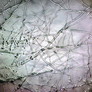
Fungi live as either single-celled organisms or multicellular organisms. Single-celled fungi are referred to as yeasts. The vast majority of fungi are multicellular. Most of the body of a fungi is made from a network of long, thin filaments called ‘hyphae’. Hyphae filaments are made from tubular cells that connect end on end.

What is the role of hyphae in fungi?
Hyphae play a vital role in the growth and expansion of fungi. Without hyphae, fungi wouldn’t be able to thrive and multiply. Each hypha is tasked with bringing in food for the collective fungi organism.
How many hypha do fungi produce?
Each fungi issues thousands of hypha, much like a plant producing roots that stretch out into the ground. The strands of hypha are so thin and small that they often can’t be seen in their individual form. However, since a fungus produces so many, they can easily be seen in masses.
Why do fungi need harsher locales?
A harsher locale requires the fungi to create even more intricate and vast mycelium networks to ensure the fungus will survive .
How do hyphae absorb nutrients?
The hyphae absorb nutrients through their cell walls and transport them to other parts of the fungus body. These nutrients are used to build more complex structures, including forming the above-ground mushroom that we are most familiar with.
How many strands of hyphae are in a hyphae?
The hyphae are so thickly combined, they form “ropes” of thousands of individual strands that work together as one structure.
What does hyphae look like?
Mycelium masses often look like thick white spider webs or an array of dense white clumps or swathes of material around a mushroom.
Why is mycelium mass not developed?
In some cases, a fungus’ mycelium mass isn’t that developed because it has easily accessible food sources. The robustness and hardiness of the mycelium depend on the environment; a rich forest floor with trees and plants nearby means less work for most fungi.
What are hyphae used for?
In some fungal species, hyphae have evolved into specialized nematode-trapping structures, using nets and ring structures to trap nematode species.
How does hyphae grow?
Hyphae growth occurs by extending the cell walls and internal components from the tips. During tip growth, a specialized organelle called the spitzenkörper, assists in the formation of new cell wall and membrane structures by harboring vesicles derived from the golgi apparatus and releasing them along the apex of the hypha.
How are hyphae classified?
Hyphae can be classified based on the presence of internal septa (septate versus aseptate species). Hyphae can also be distinguished from species which produce pseudohyphae via cell division. Pseudohyphae is a form of incomplete cell division, in which the dividing cells do not separate. There are several yeast species which produce such pseudohyphae.
What are the characteristics of hyphae?
Hyphae characteristics are an important method of classifying various fungal species. There are three main hyphae characteristics: 1 Binding: Binding hyphae have a thick cell wall and are highly branched. 2 Generative: Generative hyphae have a thin cell wall, a large number of septa, and are typically less differentiated. Generative hyphae may also be contained within other materials (e.g., gelatin or mucilage) and can also develop structures used in reproduction. All fungal species typically contain generative hyphae. 3 Skeletal: Skeletal hyphae contain a long and thick cell wall with few septa. Skeletal hyphae can also be of a fusiform subtype, with a swollen midsection surrounded by tapered ends.
What subtype of hyphae has a long and thick cell wall?
Skeletal: Skeletal hyphae contain a long and thick cell wall with few septa. Skeletal hy phae can also be of a fusiform subtype, with a swollen midsection surrounded by tapered ends.
What are the different types of hyphae?
There are four general subtypes: Monomitic: While virtually all fungal species contain generative hyphae, those with only exhibit this type are referred to as monomitic (e.g., agaric mushrooms). Dimitic: A species that contains generative hyphae in addition to one other type of hyphae. The most common combination of dimitic fungi is generative ...
What is the result of the formation of a new tip from a hypha?
As the hypha extends, new septa can be created to internally divide the cells. The characteristic branching of hyphae is the result of the formation of a new tip from a hypha, or the division of a growing tip (see diagram below).
How do hyphae grow?
After attachment to the root, the infective hyphae start to grow from the appressorium into the cortex colonizing its outer and inner layers, but never crossing the pericycle. After establishing a network of intercellular hyphae, fungus starts to develop two types of structures: intracellular and extra-radical. The fully developed highly branched arbuscules fill nearly the whole volume in infected cortical cells.
What are the hyphae of mycorrhizae?
By far the most common and most important mycorrhizae, endomycorrhizae externally appear similar to nonmycorrhizal roots in shape and color, but internally the fungus hyphae grow into the cortical cells of the feeder root either by forming specialized feeding hyphae (haustoria), called arbuscules, or by forming large, swollen, food-storing hyphal swellings, called vesicles. Most endomycorrhizae contain both vesicles and arbuscules and are, therefore, called vesicular–arbuscular mycorrhizae ( Fig. 11-164 ). Endomycorrhizae are not surrounded by a dense fungal mantle but by a loose mycelial growth on the root surface from which hyphae and large pearl-covered zygospores or chlamydospores are produced underground. Endomycorrhizae are produced on most cultivated plants and on some forest trees mostly by zygomycetes, primarily of the genus Glomus, but also by other fungi, such as Acaulospora. Endomycorrhizae are also produced by some basidiomycetes.
How do AMF fungi affect soil?
The ability to connect to an AMF network can affect nutrient capture by roots. Plants colonised by AMF may be more efficient at extracting soil phosphate and/or nitrogen than plants grown in sterile soil. Roots typically form mycorrhizal associations under nutrient limitation. Plant roots containing abundant phosphate inhibit the formation of AMF arbuscules, the plant thereby avoiding an unnecessary drain on its carbon resources. Soil minerals accumulated by the extraradical hyphae are partitioned between the fungus and the plant.
How do AMF hyphae translocate nutrients?
AMF hyphae translocate nutrients not only from soil to root, but also between roots of adjacent plants. Using growth chambers divided by partitions either permeable or non-permeable by growing hyphae, it was shown that a plant can acquire nitrogen from dead leaf litter via its AMF hyphae, when these were allowed to colonise plant litter enriched with the stable isotope 15 N ( Figure 7.6 ). Nitrogen import through AMF hyphal connections enhanced host plant growth as well as supplying the fungus. Some aspects of the mechanism by which AMF acquire soil nitrogen remain unclear. Readily-available forms of nitrogen are scarce in natural soils, in particular those of boreal forest, where most nitrogen is in the form of complex organic compounds resistant to microbial attack. Mycorrhizal fungi are probably the main route by which trees acquire nitrogen from these compounds in the soil. The ability of AM fungi to take up both labile and recalcitrant organic nitrogen compounds under field conditions in boreal forest has been recently demonstrated, using an ingenious technique involving test substances (glycine and chitosan as labile and recalcitrant nitrogen compounds, respectively) bound to the surface of nanoparticles called ‘quantum dots’.
What is the color of fungus in H&E?
In disease outside the CNS, these organisms may not always appear dark walled on standard histopathologic stains. Cell wall melanin may be visible as a brownish-yellow color on H&E stain ( Fig. 270-8 ). If melanin is not evident on fresh preparations of H&E stain, it can be stained by the Fontana-Masson method, which better enables diagnosis, especially if culture results are negative or if culture is not performed. 68 Fontana-Masson stain is, however, not 100% specific for the dark-walled fungi because the cell walls of some Aspergillus and other fungi with hyaline hyphae have been shown to stain dark with this method. 69
What are ectomycorrhizal fungi?
Ectomycorrhizal fungi (EM fungi) are phylogenetically very diverse and more than 2000 species of EM fungi worldwide have been identified, primarily from Basidiomycotina and Ascomycotina. These EM fungi form characteristic mycorrhizal associations, almost entirely with woody perennials, including Pinaceae, Betulaceae, Fagaceae, and Diperocarpaceae in tropical, subtropical, and arid environments, and are regarded as key organisms in nutrient and carbon cycles in forest ecosystems ( Agerer, 2003; Becerra et al., 2005; Jakucs, Kovacs, Agerer, Romsics, & Eros-Honti, 2005 ). Unlike AM fungi, hyphae of EM fungi do not penetrate into the root cells but are intercellular. The hyphae penetrate into the root cortex where they form a hyphal network (“Hartig net”; see Fig. 3.2) in the intercellular space through which minerals and nutrient materials are exchanged between the fungus and the plant. The fungus forms a mantle of hyphae on the outside of the plant root that extends into the surrounding soil ( Anderson & Cairney, 2007; Smith & Read, 2008 ). The structure of ectomycorrhizal extramatrical mycelium (extraradical mycelium) varies considerably between ectomycorrhizal species, ranging from a weft of undifferentiated mycelium around the root to a highly differentiated mycelium comprising a foraging fungal front connected to roots via rhizomorphs ( Bonfante & Anca, 2009; Cairney, 2000 ). In angiosperm tree roots, the ectomycorrhizal hyphae penetrate the epidermal layer and spread as hyphal network (Hartig net) intercellularly (one cortical cell deep: Fig. 3.2, panels a and b), but in the case of conifers and gymnosperms the hyphal penetration reaches up to a depth of 3–4 cortical cells ( Agueda, Parlade, de Miguel, & Martinez-Pena, 2006; Lupatini, Bonnassis, Steffen, Oliveira, & Antoniolli, 2008; Massicotte, Melville, & Peterson, 2005 ). Similar to AM fungi, ectomycorrhizae also exhibit synergistic interactions with other plant-beneficial organisms such as symbiotic N 2 -fixers. For example, ectomycorrhizal symbiosis enhanced the efficiency of inoculation of two Bradyrhizobium strains on the growth of legumes ( Andre et al., 2005 ). It is also of interest that similar synergies were seen when AM fungus (Glomus mosseae), EM fungus (Pisolithus tinctorius), and Bradyrhizobium sp. were used together to inoculate Acacia nilotica, enhancement of N 2 fixation, growth, and dry biomass were observed when all three organisms were present ( Saravanan and Natarajan, 1996, 2000 ). Moreover, Bradyrhizobium sp. when co-inoculated with either the AM fungus or the EM fungus, gave enhancement of N 2 fixatioin as compared to the control with Bradyrhizobium sp. only ( Fig. 3.3 ).
How do fungi develop in the root?
The development of AM fungi within the root is strictly controlled by the host and the mycorrhizal development should be considered as a result of complex dialogue between the fungus and the host defense systems. The defence-like reactions induced within the root cortex during AM development include: modification of the cell walls, synthesis of phytoalexins, accumulation of callose and of some pathogen-regulated (PR) proteins, including peroxidases and lytic enzymes ( Table 14.10 ). However, the plant defence from fungal pathogens differs significantly from the reactions for AM development. During Glomus growth the intensity of plant reactions inside roots is low, they are less prolonged and highly differentiated in time and space compared to pathogenesis. It looks like the plant defence reactions are repressed by some fungal signals required for the stable co-existence of partners.
How are hyphae formed?
To understand how hyphae are formed in fungi, it's important to understand the life cycle of fungi. The life cycle of fungi starts with the production of spores, which are produced in the fruiting bodies of the organism. Once the spores are released/dispersed into the surrounding environment (by wind, animals etc), ...
Where do hyphae grow?
Hyphae, as mentioned, grow from the spore/germ. Here, the first hyphae cell is produced and continues growing out at the apex. While some of these tubular structures can be seen with the naked eye (in large numbers) an individual hypha is a microscopic tube like structures that contain a cytoplasm (multinucleate cytoplasm) that is surrounded by a plasma membrane.
Why are septate hyphae called septate hyphae?
Septate Hyphae. Septate hyphae are termed septate because they form structures known as septa between the cells. Unlike the non-septate hyphae, the septate hyphae, found in organisms such as Aspergillus species, divide the hyphae into several cells along the hyphae thread.
Why are hyphae modified?
These types of hyphae are modified differently in a manner that allows them to access and obtain nutrients more effectively.
What happens to the cell wall during hyphae?
During elongation, where cells are added to the tip of the hyphae, the cell wall undergoes lysis (degradation) allowing for cells to be added at the apex for hyphal elongation. At the same time, new cell wall is created to protect the new cells as the hyphae continue growing.
What is a hypha?
Essentially, hyphae (singular; hypha) are the long, tubular branching structures produced by fungi. However, they can also be found in a number of other organisms such as oomycetes. Hyphae in fungi vary in structure and serve different functions from one species to another. Some of the most common hyphae include:
Where are vesicles located in the hyphae?
For this reason, they are found near the hyphal tip during epical elongation. During this process (apical elongation) the cell's vesicles move towards the tip of the hyphae (towards the plasma membrane) where they release various enzymes and other compounds.
What is the hyphae in fungus?
Hypha (plural is hyphae) is a long, branching, filamentous, tube-like structure found in fungus, oomycete, and actinobacterium. Collectively termed as mycelium, hyphae represent the main component for vegetative growth making up the fleshy body structure called thallus, of a multicellular fungal which are also called as molds. Most common type of hyphal structure includes septate hyphae (walls present between the cells), coenocytic hyphae (do not have walls and cell membranes between the cells and hence are non-septate) and pseudohyphae (normally seen in unicellular fungi) as shown in the figure above.
How do hyphae form?
These spores are then released or dispersed into the environment with the help of wind, animals, humans, insects etc. Once settled on the surface, these spores start to germinate and produce hyphae that later form tangled, tube-like structure known as mycelium. Formation of hyphae also depends on the environment of the surface it settles on. Conditions that favor the growth of hyphae are adequate amount of nutrients, moisture and warmth. Hyphae then elongates further as the new cells continually develop at the tip of these tubular structure. The cell wall is lysed and degraded allowing new cells to be added at the tip or apex of the hyphal structure with the help of specialized organelle i.e., spitzenkorper. It harbors vesicles from the Golgi apparatus and set them along the tip/apex of the growing hypha. As the vesicle contents are released, hypha is extended and cell wall, vesicle membranes are formed giving rise to a new cell membrane. This gives the fungi their characteristic branching pattern as shown in the figure below. During this growth, new septa may be formed which internally divide the cells into compartments giving septate or non-septatehyphal structure.
What is the cell wall of a hypha?
When observed under a microscope, a single hypha looks like a tube with cytoplasm having multiple nuclei (multinucleated) and surrounded by a plasma membrane and a tough cell wall. The cell wall of fungal hypha is made of chitin which is a long chain of N-acetylglucosamine. N-acetylglucosamine is a polysaccharide containing nitrogen. Chitinous cell wall provides an advantage of being tough and flexible and therefore helps in continuous growth and elongation in any direction. Different fungus has different length of hyphae with diameter ranging from 2-30 micrometers. It also depends on the species and the stage of growth of the organism. The hyphal structure is divided into cells with the cross walls called septa which are perforated structures. Septa allow the ribosomes, mitochondria and nuclei (sometimes) to flow between the cells as shown in the figure below.
How do fungal species absorb nutrients from the soil?
Some fungal species such as mycorrhiza absorbs nutrient from the soil. For this purpose, they develop symbiotic relationship with the vascular plants and use specialized hyphal structures called arbuscules to bind to the roots or phylum of the plant species. In this way, it absorbs water and nutrients from the soil. This mechanism benefits both plant and fungal species as they both get nutrients for their growth.
What is the name of the long tubular structure that often branches off and shows structural variations depending on the species?
Hyphae, generally found in fungi, are the long tubular structure that often branches off and show structural variations depending on the species. Hyphae are filamentous structures that play an important role in fungal growth and are collectively referred to as mycelium.
What are the structures that help to trap insects and nematodes?
Hyphae form specialized net and ring structures that help to trap insects and nematodes.
Where are hyphae found?
Hyphae are found enveloping the gonidia in lichens, making up a large part of their structure. In nematode-trapping fungi, hyphae may be modified into trapping structures such as constricting rings and adhesive nets. Mycelial cords can be formed to transfer nutrients over larger distances.
What is a hypha?
Content Source ~ Wikipedia. A hypha (plural hyphae, from Greek ὑφή, huphḗ, “web”) is a long, branching filamentous structure of a fungus. In most fungi, hyphae are the main mode of vegetative growth, and are collectively called a mycelium. A hypha consists of one or more cells surrounded by a tubular cell wall.
How do hyphae branch?
Hyphae can branch through the bifurcation of a growing tip, or by the emergence of a new tip from an established hypha.
What is the cell wall of a hypha?
A hypha consists of one or more cells surrounded by a tubular cell wall. In most fungi, hyphae are divided into cells by internal cross-walls called “septa” (singular septum). Septa are usually perforated by pores large enough for ribosomes, mitochondria and sometimes nuclei to flow between cells. The major structural polymer in fungal cell walls ...
Which strand of the hypha extends and divides into individual cells?
As a hypha extends, septa may be formed behind the growing tip to partition each hypha into individual cells.
How do hyphals control their growth?
Behavior. The direction of hyphal growth can be controlled by environmental stimuli, such as the application of an electric field. Hyphae can sense reproductive units from some distance, and grow towards them. Hyphae can weave through a permeable surface to penetrate it.
What is the polymer in a fungal cell wall?
The major structural polymer in fungal cell walls is typically chitin, in contrast to plants and oomycetes that have cellulosic cell walls. Some fungi have aseptate hyphae, meaning their hyphae are not partitioned by septa. Hyphae have an average diameter of 4–6 µm. Hyphae grow at their tips.
What happens if you get hyphal fragments?
The reaction to hyphal fragment exposure depends on the length and intensity of the exposure, as well as the sensitivity of the immune system to specific fungi. It is important to note that it depends on the fungal species from which the hyphal fragments come. Fungi produce a variety of secondary metabolites (some of them can act as mycotoxins) that can have pathogenic, inflammatory, and allergenic effects. If the fungus in question produces certain mycotoxins, prolonged exposure might pose a serious health hazard (2, 9). Because of this, in case of suspected contamination by any mold, it is best to contact mold removal experts and schedule an air test to assess the situation.
Why are Aspergillus spores grouped in total particle measurements?
Aspergillus sp. and Penicillium sp. spores are often grouped in total particle measurements because of their similarities and overall regular presence in air samples (2, 6, 8). However, the presence of certain fungi in indoor samples, even in low numbers, such as Stachybotrys sp. and Chaetomium sp., indicates serious contamination because these molds are rarely present in outdoor samples (2). According to the Alberta Health Services (6), it is not necessary to culture viable mold samples if the measured levels of fungal particles are within the given limits:
What is the structure of mold?
Most molds have filamentous, branching, thread-like structures called hypha (plural: hyphae). These filaments grow from fungal spores, aggregate, and form the mycelium. Because of their structure, hyphae are often fragmented into smaller pieces, referred to as hyphal or fungal fragments, which can become airborne (1, 2). Hyphal fragments fall into bio-aerosols, which are tiny particles in the air that come from different living organisms, including molds ( 3 ).
How to determine which species of mold is growing in a contaminated area?
Measuring the total number of particles provides information on the levels of fungal spores, hyphae, and hyphal fragments in the air. However, to determine exactly which species of mold is growing in a contaminated area, how dangerous it is, and what would be the best decontamination method, it is necessary to apply discriminatory analyses. This entails sampling viable fungal materials and germinating them in a Petri dish, which is then analyzed to determine which species of mold it is (6). Some of the most commonly found species are Aspergillus versicolor, Penicillium brevicompactum, and Cladosporium cladosporioides (6, 7).
Can hyphae cause allergies?
Their presence indicates fungal growth and can potentially cause allergies and other health issues (4, 5). With that in mind, it is prudent to perform an air test in any living quarters and areas suspected of mold contamination. The following paragraphs serve as a more detailed explanation of that process and the potential dangers of hyphal fragments.
Are hyphal fragments dangerous?
The role of hyphal fragments in respiratory illnesses has not yet been thoroughly researched. However, a number of studies indicate that exposure to hyphal fragments can increase the severity of asthma. In healthy individuals, fungal spores and hyphal fragments usually get trapped in the mucus in the nose and are easily expelled from the respiratory tract. In other cases, if the immune system is functional, these foreign fragments are quickly destroyed and removed from the body by macrophages, which are a type of white blood cell (4).
Hyphae Definition

Hyphae Structure
- Each hypha is comprised of at least one cell encapsulated by a protective cell wall typically made of chitin, and contain internal septa, which serve to divide the cells. Septa are important as they allow cellular organelles (e.g., ribosomes) to pass between cells via large pores. However, not all species of fungi contain septa. The average hyphae are approximately 4 to 6 microns in size.
Hyphae Growth
- Hyphae growth occurs by extending the cell walls and internal components from the tips. During tip growth, a specialized organelle called the spitzenkörper, assists in the formation of new cell wall and membrane structures by harboring vesicles derived from the golgi apparatus and releasing them along the apex of the hypha. As the spitzenkörper moves, the tip of the hypha is …
Hyphae Function
- Hyphae are associated with multiple different functions, depending on the specific requirements of each fungal species. The following are a list of the most commonly known hyphae functions:
Quiz
- 1. Which of the following statements is TRUE regarding hyphae? A. All fungi contain skeletal hyphae. B. All hyphae contain septa. C. Fungal species can exhibit both generative and binding hyphae. D.Fusiform skeletal hyphae are a form of pseudohyphae. 2. Which of the following is NOT a primary function of hyphae: A. Nutrient absorption from the soil B. Nutrient transportation C. N…
Production

Hyphae Structure and Morphology
- Hyphae, as mentioned, grow from the spore/germ. Here, the first hyphae cell is produced and continues growing out at the apex. While some of these tubular structures can be seen with the naked eye (in large numbers) an individual hypha is a microscopic tube like structures that contain a cytoplasm (multinucleate cytoplasm) that is surrounded by a plasma membrane. In addition t…
Mechanism of Hyphal Growth: Apical Growth
- In fungi, hyphal growth is made possible by a number of organelles. These include: 1. Actin 2. Microtubules 3. Vesicles 4. Golgi body/apparatus 5. Spitzenkörper Essentially, the Spitzenkörper, which is thought to be produced from the Golgi bodies, has been shown to play an important role of organizing/regulating hyphal growth. For this reason, they are found near the hyphal tip durin…
Septate Hyphae
- Septate hyphae are termed septate because they form structures known as septa between the cells. Unlike the non-septate hyphae, the septate hyphae, found in organisms such as Aspergillus species, divide the hyphae into several cells along the hyphae thread. While the hyphae are highly divided, the septa have pores that allow for various material to pass through fr…
Non-Septate Hyphae
- Also referred to as aseptate hyphae, the non-septate lack septa and therefore tend to be elongated cells without any divisions. Because they are made up of individual, elongated cells, non-septate hyphae tend to be shorter and more erect. They are also less branched when compared to septate hyphae. Some of the organisms that have non-septate hyphae include Muc…
Functions
- For different species, hyphae are modified to serve different functions. For instance, such hypha as Appressorium, Haustoria and Mycelial strand among others play an important role of absorbing nutrients from the substrate. These types of hyphae are modified differently in a manner that allows them to access and obtain nutrients more effectively. Some of the fungi nee…
Resources
- Charles Drechsler. (1953) Botany: Morphological features of some fungi capturing and killing amoebae. Martin Tegelaar and Han A. B. Wosten (2017) Functional distinction of hyphal compartments. Scientific Reportsvolume 7, Article number: 6039 (2017). Links http://www.davidmoore.org.uk/Assets/fungi4schools/Reprints/Mycologist_articles/Isaac_answe…