Abnormal Morphologies and inclusions of RBC
- Acanthocytes or Spur cells. Description: RBC have projections of various sizes at irregular intervals all over them. ...
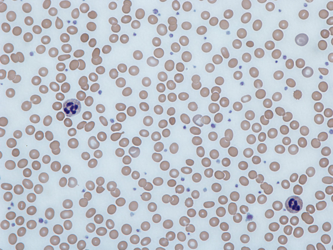
- Echinocytes or Burr Cells. ...
- Tear drop cells or Dacrocytes . ...
- Helmet cells or Schistocytes . ...
- Bite cells or Degmacytes. ...
- Elliptocytes. ...
- Spherocytes. ...
- Macro-ovalocytes
- Target Cells. ...
- Sickle Cells
What are the four most common RBC inclusions?
The four most common RBC inclusions are Howell-Jolly bodies, Pappenheimer bodies, Heinz bodies and basophilic stippling. Howell-Jolly Bodies are remnants of nuclear DNA in red blood cells. During the maturation process, erythrocytes exude their nucleus in the bone marrow just before becoming reticulocytes.
What does the presence of red blood cell inclusions indicate?
The presence of red blood cell inclusions in a peripheral blood smear can indicate an increased amount of inclusionary bodies, so much that the spleen can not keep up by removing them all. This is where the presence of red cell inclusions may indicate a disease state.
What are erythrocyte inclusions?
Erythrocyte inclusions are elements that may be present in red blood cells (RBCs). The appearance, composition, and associated physiology of the inclusions are specific for each type of inclusion. Identification and reporting of these inclusions are important because their presence may indicate diseases or disorders.
What are the Purple inclusions in red blood cells?
They are tiny, light purple, beaded inclusions found along the periphery of the red blood cells. Prussian Blue staining is often used to confirm this. Be careful not to mix these up with Gram-positive cocci bacteria, which look similar. They are associated with the following conditions:
What does RDW mean in erythrocytes?
Where are the tiny purple inclusions found?
What is sickle cell disease?
What is the central pallor zone of a red blood cell?
What is the name of the red blood cells that are spiked?
What happens when sickle cell disease causes anemia?
See more
About this website
What do you mean by cell inclusion?
Cell inclusions are considered various nutrients or pigments that can be found within the cell, but do not have activity like other organelles. Examples of cell inclusions are glycogen, lipids, and pigments such as melanin, lipofuscin, and hemosiderin.
What is the most common red cell inclusion body seen in megaloblastic anemia?
Red Blood Cell Inclusions and Associated ConditionsInclusionCommentsHowell-Jolly bodyMay see more than one inclusion per cell in megaloblastic anemia.Nucleated red blood cellTypically, circulating nucleated RBCs are at the orthochromic stage of maturation, as shown in the image to the left.4 more rows
What are RBC missing?
Explanation: Mature red blood cells lack a nucleus.
What are the types of RBC abnormalities?
There are many different types of red blood cell disorders, including:anemia.red cell enzyme deficiencies (e.g. G6PD)red cell membrane disorders (e.g. hereditary spherocytosis)hemoglobinopathies (e.g. sickle cell disease and thalassemia)hemolytic anemia.More items...
What causes RBC inclusions?
Causes include lead poisoning, hemolytic anemia, and pyrimidine 5 nucleotidase deficiency. Pyrimidine 5 nucleotidase is the enzyme responsible for degradation of the RNA material. Heinz bodies: These are denatured globin. They require supravital staining for their demonstration.
Why is it important to report red blood cell inclusions?
Erythrocyte inclusions are elements that may be present in red blood cells (RBCs). The appearance, composition, and associated physiology of the inclusions are specific for each type of inclusion. Identification and reporting of these inclusions are important because their presence may indicate diseases or disorders.
What cancers cause low red blood cell count?
Certain types of cancer. Leukemia, lymphoma, and multiple myeloma damage bone marrow. Also, cancer that spreads to the bone or bone marrow may crowd out healthy red blood cells.
Is low red blood count serious?
When you don't have enough healthy red blood cells, you have a condition called anemia. This means your blood has lower than normal hemoglobin (Hgb) levels. Hemoglobin is the part of the red blood cell (RBC) that carries oxygen to all the cells in your body. Anemia is a common side effect in patients with cancer.
What does low RBC mean?
A low red blood cell count means you have anemia, a condition that could be caused by a variety of factors like blood loss, genetic disorders, cancer treatments and other causes. Discovering anemia is often the starting point to diagnosing an underlying condition.
What are the 3 most common blood disorders?
Common blood disorders include anemia, bleeding disorders such as hemophilia, blood clots, and blood cancers such as leukemia, lymphoma, and myeloma.
What is the most common disorder of the red blood cells?
Anemia is the most common blood disorder. The body does not have enough red blood cells and is unable to deliver enough oxygen around the body.
What is the most common blood disorder?
Anemias, where there are not enough red blood cells or the cells do not work correctly, are among the most common blood disorders. According to the American Society of Hematology, anemia affects more than 3 million Americans.
Red Blood Cell Inclusions and Associated Conditions
The page below is a sample from the LabCE course Red Cell Disorders: Peripheral Blood Clues to Nonneoplastic Conditions (retired 5/26/2022).Access the complete course and earn ASCLS P.A.C.E.-approved continuing education credits by subscribing online.
Red Cell Inclusion Bodies | Blood Film - MedSchool
Common Red Cell Inclusions; Howell Jolly bodies - DNA fragments Seen post splenectomy and in functional hyposplenism; Basophilic stippling - RNA fragments Seen in haemoglobinopathies and heavy metal poisoning; Pappenheimer bodies - clumps of ferritin Seen post splenectomy, in sideroblastic anaemia and in lead poisoning; Cabot ring - strings of mitotic spindle remnants Seen in megaloblastic ...
Inclusions – eClinpath
Siderocytes are anucleate erythrocytes with iron-containing (siderotic) cytoplasmic inclusions. The inclusions can be due to aggregates of iron in the cytoplasm or within mitochondria (the latter are called Pappenheimer bodies). A nucleated erythoid cell with the same inclusions (usually within mitochondria) is termed a sideroblast. Siderocytes should be distinguished from basophilic stippling ...
Why does iron accumulate in RBC?
Iron may accumulate in RBC if hemoglobin production is inhibited. This can occur with drugs (e.g. chloramphenicol), lead poisoning, and vitamin B6 and copper deficiency in pigs (B6 is required for heme synthesis and copper is also required for release of iron from macrophages and enterocytes).
Why is it important to identify red blood cells?
Correct identification of these abnormalities is important since it can provide insights into metabolic, physiologic, and pathologic conditions affecting the red blood cells.
What is hemolytic anemia?
Diagnosis of an oxidant-induced hemolytic anemia is based on finding a regenerative anemia with characteristic red blood cell morphologic abnormalities of Heinz bodies and eccentrocytes. Some causes of oxidant injury also cause intravascular red blood cell lyse (rupturing of red blood cell membranes) and manifest with hemoglobinemia and hemoglobinuria. Some animals may also develop methemoglobinemia (brown plasma) from oxidant-induced injury to the iron in hemoglobin (reduced iron or Fe 2+ is converted to oxidized iron or Fe 3+ in the porphyrin of hemoglobin, producing methemoglobin). Note, that methemoglobemia, Heinz bodies or eccentrocytes may dominate in a particular condition. Causes of oxidant-induced hemolytic anemia are: 1 Inherited disorders: Enzyme deficiencies (necessary for enzyme function) in pathways that guard against oxidant injury can result in oxidant-induced hemolytic anemia, e.g. Glucose-6-phosphate dehydrogenase (G6PD) in horses, or methemoglobinemia, e.g. flavin adenine dinucleotide (FAD) deficiency in horses. 2 Toxins: Wilted red maple leaves (horses, camelids), Pistachia species (horses), Brassica spp. (kale, turnips; cattle), zinc (dogs ingesting pennies minted after 1982), copper (sheep), skunk musk (dogs, red panda), naphthalene (dogs), nitrate (cattle), onions. 3 Drugs : Acetaminophen, vitamin K1, phenothiazine drenches, benzocaine, propofol. These act as oxidants. 4 Mineral deficiency: Selenium is necessary for the function of enzymes that protect against oxidant injury. Selenium deficiency in cattle can result in oxidant injury to red blood cells.
Why are red and white cells important?
Red and white cell inclusions are important to report as part of the patient’s peripheral smear evaluation. You may need to submit the slides for review by the Pathologist to confirm if you are unsure of what you are seeing.
What is the purpose of reviewing a peripheral blood smear?
Reviewing a peripheral blood smear requires active searching and documentation of morphological characteristics of red and white blood cells. In addition to quantity, size, and shape, the laboratory scientist must identify unusual cellular inclusions observed on the slide, and note them on the laboratory report.
What does RDW mean in erythrocytes?
Anisocytosis means that a patient's erythrocytes are not of equal size. Red Cell Distribution Width (RDW) is a measurement of anisocytosis. If the RDW is >14.5%, this indicates a heterogenous population of RBC's, which means you will likely see a variety of sizes of RBC's on the slide.
Where are the tiny purple inclusions found?
They are tiny, light purple, beaded inclusions found along the periphery of the red blood cells. Prussian Blue staining is often used to confirm this.
What is sickle cell disease?
Sickle cell disease is actually a group of genetic blood disorders. Sickle cell anemia is the most common type of sickle cell disease. Oxygen-carrying hemoglobin found in red blood cells is abnormal and misshapen, resulting in the rigid sickled red blood cells.
What is the central pallor zone of a red blood cell?
Hypochromia means that the central pallor zone of the red blood cell is pale . This central area must be > than 1/3rd of the diameter of the cell before it is termed hypochromic . The MCHC is a gage of hypochromia unless only a few hypochromic cells are seen in the slide. It is associated with: iron-deficiency anemia.
What is the name of the red blood cells that are spiked?
For this reason, they are also called Burr cells. They have short, evenly spaced projections. This condition is reversible, and more often than not, it is a side effect of the EDTA anticoagulant coating in the vacutainer used to collect to blood to prevent it from clotting. These cells are crenated (shrunken). Sometimes, they are associated with disorders such as:
What happens when sickle cell disease causes anemia?
Sickle Cell Crisis: Sickle cell crisis results in pain, anemia, edema, organ problems or failure, infections, and can also lead to stroke due to clogging up of the blood vessels with the sickled cells. A shortened life span may occur in individuals with sickle cell disease.