Retroperitoneal Tumor
- Vascular Tumors. ...
- The abdominal wall, peritoneum and retroperitoneum. ...
- Retroperitoneal Diseases. ...
- Diagnostic and Interventional Uroradiology. ...
- Intestinal Regeneration and Adaptation Models. ...
- Hypoglycemia. ...
What is the most common retroperitoneal tumor?
Pathology. Liposarcomas are the most common primary retroperitoneal tumor. They comprise 10–15% of all soft tissue sarcomas. They are most commonly seen during the fifth to sixth decade of life and can grow to be quite large in size.
Where is a retroperitoneal tumor?
Sarcoma is a rare type of cancer that develops from the body's connective tissues, such as fat, muscle, blood vessels and fibrous tissue. About 20% of sarcomas develop in the back of the abdomen, also known as the retroperitoneum, next to the kidneys.
What are the symptoms of retroperitoneal cancer?
Retroperitoneal may cause the following symptoms:Abdominal mass.Loss of appetite or weight loss.Early satiety (feeling full after eating only a small amount)Blood in stools.Lower extremity swelling.Pain.
What does retroperitoneal mass mean?
Retroperitoneal masses constitute a heterogeneous group of lesions, originating in the retroperitoneal spaces, that pose a diagnostic challenge for radiologists(1). The majority of cases are malignant tumors, of which approximately 75% are mesenchymal in origin(2-4).
How long can you live with retroperitoneal?
Approximately 10–20% of these cases are derived from the retroperitoneum with a prevalence of ∼0.3–0.4 cases/100,000 inhabitants [3]. The prognosis for patients with retroperitoneal sarcomas (RPS) is relatively poor with a 36–58% 5-year overall survival (OS) and a natural history characterized by late recurrences [4].
Is retroperitoneal tumors cancerous?
Most retroperitoneal tumors are mesodermal in origin and can arise from any tissue type present in the retroperitoneum. They can be benign or malignant (4).
Can a retroperitoneal mass be removed?
The surgical approach for the treatment of benign retroperitoneal tumors presenting with vascular invasion should be successful in fully removing the tumors, whilst effectively protecting blood vessels; therefore, the surgical approach may significantly lower the incidence of post-operative complications and reduce the ...
Is retroperitoneal mass curable?
Abstract: Retroperitoneal sarcomas (RPS) are rare cancers that often reach massive size before detection. The mainstay of treatment for RPS is surgical resection, and complete resection is the only chance for potential cure.
How is retroperitoneal mass treated?
Complete surgical resection is the only potential curative treatment modality for retroperitoneal sarcomas and is best performed in high-volume centres by a multidisciplinary sarcoma team. Local recurrence occurs in a large proportion of patients.
What causes retroperitoneal masses?
Most primary retroperitoneal neoplasms arise from the mesodermal system with liposarcoma, leiomyosarcoma and malignant fibrous histiocytoma together, accounting for greater than 80 % of primary retroperitoneal sarcomas. The remaining primary retroperitoneal masses arise predominantly from the nervous system [1].
What does retroperitoneal mean in medical terms?
(REH-troh-PAYR-ih-toh-NEE-ul) Having to do with the area outside or behind the peritoneum (the tissue that lines the abdominal wall and covers most of the organs in the abdomen).
What is the value of a biopsy in a retroperitoneal tumor?
Image-guided retroperitoneal biopsy has proven to be low cost, accessible, and a reliable procedure (in terms of diagnostic accuracy), usually associating with a low rate of complications and a low risk of tumor seeding.
Can a retroperitoneal mass be removed?
The surgical approach for the treatment of benign retroperitoneal tumors presenting with vascular invasion should be successful in fully removing the tumors, whilst effectively protecting blood vessels; therefore, the surgical approach may significantly lower the incidence of post-operative complications and reduce the ...
Is retroperitoneal mass curable?
Abstract: Retroperitoneal sarcomas (RPS) are rare cancers that often reach massive size before detection. The mainstay of treatment for RPS is surgical resection, and complete resection is the only chance for potential cure.
Where are the retroperitoneal lymph nodes?
The lymph nodes in the back of the abdomen are called retroperitoneal lymph nodes. An RPLND is also called a retroperitoneal lymphadenectomy. The lymph nodes in the retroperitoneum lie around the large blood vessels at the back of the abdomen. The lymph nodes are part of the lymphatic system.
How fast does retroperitoneal liposarcoma grow?
The CT features of recurrent liposarcoma are similar to those of the initial manifestation; recurrent liposarcoma shows rapid growth, with a mean tumor volume doubling time of 98 days (range, 46–151 days; median, 104 days).
What is retroperitoneal tumor?
The presence of a retroperitoneal tumor can compress the lymphatic drainage from the splanchnic bed, causing obstruction, increased pressure in the system , and leakage of protein from intestinal lymphatics into the gut lumen. This phenomenon has been reported as a complication of several tumors, including neuroblastoma,62-64 metastatic melanoma,65,66 and others. Rarely, intestinal tumors, both benign and malignant, can present with PLE, including lymphoma,67-70 leukemic infiltration,71 lymphangioma,72 hemangioma, 73,74 Kaposi sarcoma,75,76 melanoma, 77 polyps, 78-80 and posttransplant lymphoproliferative disease. 81 The diagnosis of these tumors may require a high index of suspicion, because their manifestation may be primarily gastrointestinal. PLE secondary to lymphangiectasia induced by chemotherapy has also been reported.82
What are the pathologic features of retroperitoneal tumors?
Retroperitoneal tumors are often very infiltrative and may invade the pancreas, spleen, lymph nodes, intestines, and even the deep soft tissue of the abdominal wall.360 ,361 Deep-seated somatic soft tissue tumors have similarly infiltrative margins, whereas those involving only the skin and subcutaneous tissue are often ...
Why do you need to reduce blood flow through vascular renal or retroperitoneal tumors prior to surgery?
Decrease blood flow through vascular renal or retroperitoneal tumors prior to surgery to facilitate surgical removal and minimize blood loss.
What is RL lymphoma?
RL is the most common retroperitoneal malignancy, with either Hodgkin's or non-Hodgkin's lymphoma invading the retroperitoneal lymph nodes. Hodgkin's lymphoma tends to involve the spleen and retroperitoneum with a contiguous spread pattern. Non-Hodgkin's lymphoma more often involves a variety of nodal groups, with a predilection for mesenteric nodes and extranodal sites. Overall, RL most commonly involves para-aortic, aortocaval, and retrocaval nodal groups. Nodes tend to measure greater than 1.5 cm in short-axis diameter with bilateral involvement. Often, a confluent nodal mass encircles the aorta and IVC, displacing the aorta from the spine (Fig. 4-71 ). Monotonous features with mild homogeneous hyperintensity on fluid-sensitive sequences, mild enhancement with relative lack of mass effect (even despite large size), and extensive retroperitoneal involvement typify lymphoma.
What is the effect of retroperitoneal tumors on the gut?
Tumors. The presence of a retroperitoneal tumor can compress the lymphatic drainage from the splanchnic bed , causing obstruction, increased pressure in the system, and leakage of protein from intestinal lymphatics into the gut lumen.
What is RL in a neoplasm?
RL is the most common retroperitoneal malignancy, with either Hodgkin's or non-Hodgkin's lymphoma invading the retroperitoneal lymph nodes.
What is a malignant fibrous histiocytoma?
Malignant fibrous histiocytoma (MFH) was first described in 1963 and is now the most common contemporary reported soft tissue sarcoma. These lesions are most frequently seen on the extremities, less commonly occurring in the retroperitoneum. There are several subtypes of MFH including storiform-pleomorphic, myxoid, giant cell, and inflammatory. Several histologic subtypes can occur in the same lesion. The storiform-pleomorphic pattern is most common (40–60% of all MFH) and is histologically identified by its collagen pattern of curling fascicles of cells. Fibrosarcomas, as their name implies, are malignant tumors of fibroblast origin. They are less frequently reported than in past series because they are now often classified as MFH.
Is surgical resection a curative treatment?
Conclusions: Complete surgical resection is the only potential curative treatment modality for retroperitoneal sarcomas and is best performed in high-volume centres by a multidisciplinary sarcoma team. The ability completely to resect a retroperitoneal sarcoma and tumour grade remain the most important predictors of local recurrence and disease-specific survival.
Is retroperitoneum a benign tumor?
Introduction: The retroperitoneum can host a wide spectrum of pathologies, including a variety of rare benign tumours and malignant neoplasms that can be either primary or metastatic lesions. Retroperitoneal tumours can cause a diagnostic dilemma and present several therapeutic challenges because of their rarity, relative late presentation and anatomical location, often in close relationship with several vital structures in the retroperitoneal space.
How to classify a mass as retroperitoneal?
In order to classify a mass as primary retroperitoneal, the location should be determined as within the retroperitoneal space and an organ of origin is excluded. Displacement of normal retroperitoneal organs or large vessels in the space strongly suggests that a mass is retroperitoneal in location (Fig. 6.2) [ 3 ]. Several radiological signs have been described to assist in determining the organ of origin. They include claw or beak sign, invisible or phantom organ sign, and embedded organ sign (Fig. 6.3) [ 3, 12, 13 ]. The claw or beak sign is positive when a mass causes the edge of an adjacent organ into a beak shape, meaning that the mass originates from that organ. Conversely, if a mass originates from a structure adjacent to the organ, it will form obtuse angles to abut and compress the organ. The invisible or phantom organ sign is positive when a large mass arises from a small organ that then becomes undetectable. The embedded organ sign is positive when a mass that arises from a given organ often appears embedded within it and the interface between the two may be difficult to appreciate [ 3 ]. Conversely, a mass that abuts but does not originate from a hollow structure compresses it and produces a crescentic deformity. Rounded rather than beaked edges of an adjacent organ (negative beak sign) with a crescentic deformation (negative embedded organ sign) by the tumor suggest a primary retroperitoneal tumor [ 3 ].
What is extraskeletal osteosarcoma?
Extraskeletal osteosarcoma is a rare malignant mesenchymal tumor characterized by the direct production of osteoid or bone by tumor cells without primary bone or periosteal involvement . The most common location of these tumors is the lower extremity, especially the thigh, followed by the upper extremity and the retroperitoneum [ 38 ]. Imaging shows a large soft tissue with areas of amorphous calcifications, necrosis, or old hemorrhage (Fig. 6.10 ). The imaging findings of extraskeletal osteosarcoma are nonspecific, with considerable overlap with other retroperitoneal masses. A wide range of differential diagnoses include several benign and malignant conditions that show mineralization, which include nontumoral benign conditions such as calcified hematoma or myositis ossificans and numerous benign and malignant retroperitoneal tumors.
What is a leiomyosarcoma?
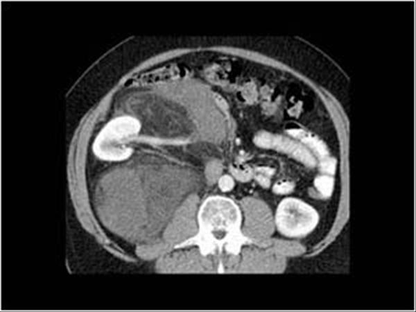
Leiomyosarcomas are the second most common primary retroperitoneal sarcoma, accounting for 28 % of all cases [ 22 ]. They arise from smooth muscle elements within the retroperitoneal muscle tissue, blood vessels, or Wolffian duct remnants [ 1, 19 ]. Leiomyosarcoma is more common in women, in the fifth to sixth decades of life [ 32 ]. They can grow to a large size (>10 cm) before compromising adjacent organs and precipitating clinical symptoms such as venous thrombosis [ 1 ]. Histopathologically, this tumor has large areas of necrosis and cystic degeneration, but calcification is uncommon [ 1 ]. They are large, well-circumscribed masses, which is isoattenuating to muscle on CT scan (Fig. 6.6 ). Contrast enhancement is often heterogeneous and predominantly peripheral. Small tumors may be homogeneously solid, but large tumors have extensive areas of low attenuation and represent necrosis and cystic degeneration. Rarely, leiomyosarcoma may appear as mostly cystic. At MR imaging, these tumors have intermediate to low signal intensity on T1-weighted images and intermediate to high signal intensity on T2-weighted images, depending on the amount of necrosis [ 1 ]. Mixed signal intensity and a fluid–debris level can be seen in hemorrhagic lesions [ 1 ]. The presence of extensive necrosis in a retroperitoneal mass, with contiguous involvement of a vessel, is highly suggestive of leiomyosarcoma [ 1 ]. Metastasis to the liver, lungs, or lymph nodes occurs late in the course of the disease [ 22, 32 ].
What is a dedifferentiated liposarcoma?
Dedifferentiated liposarcomas are high-grade tumors with poor prognosis [ 29 ]. This subtype is defined by the presence of sharply demarcated regions of non-lipogenic sarcomatous tissue within a well-differentiated tumor [ 16 ]. At CT and MR imaging, these dedifferentiated tumors appear as more heterogeneous tumors with both fat and solid components (Figs. 6.4b and 6.5) [ 1 ]. There may be no evidence of fat in up to 20 %, which makes the diagnosis difficult based on imaging alone [ 28 ]. Calcification is seen in as many as 30 % of these tumors and is an important sign of dedifferentiation [ 1, 30 ]. Variable signal intensity and enhancement of the solid portion may be seen [ 30 ].
What is the most common soft tissue sarcoma in children?
Rhabdomyosarcoma is the most common soft tissue sarcoma in children. The tumor is believed to arise from primitive muscle cells, but tumors can occur anywhere in the body. The retroperitoneum is involved in 7 % of cases diagnosed with rhabdomyosarcoma [ 36 ]. Cross-sectional imaging shows a mass with heterogeneous attenuation or signal intensity, focal calcifications, necrosis, and significant contrast enhancement.
Can retroperitoneal tumors be seen on radiographic imaging?
Although there are significant overlaps in the imaging characteristics of retroperitoneal tumors, the radiographic appearances can offer clues as to the histologic subtype and grade, which may guide decisions. Some lesions have distinctive characteristics and can be diagnosed with some accuracy on imaging. It is also possible to narrow the differential diagnosis of a retroperitoneal mass based on certain imaging characteristics in combination with the pattern of involvement and demographics [ 5 ].
Where can a solitary fibrous tumor occur?
Visceral pleura is the most common site of solitary fibrous tumor occurrence, but it can occur outside of the thoracic cavity. Retroperitoneal solitary fibrous tumors are rare, and their clinical manifestations depend on the location and size of the tumor. They grow slowly in an expansive way and displace adjacent structures by compression. For extrapleural solitary fibrous tumor, there is now complete omission of the prior synonym “hemangiopericytoma” according to the WHO classification of soft tissue tumors [ 16, 17 ].
Retroperitoneal Tumors
Retroperitoneal tumors are relatively rare lesions yet of interest to the urologist because they present in a key anatomic location and often involve urologic organs. Because of their location these lesions usually demonstrate indolent growth and present as relatively large lesions.
Incidence and Etiology
Retroperitoneal sarcomas are relatively rare lesions that comprise nearly 15% of the soft tissue sarcomas that occur annually and thus account for approximately 2000 cases per year (Jemal, 2009). Surveillance, Epidemiology, and End Results (SEER) data from the U.S.
Pathology
Retroperitoneal sarcomas arise primarily from soft tissues of fibrous and adipose origin as well as muscle, nerve, and lymphatic tissue. These tissues are derived from primitive mesenchyme from the mesoderm with some contribution from neuroectoderm (Economou, 1987).
Benign Lesions
Lipomas consist almost entirely of mature fat and are uncommonly found in the retroperitoneum. They are probably the most common soft tissue tumor in humans. Most of these lesions occur superficially, but they may occur in other areas, such as the retroperitoneum.
Malignant Lesions
Liposarcomas are among the most common of primary retroperitoneal tumors and are distinguished by their often large dimensions and range of subtypes. These lesions have their peak incidence between ages 40 and 60 (Kindblom et al, 1975). They account for 10% to 15% of sarcomas, and approximately 20% of these lesions arise in the retroperitoneum.
Diagnosis
Because of their slow growth and anatomic location, retroperitoneal tumors (usually sarcomas) tend to grow to a large size before they are detected.
What Are The Types of Retroperitoneal Mass or Tumor?
Retroperitoneal diseases and types of retroperitoneal mass or tumor are as follows-
Which malignant neoplasm is most common in retroperitoneal space?
a) Mesenchymal Neoplasm- Most common mesenchymal malignant neoplasm observed in retroperitoneal space is Liposarcoma and Leomyosarcoma.
What Are The Investigations to Diagnose Retroperitoneal Mass or Tumor?
CT Scan- Provides better picture than plain X-Ray and understanding of the tumor mass.
What is the purpose of ultrasound for a tumor?
Ultrasound- Ultrasonography helps to diagnose visualized retroperitoneal tumor mass and evaluate external damage of retroperitoneal organ like pancrease, kidney, ureter and bladder.
What is the space behind the peritoneum called?
Space behind the peritoneum in abdominal cavity is known as retroperitoneal space or retroperitoneum. Retroperitoneal organs are covered anteriorly (in front) by peritoneum and posteriorly by posterior (back) abdominal wall. Retroperitoneal space is divided in to upper and lowers retroperitoneal space. Organs and viscera are confined in either one ...
Where does a malignant tumor come from?
Malignant tumor derives from fibrous connective tissue of retroperitoneal space. The grades of neoplasm or malignancy is divided as low grade, intermediate or high grade malignancies. Intermediate and high grade tumor metastasize through blood vessels to distant tissue or organs.
What is benign tumor?
Benign tumor is covered by capsule. Growth of the tumor is slow and most of the symptoms are related to complications caused by tumor pressing on surrounding soft tissue. Compression and displacement of viscera causes symptoms of visceral obstruction, ischemia and nerve lesions. Schwannoma- Benign nerve sheath tumor.
What is the most common type of retroperitoneal neoplasm?
Primary retroperitoneal neoplasms are an extremely rare group of tumors ( lymphoma is not included in this definition). The most common type is soft tissue sarcoma (90%).
What is the prognosis of a tumor?
Prognosis depends on the histological type of a tumor and the potential for resection.
How big is a tumor?
Frequently tumors have relatively unimpeded growth where symptoms develop late and the tumor at presentation tending to be extremely large (average size 11-20 cm).
Is lymphoma a primary retroperitoneal neoplasm?
Primary retroperitoneal neoplasms are an extremely rare group of tumors ( lymphoma is not included in this definition). The most common type is soft tissue sarcoma (90%).
Abstract
The retroperitoneum is a large compartmentalized space bounded anteriorly by the posterior parietal peritoneum and posteriorly by the transversalis fascia (Fig. 6.1). The abdominal retroperitoneum is divided by the fascial planes into three distinct compartments: the anterior pararenal, the perirenal, and the posterior pararenal spaces.
Keywords
These keywords were added by machine and not by the authors. This process is experimental and the keywords may be updated as the learning algorithm improves.
