What are siliceous spicules made out of?
Siliceous spicules, found in the Demospongiae and in the Hexactinellida, are made essentially of silicic acid; they also contain some water, a small quantity of other compounds containing sodium, potassium, iron, and chlorine, and a small quantity of organic matter, called spiculin, which forms an… Read More
What are siliceous sponges?
What are siliceous spicules? Siliceous sponge. They are characterized by spicules made out of silicon dioxide, unlike calcareous sponges. Individual siliachoates (silica skeleton scaffolding) can be arranged tightly within the sponginocyte or crosshatched and fused together. Siliceous spicules come in two sizes called megascleres and microscleres.
What are some examples of spicules in plants?
Silica deposition is a fundamental process in sponges. Most sponges in the Classes Demospongiae and Hexactinellida secrete siliceous elements, which can subsequently fuse, interlock with each other, or form three-dimensional structures connected by spongin. The resulting skeletal frameworks allow sponges to grow upwards and facilitate water exchange …
What are the three types of spicules in a calcareous sponge?
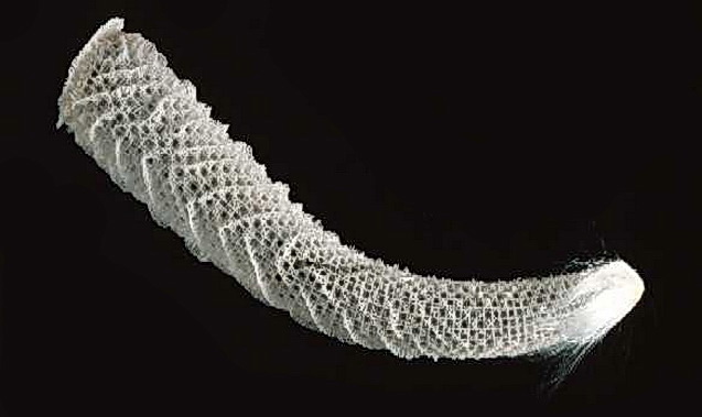
They had been described by Schulze and represent the phylogenetically oldest class of siliceous sponges (phylum Porifera); they are eye-catching because of their distinct body plan, which relies on a filigree skeleton. It is constructed by an array of …

What is calcareous or siliceous spicules?
Calcareous spicules are formed from crystalline calcite, and siliceous types from opaline silica. Calcareous spicules are needle-like, or stellate with three or four radiating needle-like rays. Siliceous spicules are needle-like, stellate with three to many rays, or of special symmetrical or irregular shapes (cf.
What class has siliceous spicules?
DemospongiaeThe siliceous sponges form a major group of the phylum Porifera, consisting of classes Demospongiae and Hexactinellida. They are characterized by spicules made out of silicon dioxide, unlike calcareous sponges.
What is siliceous Porifera?
siliceous sponge, any sponge in which the main skeletal component is silica as opposed to calcium carbonate or fibrous organic materials only. More than 95 percent of all known sponge species have a siliceous skeleton and belong to the class Demospongiae (phylum Porifera).
What are the different types of spicules?
According to the size, spicules are classified into two major types: Megascleres, which are large-sized and constitute main supporting framework of sponge body and Microscleres, which are smaller in size and occur in the mesenchyme.
What is the Pinacocytes function?
Pinacocytes are part of the epithelium in sponges. They play a role in movement (contracting and stretching), cell adhesion, signaling, phagocytosis, and polarity. Pinacocytes are filled with mesohyl which is a gel like substance that helps maintain the shape and structure of the sponge.
What are porocytes and their functions?
In the Calcarea, the outer surface of the body also contains flattened granular cells called porocytes because they contain the pores needed to allow water into the sponge. The porocytes can contract, thus closing the pores during unfavourable environmental conditions.
Which amongst organism has siliceous skeleton over?
Marine Silica Cycle* As mentioned above, the primary biota that construct siliceous skeletons are diatoms, radiolaria, silicoflagellates, and siliceous sponges. Diatoms are marine algae.
What example of sponge has an intricate network of siliceous spicules?
hexactinellid spongesThe hexactinellid sponges are characterized by siliceous spicules that display hexactinic, triaxonic (cubic) symmetries, or morphologies derived by reduction from the basic building plans of the spicules.
Why did sponges develop skeletons?
Sponges have an internal skeleton that gives them support and protection. An internal skeleton is called an endoskeleton. A sponge endoskeleton consists of short, sharp rods called spicules (see Figure below). Spicules are made of silica, calcium carbonate, or spongin, a tough protein.Jul 3, 2019
Are sponges microscopic?
Coming in many sizes and shapes, sponge bodies are a loose assemblage of cells held together by a special protein called collagen which is present in all animals. In addition, sponges have microscopic crystalline spicules that act as a skeleton.
What is the difference between sponges and spicules?
Generally, species are identified based on the presence or absence of spongin in a sample. Spicules are the structural components of a sponge, or the "bricks," and the shapes, sizes, and composition are unique for each species.Sep 28, 2016
What are 3 types of spicules in Porifera?
Based on the number of axis present in the rays spicules may be of three types: monoaxon, triaxon and polyaxon. Monaxon: These spicules grow along a single axis. These may be straight needle-like or rod-like or may be curved. Their ends may be pointed, knobbed or hooked.
Abstract
Pottery was a traditional art and technology form in pre-colonial Amazonian civilizations, widely used for cultural expression objects, utensils and as cooking vessels. Abundance and workability of clay made it an excellent choice. However, inferior mechanical properties constrained their functionality and durability.
Introduction
The concept of combining materials with different properties such as composites goes back to prehistoric times. Given its natural abundance and workability, clay has been the material of choice for producing utilitarian pottery and cultural expression ornaments, as extensively documented by archaeological records 1.
Methods
Spicules were kindly provided by Dr. Renato Batel (Ruđer Bošković Institute, Rovinj, Croatia) and cleaned from the tissue by using a previously described procedure 16. Infrared analysis was performed on a Nicolet Nexus spectrometer fitted with a Golden Gate attenuated total reflection (ATR) accessory (Thermo Nicolet).
Additional Information
How to cite this article: Natalio, F. et al. Siliceous spicules enhance fracture-resistance and stiffness of pre-colonial Amazonian ceramics. Sci. Rep. 5, 13303; doi: 10.1038/srep13303 (2015).
Acknowledgements
This work was partially supported by the DFG within the SPP 1420 and Forschungsschwerpunkt Nano-strukturierte Materialien (NWG IV: Bioanorganische Chemie) Ministerium für Wissenschaft und Wirtschaft des Landes Sachsen-Anhalt. S.W. was supported by Boehringer Ingelheim (Ingelheim, Germany).
Rights and permissions
This work is licensed under a Creative Commons Attribution 4.0 International License.
About this article
Natalio, F., Corrales, T., Wanka, S. et al. Siliceous spicules enhance fracture-resistance and stiffness of pre-colonial Amazonian ceramics. Sci Rep 5, 13303 (2015). https://doi.org/10.1038/srep13303
How many phases are there in spicule formation?
Based on the electron microscopical studies, and especially using the primmorph system, it can be deduced that the process of spicule formation can be divided into two phases; the intracellular initial steps and the extracellular final and shaping phase.
When were sponge spicules first described?
First descriptions of sponge spicules were given by Donati (1753) . He isolated them from a species belonging to the genus Geodia ( Alcyonium) ( Fig. 2 A). Distinct cytological studies on the spicule formation were first published in 1856 ( Lieberkühn, 1856) with the freshwater sponge S. fluviatilis (Demospongiae) as a model. This work gave even detailed analyses on the development and differentiation of fertilized eggs, together with the differentiation stages of somatic sponge cells from the ‘Schwärmsporen’ to the spicule-forming sclerocytes ( Fig. 2 B). Spicule formation starts intracellularly mainly within the ‘Schwärmsporen’ but to a smaller extent also in other cell types ( Fig. 2 C). It is interesting to note that spicule-forming cells have a large nucleus and nucleolus ( Fig. 2 C). A few years later, DeLage (1892) observed likewise precisely that the spicules are formed intracellularly. Very notably he mentioned that the spicule-containing cell ‘est de même nature que les amoeboïdes’. This remark can also be taken as an indication that he had been aware of the new stem cells concept that originates from this time ( Weismann, 1892 ).
Why are skeletal elements formed in all metazoan phyla?
Skeletal elements are formed in all metazoan phyla to stabilize the body and to allow growth. In triploblasts, two different strategies have been followed. While in Protostomia an exo-skeleton (integument) which is made of organic molecules (chitin), especially well developed in Arthropoda/Antennata, is formed, Deuterostomia form their the skeletal structures (bones) from the somatic mesoderm. In diploblasts, in coelenterates, the basic inorganic biomineral is calcium carbonate.
What are the body plans of all metazoan animals?
Common to all metazoan body plans is the formation of at least one axis that runs from the apical to the basal region; examples for this type of organization are the Porifera and the Cnidaria (diploblastic animals). It seems conceivable that the basis for the formation of the Bauplan in sponges is the construction of their skeleton by spicules. In Demospongiae (we use the model species Suberites domuncula) and Hexactinellida, the spicules consist of silica. The formation of the spicules as the building blocks of the skeleton, starts with the expression of an enzyme which was termed silicatein. Spicule growth begins intracellularly around an axial filament composed of silicatein. When the first layer of silica is made, the spicules are extruded from the cells and completed extracellularly to reach their the final form and size. While the first steps of spicule formation within the cells are becoming increasingly clear, it remains to be studied how the extracellularly present silicatein strings are formed. The understanding of especially this morphogenetic process will allow an insight into the construction of the amazingly diverse skeleton of the siliceous sponges; animals which evolved between two periods of glaciations, the Sturtian glaciation (710–680 MYA) and the Varanger-Marinoan ice ages (605–585 MYA). Sponges are—as living fossils—witnesses of evolutionary trends which remained unique in the metazoan kingdom.
What is the oldest phylum of the metazoan?
Sponges, as the oldest still extant metazoan phylum, are characterized by a simple Bauplan (reviewed in Müller et al., 2004; Müller, (2005) [these reviews give more detailed literature reference]). They are filter-feeding organisms that are sessile. Their body is composed of an epithelial layer which surrounds a mesogleal compartment, the mesohyl; this is reticulated in a highly organized manner by a canal system. The major structural and functional novelties, which evolved during the major transitions to the Porifera and Cnidaria are summarized in Fig. 3.
What are the three classes of homeobox genes?
The genes which are involved in the establishment of the head organizer during embryogenesis have been grouped into three classes of homeobox genes, the Paired-class, the Antennapedia-class and the Lim-class genes. Also in this area a strong progress was made in sponges in the last few years. A paired-class ( Pax-2/5/8 )-gene had been isolated from the freshwater sponge E. fluviatilis, which encodes a complete although substantially degenerated homeodomain. In S. domuncula a cDNA encoding a LIM/homeobox protein has been isolated which comprises high sequence similarity to the related LIM homeodomain proteins in other animals.
What is the WNT pathway?
The Wnt signaling pathway is a cell communication system which regulates cell-fate decisions, tissue polarity and morphogenesis. The Frizzled protein is the membrane receptor for the Wnt secreted glycoproteins. Through the canonical Wnt signaling pathway, the activated Frizzled binds to Dishevelled (Dsh), which leads to the stabilization and accumulation of ß-catenin in the nucleus, where it activates the TCF/LEF transcription factor. Very recently we isolated the gene encoding the Frizzled receptor from S. domuncula, suggesting that a Wnt-pathway involved in cell-fate determination and morphogenesis was already established in sponges.
How are spicules formed?
Spicules are formed by sclerocytes, which are derived from archaeocytes. The sclerocyte begins with an organic filament, and adds silica to it. Spicules are generally elongated at a rate of 1-10 μm per hour. Once the spicule reaches a certain length it protrudes from the sclerocyte cell body, but remains within the cell’s membrane. On occasion, sclerocytes may begin a second spicule while the first is still in progress.
What are sponge spicules made of?
Sponge spicule. For other uses, see Spicule (disambiguation). Spicules are structural elements found in most sponges, made of calcium carbonate or silica. Large spicules visible to the naked eye are referred to as megascleres, while smaller, microscopic ones are termed microscleres.
What are megascleres?
Megascleres are large spicules meas uring from 60-2000 µm and often function as the main support elements in the skeleton. Acanthostyles are spiny styles. Anatriaenes, orthotriaenes and protriaenes are triaenes - megascleres with one long and three short rays. Strongyles are megascleres with both ends blunt or rounded.
What are the two types of biota that make up siliceous skeletons?
As mentioned above, the primary biota that construct siliceous skeletons are diatoms, radiolaria, silicoflagellates, and siliceous sponges. Diatoms are marine algae. These phytoplankton account for 20–40% of the primary production in the ocean and an even greater percentage of the export production from the photic zone. Diatom skeletons are the primary form of biogenic silica in deposits associated with coastal upwelling areas, high-latitude oceans (predominantly in the Pacific and the Southern Oceans), and the continental margins (Figure 4 ). In equatorial upwelling areas radiolarian skeletons commonly occur in marine sediments along with the diatom frustules. Radiolaria are zooplankton that live in the upper few hundred meters of the water column. Their skeletons are larger and more robust than many diatoms; consequently their preservation in marine sediments is greater than that of most diatoms. Silicoflagellates account for a very small fraction of the biogenic silica in marine sediments because most of them dissolve up in the water column or in surface sediments. They have been used in some continental margin sediments as a paleo-indicator of upwelling intensity. Siliceous sponge spicules can make up a significant fraction of the near-interface sediments in areas in which the sediment accumulation rate is low (<5×10 −3 cm y −1 ). For example, on the Ross Sea continental shelf, fine sediments accumulate in the basins, whereas the topographic highs (<400 m water depth) have minimal fine-grained material (because of strong currents and turbulence). As a result, mats of siliceous sponge spicules occur in high abundance on some of these banks.
Where is silicatein found in sponges?
In siliceous sponges (phylum Porifera; class Demospongiae), the silicatein is located inside the spicules as axial filament in a distinct canal, termed axial canal (see Uriz, 2006 ). There, several isoforms of silicatein have been identified, with silicatein-α, -β and -γ as the main forms in marine sponges ( Shimizu et al., 1998 ), while in the freshwater species, several silicatein-α isoforms exist in the axial filament ( Wiens et al., 2009b ). As mentioned above, the individual silicatein molecules undergo self-assembly to oligomers ( Murr and Morse, 2005 ), a process that proceeds and results in the formation of filaments via fractal intermediates ( Murr and Morse, 2005; Müller et al., 2007a ). The association of the different isoforms is stoichiometrically controlled; four silicatein-α molecules associate and allow one silicatein-β molecule to stick inside this pentamer ( Müller et al., 2007a) in a way that the active centres of the enzyme are exposed to the environment. This association sequence, starting from a chaotic aggregation, leads to fractal-like structures that result in the building of filaments of higher-ordered pattern. Based on diffraction experiments, using a small-angle X-ray scattering approach, it had been found that those patterns are not identical in the different sponge species ( Croce et al., 2003 ). The protein units within the filaments are highly organized and show two different orientations of the lattices formed from silicatein molecules that are spaced by distances of 5.8 nm. The orientations of the lattices are either regular in 2D hexagonal arrangements of protein units in perpendicular direction along the spicule axis (like in Geodia cydonium ), or two different 2D lattices exist in which the repeating protein units are inclined by a defined angle (e.g. in the hexactinellid Scolymastra joubini) ( Croce et al., 2003 ). This view is supported by the fact that the morphologies of the spicules are manifold and filigree and they are characteristic in the appearance for each sponge species (see Uriz, 2006 ).
What are biota made of?
Biota that make their skeletal material out of amorphous silica have occurred in the geologic record since Cambrian time. Siliceous sponges first appeared in the lower Cambrian, whereas radiolaria and diatoms first appeared in the Ordovician and Jurassic, respectively (Blatt et al., 1980 ). Silicoflagellates also form their skeletal material out of amorphous silica (commonly called biogenic silica), but their delicate skeletons are rarely preserved to a significant extent in the sedimentary record ( Treppke et al., 1996 ). Siliceous biota have been studied extensively from biological, paleontological, and geochemical perspectives. Diatoms, a type of unicellular algae ( Figure 1), are responsible for as much as 30–40% of the primary production occurring in the surface ocean, and even a greater percentage of the organic-carbon flux exported from the euphotic zone ( Buesseler, 1998 ). Radiolaria, a unicellular zooplankton, as well as diatoms commonly are used to establish time horizons in paleoceanographic studies of sediment stratigraphy. Geochemists study the burial of biogenic silica in the oceans because it offsets the supply of riverine-dissolved silicate to the ocean that results from continental-rock weathering reactions. Approximately 400 Mt of biogenic silica are buried each year in marine sediments, making this phase second only to calcium carbonate in terms of biogenic sediment burial rate. Prior to the evolution of siliceous biota in the Cambrian, dissolved silicate was likely to have been removed from the oceans via inorganic precipitation of opal-A and opal-CT. Dissolved silicate concentrations in the ocean during the Precambrian may have been as high as several 1,000 μM ( Siever, 1992 ). As siliceous biota became more prevalent in oceanic environments hundreds of millions of years ago, the primary mode of silica precipitation became biogenic and surface-ocean waters (where most of the siliceous biota live) became depleted in dissolved silicate. In the modern ocean, siliceous biota commonly precipitate their skeletal material from surface waters that are 10–100 fold undersaturated with respect to amorphous silica. This situation is very different than that for calcareous biota, which precipitate their skeletons from surface waters that typically are supersaturated by a factor of 2–4.
Where are glass sponges found?
Hexactinellida (glass sponges) are exclusively marine and siliceous sponges largely restricted to the deep sea, with a few notable exceptions, such as massive glass sponge reefs found in SCUBA-accessible depths off the Canadian west coast (e.g. Conway et al., 2001; Krautter et al., 2001; Cook et al., 2008) and population of sublittoral caves in the Mediterranean by one species ( Oopsacas minuta: Vacelet et al., 1994; Bakran-Petricioli et al., 2007 ). Currently, 623 extant species are considered valid according to the World Porifera Database ( Van Soest et al., 2011 ), but because the deep sea is still to a large extent unexplored and vast museum collections await revision by a limited number of experts, this is probably a gross underestimate of the actual diversity of this group ( Reiswig, 2002 ). Glass sponges are remarkably distinct from other sponges in many aspects of their biology (reviewed in Leys et al., 2007 ). In particular, their syncytial tissue organization and triaxonic spicule symmetry clearly distinguish them from the other three major sponge groups and make them one of the best-supported higher-level metazoan monophyla ( Mehl, 1992 ). They also differ from other sponges because they generally have a richer set of morphological characters, displaying a complex skeletal anatomy and a vast array of different spicule types that provide a wealth of information for the taxonomy of the group.
What is the process of inversion of the blastula?
This phenomenon was first discovered in G. compressa and S. raphanus by Duboscq and Tuzet (1935, 1937). They called this process ‘inversion des surfaces’ . It has been described only in the Calcaronea ( Ereskovsky, 2010 ). This process ( Figs 4.16 A (h)– (k) and 4.18 C) begins with the reopening of the blastula mouth, which enlarges to expose the flagellated cells. The embryo then turns inside out through this new opening by the detachment of the two lips of the opening away from each other. The flagellated region invaginates inwards and the lips extend upwards to meet in the opposite pole of the mouth. The result is a volvox blastula, with flagellae directed outwards. This is a growing amphiblastula larva. In L. complicata, before release, the embryo attains the pseudogastrula stage, in which granular cells enter and fill the larval cavity ( Fig. 4.16 A (l)). This stage is transitory, and this temporary invagination has nothing to do with gastrulation. Pseudogastrulation is merely a side effect of the separation of larval cells from the parent choanoderm prior to larval release ( Ereskovsky, 2010 ).
What is the frequency of K strategy?
The frequency of K strategy and the brooding that accompanies it is not restricted only to sea urchins, and nearly all groups have species that adopt strategy. In demosponges (siliceous sponges), more than 80% of southern species are brooders, whereas the proportion is only about 50% in other seas, except the Arctic Ocean, where there is a similar percentage to that of the Southern Ocean [ARN 74b ]. In the solitary coral, Flabellum, the three Antarctic species are brooders, whereas other species of the genus living in the cold North Atlantic waters are broadcast spawners that produce lecithotrophic eggs. As a corollary, Antarctic species produce oocytes about five times larger than those of other species [ WAL 08 ]. Numerous bivalve species protect their young in their mantle cavity or between the gill lamellae. Some are very marked K strategists, with the number of descendants reduced to a few dozen eggs or embryos. Earlier in this chapter ( section 6.4.1 ), the incredibly long incubation periods observed in the isopod Glyptonotus antarcticus and the gastropod Neobuccinum eatoni were mentioned. A very similar situation to that observed in sea urchins is found in other echinoderms, like starfish and brittle stars: around 50% are brooders, a much higher percentage than that observed in other seas, including the Arctic. Many other examples could also be given.
What are calcareous sponges made of?
A common feature is the supporting skeleton, made of calcareous, star shaped structures - or spicules.
What are the characteristics of sponges?
Other, more complex sponges have folded body walls, and many contain branched water canals and several oscula. 1)No definite symmetry. 2)Body multicellular, few tissues, no organs. 3)Cells and tissues surround a water filled space but there is no true body cavity.
What is sponge biology?
Animals in the phyla Calcarea and Silicea are most commonly. known as "sponges". Sponge are among the simplest of animals and are sedentary. Ancient Greeks had actually mistaken them for plants.
How many points does a calcareous sponge have?
These usually have three points, but some species have two or four pointed spicules. The class of the calcareous sponges, Calcarea, includes 400, strictly marine species. Although most of the species are found in tropical waters, there are approximately 10 species along the coast of Norway.
What phylum are sponges in?
Almost all sponges function first as one sex and then as the other. The calcareous sponges of class Calcarea are members of the animal phylum Porifera, the cellular sponges. These organisms are characterized spicules made out of calcium carbonate in the form of calcite or aragonite.
How do sponges reproduce?
In Calcareous sponges, reproduction can be both sexual and asexual, by budding. The eggs hatch to free swimming larvae, which attach themselves to the bottom after a few days. In the case of budding, the sponges produce a cluster of cells known as a gemmule that is covered in a hard coating.
How big are sponges?
Sponges range in size from a few millimeters to a few meters. They live in both fresh and marine waters. Sponges are suspension feeders, meaning that they capture food particles suspended in the water that passes through their body, which in some species resembles a sac perforated with pores.
What is a siliceous sponge?
Siliceous sponge. The siliceous sponges form a major group of the phylum Porifera, consisting of classes Demospongiae and Hexactinellida. They are characterized by spicules made out of silicon dioxide, unlike calcareous sponges .
What phylum are sponges in?
The siliceous sponges form a major group of the phylum Porifera, consisting of classes Demospongiae and Hexactinellida. They are characterized by spicules made out of silicon dioxide, unlike calcareous sponges .