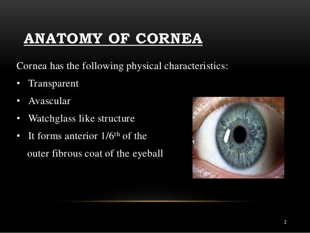
- outer layer – made up of the sclera and cornea (called the fibrous tunic)
- middle layer – made up of the uvea (called the vascular tunic)
- inner layer – made up of the retina (called the neural tunic)
What are the 3 layers of the eyeball and give a description of each?
The eye is made up of three layers: the outer layer called the fibrous tunic, which consists of the sclera and the cornea; the middle layer responsible for nourishment, called the vascular tunic, which consists of the iris, the choroid, and the ciliary body; and the inner layer of photoreceptors and neurons called the ...
What are the three layers of the eyeball quizlet?
What are the three layers of the eye? The sclera, the choroid layer, and the retina.
What are the 3 layers of the eye from superficial to deep?
From superficial to deep, they include:The fibrous layer, which consists of the sclera and cornea. ... The vascular layer, also known as the uvea or uveal tract. ... The nervous layer, also known as the retina, which is the innermost layer of the eyeball.
What are the internal layers of the eye quizlet?
internal anatomy of the eyefibrous layer (outermost)Uvea; vascular layer (middle layer)retina;sensory (innermost)
What are the three layers of the neural tunic of the eye wall?
The eyeball has three layers: the outer fibrous tunic, the middle vascular tunic, and the inner sensory tunic. The part of the fibrous tunic shown here is the sclera (top arrow bar). Just below it, but not labeled, is the choroid (part of the vascular tunic).
How many layers does the eyeball have?
threeThe eye has three main layers. These layers lie flat against each other and form the eyeball. The outer layer of the eyeball is a tough, white, opaque membrane called the sclera (the white of the eye).
How many layers are in the sclera?
four layersThe sclera has four layers, from the outside to the inside: Episclera, clear, thin tissue resting on top of the whites of your eyeballs. Stroma, made up of fibroblasts and collagen fibers, blending into the episclera. Lamina fusca, a transitional layer between the sclera and the choroid and ciliary body outer layers.
What are the three parts of the eyeball?
It consists of three parts that are continuous with each other. From posterior to anterior, they are the choroid, ciliary body, and iris. The nervous layer, also known as the retina, which is the innermost layer of the eyeball.
What are the layers of the retina?
The retina itself is divided into two layers; an outer, pigmented layer, and an inner neurosensory layer. These three layers comprise the circular outline of the eyeball. The inside of the eye contains the two refractive structures of the eye called the lens and vitreous body.
What is the outer layer of the eyeball called?
Sclera. The sclera is an opaque, white, outer layer that surrounds the posterior five-sixths of the eyeball. The sclera is thickest posteriorly, becoming progressively thinner anteriorly. The posterior pole of the sclera is perforated by the optic nerve and this site is marked as the posterior scleral foramen.
Where is the retina located?
The retina is the innermost layer of the eyeball that extends from the site of exit of the optic nerve to the posterior margin of the ciliary body. It is a site where the image of the environment is converted to the neural impulses that are transmitted to the brain via optic nerve for interpretation and analysis.
Where is the anterior chamber of the eyeball located?
The anterior chamber of eyeball is found between the cornea and iris. The posterior chamber of eyeball is more of a slit-like cavity, found between the iris and lens.
What happens when you get injured in your eyeball?
Injuries to the most parts of the eyeball or structures related to it, such as arterial or nerve supply, may lead to different forms of visual impairments or total blindness.
What is the function of the eyeball?
The main function of the eye is to detect the visual stimuli (photoreception) and to convey the gathered information to the brain via the optic nerve (CN II). In the brain, the information from the eye is processed and ultimately translated ...
How many layers are there in the retina?
The retina is a complex, multilayered tissue at the back of the eye. It covers the back two-thirds of the inner eye and is composed 10 distinct layers Each layer is made up of different cell types and serves a different function.
What is the white layer of the eye?
The sclera is the dense, white layer of tissue that coats the sides and back of the eyeball and acts, like the cornea, as a protective layer. It is a fibrous layer that is made up mainly of collagen and is what is often referred to as the "white of the eye.".
What is the ciliary body?
The ciliary body is a triangularly-shaped structure that extends from the front end of the choroid toward the iris. The ciliary body itself is composed of several structures, including ciliary processes, that form the clear liquid filling the eyeball, known as the aqueous.
What is a macro human eye?
macro human eye image by Anatoly Tiplyashin from Fotolia.com. The human eye is a marvel of anatomy, providing us with the ability to see the world in all its textures, colors, and sizes. While dividing the eye into multiple layers can be done in a variety of ways, one way to think of layers of the eye is to consider the eyeball as being composed ...
Which muscle controls the amount of light that passes through the lens of the eye?
The iris is a muscular structure--actually three muscles working together--that controls the amount of light that passes through the lens of the eye; the pupil of the eye is simply the opening that is controlled by the muscles of the iris and allows light to pass through the lens to the retina at the back of the eye.
Which layer of the eye contains the most light sensitive cells?
The photoreceptor layer contains special light-sensitive cells, called rods and cones because of their characteristic appearance.
Why is the cornea important?
First, because it has no blood vessels in it, the cornea is a perfectly transparent membrane and allows light to enter the pupil and hit the back of the eye (the retina), which is what enables us to have the sense of sight. Second, the cornea protects the iris and pupil from potential harm ...
How many layers are there in the eyeball?
Layers of the Eyeball. The eyeball is formed by three layers - fibrous, vascular and inner. Each of these layers has a specialised structure and function.
What are the three parts of the eyeball?
Anatomically, the eyeball can be divided into three parts - the fibrous, vascular and inner layers. In this article, we shall consider the anatomy of the eyeball in detail, and its clinical correlations. The eyeball is formed by three layers - fibrous, vascular and inner.
What is the term for swelling of the optic nerve?
Papilloedema refers to swelling of the optic disc that occurs secondary to raised intracranial pressure. The optic disc is the area of the retina where the optic nerve enters and can be visualised using an ophthalmoscope.
What are the two parts of the ciliary body?
Ciliary body - comprised of two parts - the ciliary muscle and ciliary processes . The ciliary muscle consists of a collection of smooth muscles fibres. These are attached to the lens of the eye by the ciliary processes. The ciliary body controls the shape of the lens, and contributes to the formation of aqueous humor.
What is the ciliary muscle?
The ciliary muscle consists of a collection of smooth muscles fibres. These are attached to the lens of the eye by the ciliary processes. The ciliary body controls the shape of the lens, and contributes to the formation of aqueous humor. Iris – circular structure, with an aperture in the centre (the pupil).
Which artery is responsible for the blood flow to the eyeball?
The eyeball receives arterial blood primarily via the ophthalmic artery. This is a branch of the internal carotid artery, arising immediately distal to the cavernous sinus. The ophthalmic artery gives rise to many branches, which supply different components of the eye. The central artery of the retina is the most important branch – supplying the internal surface of the retina. Occlusion of this artery will quickly result in blindness.
What is the inner layer of the eye?
Inner. The inner layer of the eye is formed by the retina; its light detecting component. The retina is composed of two layers: Pigmented (outer) layer – formed by a single layer of cells. It is attached to the choroid and supports the choroid in absorbing light (preventing scattering of light within the eyeball).
What is the outer layer of the eye?
Outer coat (fibrous tunic) : The eye’s outer layer is made of dense connective tissue, which protects the eyeball and maintains its shape. It is also known as the fibrous tunic. Middle coat (vascular tunic): The middle layer of tissue surrounding the eye, also known as the vascular tunic or „uvea“, is formed – from behind forward – by the choroid, ...
What organ collects light from the visible world around us?
The eye is a sensory organ. It collects light from the visible world around us and converts it into nerve impulses. Human eyes primarily consist of two globe-shaped structures, the eyeballs, which are surrounded by the the bony sockets of the skull, the orbits.
