The prefrontal cortex is the association cortex of the frontal lobe. It is one of the last brain structures to develop in the course of evolution. In the human, it constitutes more than one-quarter of the entire cerebral cortex.
- Limbic association area. Located in the anterior-ventral portion of the temporal lobe, the parahippocampal gyrus. ...
- Posterior association area. ...
- Anterior association area.
What is the function of the Association area?
The association areas are not involved in primary motor or sensory functions. Rather, they interpret, integrate, and act on information processed by the sensory areas. They are involved in higher mental functions, such as learning, remembering, thinking, and speaking.
What is the definition of association areas?
parts of the cerebral cortex that receive inputs from multiple areas; association areas integrate incoming sensory information, and also form connections between sensory and motor areas. Because they are involved in organizing information that comes from various other areas of the brain, association areas are often linked to complex functions.
What is the function of the somatosensory association area?
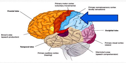
The somatosensory and somatomotor cortexes are two adjoining narrow bands of the topmost central area of the brain, stretching roughly from ear to ear. The motor cortex is partly responsible for the body’s voluntary muscle movements. The function of the somatosensory cortex is to receive and interpret most of the human sense of touch.
What is the function of the posterior association area?
The posterior association area is where visual, auditory and somatosensory association areas meet. This is what gives us our spatial awareness of our body. It is the kinesthetic sense that is very strong in professional dancers or athletes for example.
What are association areas of the brain and their functions?
parts of the cerebral cortex that receive inputs from multiple areas; association areas integrate incoming sensory information, and also form connections between sensory and motor areas.
What are some examples of association areas?
Association CortexAmygdala.Hippocampus.Thalamus.Sensory Cortex.Visual Cortex.
What are association areas in the cerebral cortex?
The term 'association cortex' refers to cerebral cortical regions other than primary motor and sensory areas. Association cortexes differ from the primary cortexes in terms of their laminar organization and their afferent and efferent connections.
What are the association areas of the brain quizlet?
Terms in this set (6)association areas. ... prefrontal cortex (frontal lobes) ... parietal lobes. ... right temporal lobe. ... Broca's area. ... Wernicke's area.
What is an association area?
Definition of association area : an area of the cerebral cortex that functions in linking and coordinating the sensory and motor areas.
Is Broca's area an association area?
Broca's area is the association area of the primary motor cortex for speech.
What is the difference between a primary cortex and an association area?
Specific parts of the cortex is specialized for specific functions. Primary = direct processing of primary sensory or motor info. Performs the actual task of the region. Secondary/Association = plans & integrates info for the primary area.
What does the anterior association area do?
The posterior multimodal association cortex is highly connected to the anterior association areas which in turn are responsible for conceptual cognitive functions and planning motor actions.
Where are the association areas?
Association areas can be located in the four cortical lobes of the Cerebral cortex. They are primarily involved in processing and integrating information from the senses and relate to higher mental abilities such as [[[thinking]] and reasoning.
What are association areas quizlet?
Association Areas. Association areas are all the areas in cerebral cortex except primary sensory area and primary motor area. It receives information from sensory areas and it is involved in "higher" functions such as perception, thoughts and decision-making, etc.
Where are association areas found?
The anterior association area is in the frontal lobes. It is rostral to the postcentral gyri, Rolandic fissure, and premotor areas. It has Sylvian fissure as its posterior boundary. It is referred to as prefrontal cortex.
What are the three types of association?
The three types of associations include: chance, causal, and non-causal.
What is an example of a business association?
Leading business associations in the United States include the U.S. Chambers of Commerce, the Better Business Bureau, the National Restaurant Association, the National Retail Federation, and the National Manufacturers Association, but there are tens of thousands more that operate at local, state, regional, and national ...
How do you organize an association?
Key steps facilitate formation of associationsVisit neighbors and/or other landowners in the local area to determine level of interest.Contact people involved with similar associations.Organize an informal meeting and meal to discuss interest and potential. ... Generate rough ideas for goals and objectives.More items...
What is association in sociology?
An association is a group of people organised for the pursuit of a specific purpose. Institutions, on the other hand, are the rules of procedure. Family is an association organised for the preparation of children, while marriage is its main institution. Political party is an institution, State is an association.
What is the association area of the brain?
Association areas of brain are the parts of cerebral cortex region of brain that receive information from multiple areas, integrate sensory inputs and establish connections between sensory and motor areas. Association areas are involved in collection of information from different regions of the brain and thus perform a major role in all the complex functions of brain.
What are the parts of the brain?
The human brain consists of the forebrain, midbrain, and the hindbrain. The main parts of the forebrain are the cerebrum, thalamus, and the hypothalamus. Association areas are located in cerebral cortex region of forebrain.
Where is the auditory cortex located?
Auditory Cortex: Auditory Cortex is located in the temporal lobe above ear. Function of auditory cortex is to receive and process auditory stimulus such as sound location, pitch and volume of sound.
Which lobe is the visual cortex located in?
Visual Cortex: This region is located on posterior tip of occipital lobe and involved in the processing of visual information. It receives visual stimulus from retina and help in detection of colour, form and movement.
Which lobe of the brain is responsible for processing somatic sensations such as touch, pain, and temperature?
Primary somatosensory cortex: It is located in parietal lobe and responsible for processing of somatic sensations such as touch, pain and temperature.
What are the two types of tissues in the cerebral hemisphere?
The cerebral hemisphere consists of two types of tissues: Grey matter and White matter.
Which lobe controls complex actions and thoughts?
Frontal Lobe: The frontal lobe controls complex actions and thoughts. It is associated with functions like parts of speech, self-control, planning, reasoning, problem solving mental abilities movements, and abstract thoughts.
What is the largest area of the brain?
Different areas of the cerebral cortex control different functions. The largest area of the human brain is the cerebrum. It’s also known as the cerebral cortex and controls many of the thoughts and processes we equate to the human mind. It sets us most apart from other creatures.
What is the main part of the brain?
The cerebral cortex is what sets apart human brains from other organisms. It’s organized at many physical scales starting at the level of single neurons and expanding up to functional systems.
What is the difference between left and right brain thinking?
There, however, some important differences between these areas. The left brain contains regions involved in speech and language (Broca’s area and Wernicke’s area). It also performs mathematical calculation and fact retrieval. It’s considered the logical or academic side of the brain.
What is the right brain?
The right brain plays a role in visual, auditory processing, and spatial skills. These generally are thought to equate to artistic ability considered more instinctive or creative in nature. However, these functions involve both hemispheres.
What is the command center of the nervous system?
The classical idea of the human brain is the command center of your nervous system. However, research is revealing that it is far more complex than just a switchboard between your ears. Your brain is so powerful and so diverse, it’s a big task to name all its different functions. Neuroscientists have attempted to break down ...
How many neurons are in the human brain?
Human brain structure is a hierarchy made up of about 85 billion neurons. Those neurons send signals across 18-32 trillion tiny spaces called synapses. Each synapse can convey a signal at 0.1–2 times per second. That equates to between 18 and 640 trillion signals that your brain sends and receives every second.
What is the function of the brain?
The Functional Areas of the Brain. Your brain relays between 18 and 640 trillion signals every second. The mind blowing amount of data is sent and decoded in the functional areas of the brain. The classical idea of the human brain is the command center of your nervous system.
Where do neurons get their information?
Neurons in this gyrus receive info from the general sensory receptors in the skin and proprioceptors in muscles, joints, etc. Neurons then identify the body region being stimulated
Which cells in the gyri allow us to consciously control the precise/ skilled voluntary movements of our?
Pyramidal cells in the gyri allow us to consciously control the precise/ skilled voluntary movements of our skeletal muscles
Where does the eye receive visual information?
Receives visual info that originates on the retina of the eye
Why Am I What I Am?
The association cortex consists of extensive territories of gray matter that surround and overshadow the primary and secondary sensory and motor areas. It is concentrated in three major regions: the parietal, occipital and temporal lobes; the territory near the temporal pole (under which the amygdala and hippocampus lie); and the forward part of the frontal lobe (including the frontal pole). We have already discussed these three regions to some extent: The first in relation to gnostic or knowing functions; the second as it relates to emotion, perceptual matters and memory; and the third as a powerful component of the limbic system. But we have not yet said anything about speech, except that it is not possible until many syntheses of information have taken place. Such syntheses are the major business of the association cortex, and many (if not all) of the above regions are drawn on. Several stand out, as we shall see.
What is transcortical aphasia?
The word transcortical refers to disruption of areas of the association cortex (Lichtheim called these the "area of concepts") that project onto the perisylvian language cortex, rather than of the language cortex itself. Transcortical motor aphasia (TCMA) resembles Broca's aphasia in that there is marked dysfluency or difficulty initiating speech, but the patient with TCMA repeats normally. The lesions of TCMA spare Broca's area but involve the adjacent left frontal cortex, medial frontal cortex, or subcortical white matter. Strokes causing transcortical motor aphasia generally are within the territory of the anterior cerebral artery, so the syndrome is relatively specific. Transcortical sensory aphasia resembles Wernicke's aphasia except for the sparing of repetition. The lesions involve the posterior left temporo-occipital region or the temporal lobe itself; Boatman et al. found that stimulation of adjacent cortical areas in the left superior temporal region could produce either Wernicke's or transcortical sensory aphasia. This syndrome also occurs in Alzheimer's disease. Mixed transcortical aphasia, also called the syndrome of the isolation of the speech area, resembles global aphasia except that repetition is not only spared but may be excessive or palilalic. Some patients mimic and learn new song lyrics or complete poems if given the first lines. Reported cases have had large, watershed infarctions sparing the perisylvian language area or advanced dementing illnesses.
What are the two primary factors involved in the development of SC multisensory integration capabilities?
Thus, the two primary factors involved in the development of SC multisensory integration capabilities appear to be experience with multisensory events and functional inputs from association cortex. This is unlikely to be coincidental. One possibility is that the tectopetal projections from association cortex (i.e., AES, rLS) serve as the portal through which multisensory experience affects the multisensory circuit. Thus, projection patterns of these afferents form a crucial substrate for the expression of multisensory integration capabilities. There are several additional points of evidence consistent with this idea.
What are the brains that are responsible for reasoning?
Species that possess large amounts of cortex and especially association cortex tend to show advanced reasoning skills. The relationship of large brains to complex behaviors may be driven by whether an organism is a predator. Many of the early neuroimaging studies of reasoning suggested that the frontal lobes were particularly important. Some network studies indicate that the frontal lobes may serve a coordinating or control function, possibly integrating wide-scale activity across the brain, rather than operating as a relational module. There is a strong impact of materials on reasoning studies in the brain. Materials invoke our semantic memories, which tend to be supported by temporal lobe regions. The integrated sets of information that we can use in reasoning are called schemas or scripts. Brain network interconnectivity looks to be an especially promising area toward capturing the complexity of neural processing in reasoning and may further clarify some of the roles of specific brain areas that have been linked to reasoning in various forms.
How are the two hemispheres coordinated?
As an alternative to a “high-level unifying structure” that might control the motor apparatus bilaterally, as suggested in the previous sections for the premotor and posterior association cortex, the two hemispheres might be coordinated essentially via the commissures, in humans mainly via the corpus callosum. Surprisingly, split-brain patients have been less well investigated from the point of view of movement execution than from that of perception. Although it was regularly found that bimanual skills like “buttoning clothes, tying shoelaces, shuffling cards, riding a bicycle, etc. are not significantly influenced by commissurotomy,” learning of new and difficult bimanual tasks was found to be almost impossible in split-brain patients ( Preilowski, 1990 ). In an early postoperative stage, the situation is different, however. As noted by Sperry (1966), “lack of coordination in activities that require close cooperation between the hands was also an early complaint …,” and “voluntary control of left arm and hand was much more severely affected than the right in the early weeks after surgery.” In fact, the dramatic disturbances, alluded to in the section on SMA lesions, were first described in split-brain patients: both “intermanual conflict” and the “alien hand sign” were considered typical for the callosal disconnection syndrome, expressed in the early postoperative period (cf. Bogen, 1985, for critical review). It is not clear to what extent the SMA or other medial frontal cortical regions contributes to the syndromes. However, it appears that only a minority of reported SMA cases developed this bimanual disorder. Although it is difficult to appreciate the exact extent of the lesions, it turns out that, in general, the lesions leading to the deficit were complex, concerning, for example, the entire territory of the anterior cerebral artery ( Figure 1 ).
Why are afferent and efferent engrams necessary?
It is necessary to preserve serial order in a time dimension that is polarized. Whatever may be the basis of that aspect of memory in which the temporal lobes are involved, if an epileptic discharge or an electrical stimulus evokes a hallucination of a tune or words, it is always in the correct serial order. It is not different in principle from the neuronal organization required to detect the direction of a visual or moving cutaneous stimulus, a property of some thalamic VPL neurones as well as in the cortex. (Indeed the necessity to move a noxious stimulus at a particular velocity and in a special direction over the plantar skin to elicit the plantar reflex suggests that a similar mechanism is present in the dorsal horn grey matter.) A possible mechanism could be that synapses sequentially excited by a moving stimulus could be spatially arranged on one dendrite so that each has a different potency, requiring them to be activated in the correct order to provide sufficient temporal and spatial summation to depolarize the spike trigger zone of the cell ( see Fig. 4.2 ). Some direction-sensitive neurones in area 5 of the parietal lobe are excited by joint movements, particularly when the movement is initiated by the animal (they have been found in the monkey) rather than by passive manipulation, a property which is probably used in ‘active touch’ ( p. 94 ).
How long does it take for ERP to respond to subsequently remembered items?
Differences in encoding processes continue beyond infancy. For example, Rollins and Riggins (2013) observed that 6-year-olds exhibited a differential pattern of neural responding during encoding, relative to adults. For children, ERP responses to subsequently remembered versus subsequently forgotten items differed at 700–900 ms, whereas for adults, differential responding was observed earlier, around 400–600 ms. Furthermore, the scalp sites at which the neural responses were observed differed between children and adults, suggesting the pattern of engagement of the underlying neural substrates changes with development ( Rollins and Riggins, 2013 ).
What are the cortices of the brain?
The association cortices include most of the cerebral surface of the human brain and are largely responsible for the complex processing that goes on between the arrival of inputin the primary sensorycortices and the generation of behavior. The diverse functions of the association cortices are loosely referred to as “cognition,” which literally means the process by which we come to know the world (“cognition” is perhaps not the best word to indicate this wide range of neural functions, but it has already become part of the working vocabulary of neurologists and neuroscientists). More specifically, cognition refers to the ability to attend to external stimuli or internal motivation, to identify the significance of such stimuli, and to plan meaningful responses to them. Given the complexity of these tasks, it is not surprising that the association cortices receive and integrate information from a variety of sources, and that they influence a broad range of cortical and subcortical targets. Inputs to the association cortices include projections from the primary and secondary sensory and motorcortices, the thalamus, and the brainstem. Outputs from the association cortices reach the hippocampus, the basal gangliaand cerebellum, the thalamus, and other association cortices. Insight into how the association areas work has come primarily from observations of human patients with damage to one or another of these regions. Noninvasive brain imaging of normal subjects, functional mapping at neurosurgery, and electrophysiological analysis of comparable brain regions in nonhuman primates have generally confirmed these clinical impressions. Together, these studies indicate that, among other functions, the parietal association cortexis especially important for attending to complex stimuli in the external and internal environment, that the temporal association cortex is especially important for identifying the nature of such stimuli, and that the frontal association cortex is especially important for planning appropriate behavioral responses to the stimuli.
What is NCBI bookshelf?
NCBI Bookshelf. A service of the National Library of Medicine, National Institutes of Health.
What are lesions of the parietal association cortex?
Lesions of the Parietal Association Cortex: Deficits of Attention
:no_upscale()/cdn.vox-cdn.com/uploads/chorus_asset/file/10096901/Screen_Shot_2018_01_25_at_12.05.40_PM.png)