What 4 bones make up the lower limb?
The femur is the single bone of the thigh. The patella is the kneecap and articulates with the distal femur. The tibia is the larger, weight-bearing bone located on the medial side of the leg, and the fibula is the thin bone of the lateral leg. The bones of the foot are divided into three groups.
How do you remember the bones of the lower limb?
0:321:25Easy Trick to remember the Bones of upper and lower limbs. - YouTubeYouTubeStart of suggested clipEnd of suggested clipIt's.MoreIt's.
What is the name of lower limb?
The thigh is between the hip and knee and makes up the rest of the lower limb. The term lower limb or lower extremity is commonly used to describe all of the leg. The leg from the knee to the ankle is called the crus....Thigh.MovementMuscles (in order of importance)Extension•Quadriceps femoris •Tensor fasciae latae*4 more rows
What are the bones of upper limb and lower limb?
The humerus is the single bone of the upper arm, and the ulna (medially) and the radius (laterally) are the paired bones of the forearm. The base of the hand contains eight bones, each called a carpal bone, and the palm of the hand is formed by five bones, each called a metacarpal bone.
How many joints are in the lower limb?
Joints and Ligaments of the Lower LimbJoint or LigamentDescriptionhip jointsynovialknee jointsynovialpatellar ligamenttendontibial collateral ligament38 more rows
How many bones are in your lower leg?
The lower leg is comprised of two bones, the tibia and the smaller fibula.
What is the largest bone of the lower limb?
Tibia. The tibia is the larger bone in the lower leg also known as the shin bone. It connects the knee with the ankle bones. The tibia is connected to the fibula by the interosseous membrane of the leg, forming a type of fibrous joint called a syndesmosis with very little movement.
Why are there 2 bones in the lower leg?
The top of the tibia connects to the knee joint and the bottom connects to the ankle joint. Although this bone carries the majority of the body's weight, it still needs the support of the fibula. The fibula, sometimes called the calf bone, is smaller than the tibia and runs beside it.
What is the meaning of lower limbs?
(LOH-er ek-STREH-mih-tee) The part of the body that includes the leg, ankle, and foot.
What are 6 bones of limbs?
question. Answer: Six bones of limbs are: Humerus,Ulna,Radius,Carpel,Metacarpal, and Phalanx bone .
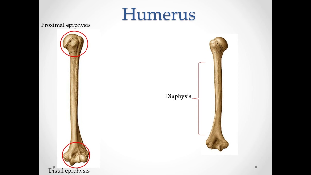
What are the bones of upper limb?
There is one bone in the upper arm region, the humerus. The forearm contains two bones, the radius and the ulna. When picturing the upper extremity in a standard anatomical position with the palm of the hand facing forward, the radius is located laterally and the ulna medially.
What is the proximal bone in the lower limb?
The two bones of the leg are the tibia and fibula. The fibula is located posterolateral to the tibia, making the tibia the more anterior of the two. The femur is located in the thigh, and the ilium forms part of the pelvis. These are both located proximal to the leg.
Why are there 2 bones in the lower leg?
The top of the tibia connects to the knee joint and the bottom connects to the ankle joint. Although this bone carries the majority of the body's weight, it still needs the support of the fibula. The fibula, sometimes called the calf bone, is smaller than the tibia and runs beside it.
How do you know what bone is your limb?
2:269:29ARPA309 Zooarchaeology Identifying Bones - YouTubeYouTubeStart of suggested clipEnd of suggested clipAgain. We can see that it's four limbs. And its hind limbs are made up of long bones similarly we goMoreAgain. We can see that it's four limbs. And its hind limbs are made up of long bones similarly we go over to the human and your arms and your legs are also long bones there the limb bones.
What are the 8 appendicular bones?
Structure and FunctionShoulder girdle:Clavicle. Scapula.Arm.Humerus.Forearm.Radius. Ulna.Wrist or carpal bones.Scaphoid. Lunate. Triquetrum. Pisiform. Trapezium. Trapezoid. Capitate. Hamate.More items...•
What are the 3 major leg bones?
The bones of the human leg, like those of other mammals, consist of a basal segment, the femur (thighbone); an intermediate segment, the tibia (shinbone) and the smaller fibula; and a distal segment, the pes (foot), consisting of tarsals, metatarsals, and phalanges (toes).
Which skeleton connects the lower limb to the axial skeleton?
The lower limbs actually have an incredibly detail-rich skeleton that can be divided into two functional components: the pelvic girdle, which connects the lower limb to the axial skeleton, and the bones of the free lower limb.
What bone connects the foot to the leg?
The foot bone’s connected to the...leg bone. And the leg bone’s connected to the... thigh bone! Alright, as a quick recap… just kidding. The lower limbs actually have an incredibly detail-rich skeleton that can be divided into two functional components: the pelvic girdle, which connects the lower limb to the axial skeleton, ...
Which bone is the anteromedial part of the hip?
Anteriorly, the ramus of the ischium joins the inferior ramus of the pubis to form a bar of bone called the ischiopubic ramus that represents the inferior border of the obturator foramen. Ok, now, finally, there’s the pubis, which forms the anteromedial part of the hip bone.
Which spine joins the pubis and ischium to form the acetabulum?
Ok, so the ala continues inferiorly with the body of the ilium, which joins the pubis and ischium to form the acetabulum. Next, let’s look at the ischium, which forms the postero-inferior part of the hip bone.
What is the opening in the hip called?
Moving on, there is a large oval opening of the hip bone called the obturator foramen, bounded by the pubis, ischium, and their rami. Normally, the obturator foramen is closed by the very strong obturator membrane, which has a tiny passageway for the obturator nerve and vessels called the obturator canal.
Which bone articulates with the head of the femur to form the hip joint?
Now that we know a bit about the bones separately, let’s talk about what they form together. First, there’s the acetabulum, which is the large socket on the lateral face of the hip bone that articulates with the head of the femur to form the hip joint.
What is the purpose of the femur?
The femur’s main purpose is to transmit body weight from the hip bone to the tibia when a person is standing, and, at the same time, to provide attachments for 23 muscles. Ok, so the proximal femur has a head, neck, and the greater and lesser trochanters.
What are the bones of the lower limb?
The leg is specifically the region between the knee joint and the ankle joint. Distal to the ankle is the foot. The lower limb contains 30 bones. These are the femur, patella, tibia, fibula, tarsal bones, metatarsal bones, and phalanges (see Figure 8.2 ). The femur is the single bone of the thigh. The patella is the kneecap and articulates with the distal femur. The tibia is the larger, weight-bearing bone located on the medial side of the leg, and the fibula is the thin bone of the lateral leg. The bones of the foot are divided into three groups. The posterior portion of the foot is formed by a group of seven bones, each of which is known as a tarsal bone, whereas the mid-foot contains five elongated bones, each of which is a metatarsal bone. The toes contain 14 small bones, each of which is a phalanx bone of the foot.
Which bones are the single bones of the thigh?
These are the femur , patella, tibia, fibula, tarsal bones, metatarsal bones, and phalanges (see Figure 8.2 ). The femur is the single bone of the thigh. The patella is the kneecap and articulates with the distal femur.
What is the longest bone in the body?
The femur , or thigh bone, is the single bone of the thigh region ( Figure 8.16 ). It is the longest and strongest bone of the body, and accounts for approximately one-quarter of a person’s total height. The rounded, proximal end is the head of the femur , which articulates with the acetabulum of the hip bone to form the hip joint. The fovea capitis is a minor indentation on the medial side of the femoral head that serves as the site of attachment for the ligament of the head of the femur. This ligament spans the femur and acetabulum, but is weak and provides little support for the hip joint. It does, however, carry an important artery that supplies the head of the femur.
Which bone articulates with the hip bone at the hip joint?
Figure 8.16 Femur and Patella The femur is the single bone of the thigh region. It articulates superiorly with the hip bone at the hip joint, and inferiorly with the tibia at the knee joint. The patella only articulates with the distal end of the femur. The narrowed region below the head is the neck of the femur.
Which part of the femur has lateral and medial expansions?
The distal end of the femur has medial and lateral bony expansions. On the lateral side, the smooth portion that covers the distal and posterior aspects of the lateral expansion is the lateral condyle of the femur. The roughened area on the outer, lateral side of the condyle is the lateral epicondyle of the femur.
How many bones are there in the foot?
The bones of the foot are divided into three groups. The posterior portion of the foot is formed by a group of seven bones, each of which is known as a tarsal bone, whereas the mid-foot contains five elongated bones, each of which is a metatarsal bone.
What is the elongated shaft of the femur?
The elongated shaft of the femur has a slight anterior bowing or curvature. At its proximal end, the posterior shaft has the gluteal tuberosity, a roughened area extending inferiorly from the greater trochanter. More inferiorly, the gluteal tuberosity becomes continuous with the linea aspera (“rough line”). This is the roughened ridge that passes distally along the posterior side of the mid-femur. Multiple muscles of the hip and thigh regions make long, thin attachments to the femur along the linea aspera.
What are the bones of the lower limb?
The leg is specifically the region between the knee joint and the ankle joint. Distal to the ankle is the foot. The lower limb contains 30 bones. These are the femur, patella, tibia, fibula, tarsal bones, metatarsal bones, and phalanges. The femur is the single bone of the thigh. The patella is the kneecap and articulates with the distal femur. The tibia is the larger, weight-bearing bone located on the medial side of the leg, and the fibula is the thin bone of the lateral leg. The bones of the foot are divided into three groups. The posterior portion of the foot is formed by a group of seven bones, each of which is known as a tarsal bone, whereas the mid-foot contains five elongated bones, each of which is a metatarsal bone. The toes contain 14 small bones, each of which is a phalanx bone of the foot.
What is the thigh bone?
The femur, or thigh bone, is the single bone of the thigh region (Figure 11.4.1). It is the longest and strongest bone of the body, and accounts for approximately one-quarter of a person’s total height. The rounded, proximal end is the head of the femur, which articulates with the acetabulum of the hip bone to form the hip joint. The fovea capitis is a minor indentation on the medial side of the femoral head that serves as the site of attachment for the ligament of the head of the femur. This ligament spans the femur and acetabulum but is weak and provides little support for the hip joint. It does, however, carry an important artery that supplies the head of the femur.
What is the smooth portion of the distal end of the femur?
On the lateral side, the smooth portion that covers the distal and posterior aspects of the lateral expansion is the lateral condyle of the femur. The roughened area on the outer, lateral side of the condyle is the lateral epicondyle of the femur. Similarly, the smooth region of the distal and posterior medial femur is the medial condyle of the femur, and the irregular outer, medial side of this is the medial epicondyle of the femur. The lateral and medial condyles articulate with the tibia to form the knee joint. The epicondyles provide attachment for muscles and supporting ligaments of the knee. The adductor tubercle is a small bump located at the superior margin of the medial epicondyle. Posteriorly, the medial and lateral condyles are separated by a deep depression called the intercondylar fossa. Anteriorly, the smooth surfaces of the condyles join together to form a wide groove called the patellar surface, which provides for articulation with the patella bone. The combination of the medial and lateral condyles with the patellar surface gives the distal end of the femur a horseshoe (U) shape.
What is the largest sesamoid bone in the body?
The patella (kneecap) is largest sesamoid bone of the body (see Figure 11.4.1). A sesamoid bone is a bone that is incorporated into the tendon of a muscle where that tendon crosses a joint. The sesamoid bone articulates with the underlying bones to prevent damage to the muscle tendon due to rubbing against the bones during movements of the joint. The patella is found in the tendon of the quadriceps femoris muscle, the large muscle of the anterior thigh that passes across the anterior knee to attach to the tibia. The patella articulates with the patellar surface of the femur and thus prevents rubbing of the muscle tendon against the distal femur. The patella also lifts the tendon away from the knee joint, which increases the leverage power of the quadriceps femoris muscle as it acts across the knee. The patella does not articulate with the tibia.
Which part of the tibia is expanded?
The proximal end of the tibia is greatly expanded. The two sides of this expansion form the medial condyle of the tibia and the lateral condyle of the tibia. The tibia does not have epicondyles. The top surface of each condyle is smooth and flattened. These areas articulate with the medial and lateral condyles of the femur to form the knee joint. Between the articulating surfaces of the tibial condyles is the intercondylar eminence, an irregular, elevated area that serves as the inferior attachment point for two supporting ligaments of the knee.
Which bone articulates with the hip bone at the hip joint?
Figure 11.4.1. Femur and patella. The femur is the single bone of the thigh region. It articulates superiorly with the hip bone at the hip joint, and inferiorly with the tibia at the knee joint. The patella only articulates with the distal end of the femur.
What is the elongated shaft of the femur?
The elongated shaft of the femur has a slight anterior bowing or curvature. At its proximal end, the posterior shaft has the gluteal tuberosity, a roughened area extending inferiorly from the greater trochanter. More inferiorly, the gluteal tuberosity becomes continuous with the linea aspera (“rough line”). This is the roughened ridge that passes distally along the posterior side of the mid-femur. Multiple muscles of the hip and thigh regions make long, thin attachments to the femur along the linea aspera.
What are the nerves that are located in the lumbar region?
Nerves : common fibular/peroneal, tibial and saphenous nerves, branches of the sciatic and femoral nerves. Bones : calcaneus, talus, navicular, cuboid, and cuneiform bones, as well as the metatarsals and phalanges.
How many muscles are in the posterior compartment?
The posterior compartment consists of seven muscles in total, divided into superficial and deep groups. The superficial muscles are the gastrocnemius, soleus, and plantaris (together forming the triceps surae ), while the deep layer consists of the popliteus, tibialis posterior, flexor digitorum longus, and flexor hallucis longus.
What are the bones of the thigh?
Muscles: anterior, medial and posterior groups. Arteries: femoral artery and its branches. Veins: femoral vein, circumflex vein, long saphenous vein, and deep vein of the thigh. Nerves: femoral and sciatic nerves, branches from the lumbar and sacral plexuses, respectively. Knee.
Where does the arterial supply come from?
The arterial supply comes from the femoral artery and its branches. The main vein draining the thigh, and actually the entire lower limb, is the femoral vein. It is part of the deep venous system, drains into the external iliac vein, and is a direct continuation of the popliteal vein.
What is the function of the hip joint?
This ball-and-socket joint is responsible for providing the lower extremity with an extensive degree of movement. Anatomy of the pelvis and hip bones.
What is the structure of the hip?
The structural framework of the hip region is provided by the pelvis, a structure composed of the pelvic girdle and the coccyx. In turn, the pelvic girdle consists of two hip bones and the sacrum, interconnected at the pubic symphysis and sacroiliac joints.
What are the parts of the lower extremity?
The lower extremity can be divided into several parts or regions, as follows: Hip. Thigh. Knee.
Which bone is the longest and strongest?
the femur is the longest and strongest bone in the body
Which bone is the site of attachment of the sacrospinous ligament?
one of three bones that form the os coxae: ilium, ischium, pubis. the greater sciatic notch is converted to the greater sciatic foramen by the sacrospinous ligament and the sacrotuberous ligament.
What is the lesser sciatic notch?
the lesser sciatic notch is converted to the lesser sciatic foramen by the sacrospinous ligament and the sacrotuberous ligament. it is the site of attachment of the sacrospinous ligament and the site of origin of the superior gemellus m. one of three bones that form the os coxae: ilium, ischium, pubis.
What is the coccyx?
the coccyx results from the fusion of the four coccygeal vertebrae; it may be a single bone or the first coccygeal vertebra may be separated from the other three; it articulates with the fifth sacral segment; coccygeal vertebrae are reduced in complexity, having no pedicles, laminae or spines
How much of the acetabulum is the pubis?
the body of the pubis forms about 1/5 of the acetabulum
How many pairs of ramus are there?
there are four pairs; each transmits the dorsal primary ramus of the respective sacral spinal nerve
Which organ articulates with the ilium and pubis?
it articulates with the ilium and the pubis at the acetabulum; the body of the ischium forms 2/5 of the acetabulum