
What are the 4 parts of the pelvis?
These include the following:Ilium. The broad, flaring portion of the hip bone (the crest of the pelvis).Pubis. The lower, posterior part of the hip bone.Ischium. One of the bones that helps form the hip.
What are the three regions of pelvis?
There are three bones of the pelvis: the hip bone, sacrum and coccyx. These bones connect the axial skeleton to the lower limbs, and therefore play a role in bearing the weight of the upper body.
What are the five types of pelvis?
Actually, the majority of pelves are of mixed types:Gynaecoid pelvis(50%): It is the normal female type. Inlet is slightly transverse oval. ... Anthropoid pelvis (25%): It is ape-like type. All anteroposterior diameters are long. ... Android pelvis (20%): It is a male type. ... Platypelloid pelvis (5%): It is a flat female type.
What are the 3 major joints of the hip pelvis?
The hip joint capsule is formed by three major ligaments: the iliofemoral, pubofemoral, and ischiofemoral ligaments. The capsular ligaments run in a spiral fashion preventing hip extension and are surrounded by thick longitudinal fibers that provide additional stability in the lateral plane.
What are the 4 functions of the pelvis?
pelvis, also called bony pelvis or pelvic girdle, in human anatomy, basin-shaped complex of bones that connects the trunk and the legs, supports and balances the trunk, and contains and supports the intestines, the urinary bladder, and the internal sex organs.
How many levels of the pelvis are there?
Three LevelsThree Levels of the Pelvis.
What are the 5 functions of the pelvis?
The pelvic floor muscles play five important roles in the body:Organ support. The pelvic floor muscles support our bladder, uterus, rectum, and important abdominal organs against gravity and any added downward pressure.Stability. ... Sphincteric function. ... Sexual Function. ... Circulation.
What are the 3 diameters of the pelvic outlet?
The diameters of the outlet of the pelvis are two, antero-posterior and transverse.
What 3 systems does the pelvis protect?
Together with the sacrum and coccyx, the pelvic girdle forms a bowl‐shaped region, the pelvis, that protects internal reproductive organs, the urinary bladder, and the lower part of the digestive tract.
Where do the 3 pelvic bones meet?
acetabulumThe hip bone is formed by three parts: the ilium, ischium, and pubis. At birth, these three components are separated by hyaline cartilage. They join each other in a Y-shaped portion of cartilage in the acetabulum.
What are the bones in the pelvis?
The pelvis consists of paired hipbones, connected in front at the pubic symphysis and behind by the sacrum; each is made up of three bones—the blade-shaped ilium, above and to either side, which accounts for the width of the hips; the ischium, behind and below, on which the weight falls in sitting; and the pubis, in front.
What is the function of the pelvis?
The pelvis provides attachment for muscles that balance and support the trunk and move the legs, the hips, and the trunk. In the human infant the pelvis is narrow and nonsupportive.
What is the pelvic girdle of a shark?
The pelvic girdle of the elasmobranch fishes (e.g., sharks, skates, and rays) consists of either a curved cartilaginous structure called the puboischial bar or a pair of bars lying transversely in the ventral part of the body anterior to the cloaca; projecting dorsally…
Which part of the pelvis is shown in an anterior view?
Anterior view of the hip and pelvis, showing attachment of ligaments to the femur, ilium, ischium, and pubis.
Which fossils have pelvis?
The pelvis of Australopithecus africanus —which lived more than two million years ago—is clearly hominin (of human lineage). Homo erectus and all later fossil hominins, including Neanderthals, had fully modern pelvises. Comparison of the pelvis and lower limbs of a chimpanzee, an australopith, and a modern human.
When does pelvic girdle pain resolve?
PGP typically resolves on its own in the weeks or months following childbirth, though full recovery may take years.
What is the pelvis?
The pelvis (plural pelves or pelvises) is either the lower part of the trunk of the human body between the abdomen and the thighs (sometimes also called pelvic region of the trunk) or the skeleton embedded in it (sometimes also called bony pelvis, or pelvic skeleton ). The pelvic region of the trunk includes the bony pelvis, ...
What is the pelvic region?
The pelvic region of the trunk includes the bony pelvis, the pelvic cavity (the space enclosed by the bony pelvis), the pelvic floor, below the pelvic cavity, and the perineum, below the pelvic floor. The pelvic skeleton is formed in the area of the back, by the sacrum and the coccyx and anteriorly and to the left and right sides, by a pair of hip bones .
How many rings are there in the pelvis?
As a mechanical structure the pelvis may be thought of as four roughly triangular and twisted rings. Each superior ring is formed by the iliac bone; the anterior side stretches from the acetabulum up to the anterior superior iliac spine; the posterior side reaches from the top of the acetabulum to the sacroiliac joint; and the third side is formed by the palpable iliac crest. The lower ring, formed by the rami of the pubic and ischial bones, supports the acetabulum and is twisted 80–90 degrees in relation to the superior ring.
What is the lower part of the body called?
Pelvis. The pelvis (plural pelves or pelvises) is either the lower part of the trunk of the human body between the abdomen and the thighs (sometimes also called pelvic region of the trunk) or the skeleton embedded in it (sometimes also called bony pelvis, or pelvic skeleton ). The pelvic region of the trunk includes the bony pelvis, ...
Why is the pelvis important to human development?
Modern humans are to a large extent characterized by bipedal locomotion and large brains. Because the pelvis is vital to both locomotion and childbirth, natural selection has been confronted by two conflicting demands: a wide birth canal and locomotion efficiency, a conflict referred to as the " obstetrical dilemma ". The female pelvis, or gynecoid pelvis, has evolved to its maximum width for childbirth—a wider pelvis would make women unable to walk. In contrast, human male pelvises are not constrained by the need to give birth and therefore are more optimized for bipedal locomotion.
Which spine is heavier, male or female?
The ischial spines and tuberosities are heavier and project farther into the pelvic cavity in males. The greater sciatic notch is wider in females. The iliac crests are higher and more pronounced in males, making the male false pelvis deeper and more narrow than in females.
Why does the pelvis widen laterally?
This is similar to the rachitic pelvis where the softened bones widen laterally because of the weight from the upper body resulting in a reduced antero posterior diameter. Giving birth with this type of pelvis is associated with problems, such as transverse arrest. Less than 3 per cent of women have this pelvis type.
What is the difference between a female and a male pelvis?
As a result, the female pelvis is generally broader and wider than the male pelvis.
What is the largest muscle in the pelvis?
The levator ani muscles are the largest group of muscles in the pelvis. They have several functions, including helping to support the pelvic organs. The levator ani muscles consist of three separate muscles: Puborectalis. This muscle is responsible for holding in urine and feces.
What is the term for the growth of the uterus outside the uterus?
Endometriosis. Endometriosis occurs when the tissue that lines the inside walls of the uterus (endometrium) begins to grow outside of the uterus. The ovaries, fallopian tubes, and other tissues in the pelvis are typically affected by the condition.
What are the conditions that affect the pelvis?
The pelvis contains a large number of organs, bones, muscles, and ligaments, so many conditions can affect the entire pelvis or parts within it. Some conditions that can affect the female pelvis as a whole include: Pelvic inflammatory disease (PID). PID is an infection that occurs in the female reproductive system.
What part of the body is the hip bone?
Hip bones. There are two hip bones, one on the left side of the body and the other on the right. Together, they form the part of the pelvis called the pelvic girdle. The hip bones join to the upper part of the skeleton through attachment at the sacrum. Each hip bone is made of three smaller bones that fuse together during adolescence:
How often does the lining of the uterus shed during menstruation?
During the reproductive years, the lining of the uterus sheds every month during menstruation if you don’t become pregnant.
What are the symptoms of pelvic pain?
Some common symptoms of a pelvic condition can include: pain in the lower abdomen or pelvis. a feeling of pressure or fullness in the pelvis. unusual or foul-smelling vaginal discharge. pain during sex.
What are the bones of the pelvis?
Each pelvic bone (hip bone) is made by the combination three bones namely, the ilium, pubis, and ischium. These three bones fuse at a cup-shaped concavity called the acetabulum which articulates with the head of the femur to form the hip joint.
Where is the pelvis located?
The pelvis is located between the fifth lumbar vertebra and the femoral heads. It forms an irregular bony girdle connecting the lower limbs to the trunk.
Why is the pelvis different between male and female?
The male pelvis is more adapted to bear the heavier build of the upper body and for stronger muscle attachment, while the female pelvis is adapted to provide enough space for the birth canal (or birth passage) in the region. Some of these differences are compared in the table below.
What is the pubis joint?
Each pubis consists of a body and two rami. The bodies are located medially, and are joined with one another by a cartilaginous joint called the symphysis pubis which lies in the midline. The anterior surface of the body has a bony ridge named the pubic crest, in its upper part.
What is pelvic bone?
Clinical significance. The pelvis is a basin shaped bony structure formed by the combination of two pelvic bones ( hip bones or innominate bones) and the sacrum. It is strengthened and supported by several joints and ligaments. It provides attachment to some important muscles in the region, and forms a cavity which accommodates several important ...
Which position of the pelvis is positioned in an anteriorly tilted manner?
The pelvis is positioned in an anteriorly tilted manner, so that in the anatomical (erect) position, the anterior superior iliac spine and the upper margin of the pubis lie in the same vertical plane. This can be demonstrated by holding the aforementioned points against a wall.
How many vertebrae are in the coccyx?
The coccyx is also a roughly triangular bone formed by the fusion of three to five vertebrae, and it articulates with the lower end of the sacrum to form the sacrococcygeal joint.
What are the three parts of the pelvis?
There are three separate parts to the pelvis: the single midline sacrum and paired innominate bones ( os coxae). The line of division between the sacrum and the innominate bones is the sacroiliac joint, while the two innominate bones are separated from one another by the pubic symphysis (Fig.1 and Fig. 2). Fig. 1.
How many joints are there in the pelvis?
The pelvis has three joints: two sacroiliac joints and the pubic symphysis. The sacroiliac joints are true synovial joints, but the symphysis is a synchrondrosis, without a synovial space. Although immobile during most of life, these joints do display some movement during pregnancy. 1,2.
Why is the middle portion of the pelvic cavity longer than the transverse?
Because of the inward inclination of the walls and protrusion of the ischial spines into the pelvic cavity (without concomitant shortening of the anterior posterior dimension), the middle portion of the pelvic cavity becomes longer in its anterposterior diameter than in the transverse.
What are the three bones that make up the sagittal section of the pelvic bones?
Sagittal section of the pelvic bones. Each innominate bone is formed from three bones: the ilium, the ischium, and the pubis. These bones have fused into a single unit before reproductive age is reached. Their individual names persist, however, in terms such as the iliac crest, ischial tuberosity, and pubic ramus.
Where does the pelvic articulation ligament come from?
It arises from the posterior iliac spines and the back and side of the lower sacrum and coccyx and ends at the ischial tuberosity. These ligaments help to stabilize the pelvis and probably undergo the same softening that the ligaments around the pelvic articulations experience.
Where is the anterior urogenital triangle?
The anterior (urogenital) triangle has its corners at the lower end of the pubic symphysis and the inner aspects of the ischial tuberosities. It is bounded laterally by the inferior ischiopubic rami. The posterior (anal) triangle has its apex at the tip of the sacrum and shares its base with the anterior triangle.
How much does the uterus weigh?
It is obvious that the uterus must undergo significant enlargement to accommodate the growing fetus (Fig. 11). Its weight increases from approximately 60 g to 1 kg. The volume of its cavity grows from 10 ml to 6 to 10 liters, and its external dimensions from 8 × 2.5 × 4 cm to 32 × 24 × 22 cm at term. 22
How many normal variants of pelvic shape are there?
The four normal variants of pelvic shape according to Caldwell-Malloy classifications[7][13]:
How does the pelvis form?
By the fifth week, the condensation of mesenchymal tissue in the limb buds form the template for bone models, and the ilium becomes recognizable by about five months prenatally. The pelvis forms via the process of endochondral ossification, where chondrocytes in cartilage stimulate mineralization and blood vessel growth. This ossification stems from three primary ossification centers termed the iliac center, ischial center, and pubic center. The primary ossification centers are well developed by birth, but the ossification process continues postnatally, slowing down after three months of age. Ontogeny of the pelvis is completed in adulthood, typically around 25 years of age. Sexual dimorphism of the pelvis is visually apparent in the ilium, the acetabulum, the iliac height, and the width of the pelvic inlet and outlet. [4][5][6]
What is the pelvic outlet?
The pelvic outlet also called the inferior pelvic aperture, defines the lower margin of the lesser (true) pelvis. The pelvic cavity (the true pelvis) predominantly contains the urinary bladder, the colon, and the internal reproductive organs. This space is enclosed between the pelvic inlet and the pelvic outlet. The pelvic outlet is the inferior opening of the pelvis that is bounded by coccyx, the ischial tuberosities, and the pubis symphysis.
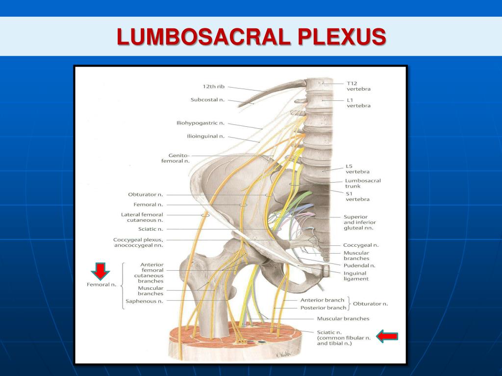
What nerves are in the sacral plexus?
The pudendal nerve (S 2,3,4) is a paired nerve that branches off of the sacral plexus. This nerve travels out of the greater sciatic foramen, into the lesser sciatic foramen, through the pudendal canal (sometimes called Alcock’s canal), and into the perineum. The terminal branches of the pudendal nerve, the dorsal nerve, and inferior rectal nerve provide sensory and motor innervation to the perineum and external genitalia. A common procedure during obstetric and anorectal procedures uses local anesthesia to block the pudendal nerve. During this procedure, the clinician will typically approach transvaginally, passing through the pelvic outlet, and use the ischial spine as a landmark to then inject a local anesthetic into the pudendal nerve. [9][10]
What is the station of the fetal head?
The station of the fetal head is a numerical scale that estimates the number of centimeters between the fetal head and the ischial spines. This metric marks the progression of labor as the fetus passes through the birth canal. The narrowest distance that the head of the child must pass through during vaginal birth is the interspinous distance, which typically measures around 10 centimeters, is called station 0. However, this space is not a fixed distance due to pubic symphysis loosening during labor. Station +3, when the leading portion of the fetal head is 5 cm inferior to the ischial spine, is approximately at the opening of the pelvic outlet. As the fetus moves through the birth canal, the widest diameter of the pelvic canal changes direction from a transverse direction in the pelvic inlet, to an anteroposterior diameter in the pelvic outlet. For this reason, the fetal head must rotate as it passes through the birth canal during labor.
What nerves pass through the pelvis?
The lumbosacral trunk (L4-S3), the sacral plexus, and coccygeal plexus all pass through the pelvic cavity. Numerous branching nerves innervate pelvic and perineal muscles. Additionally, there are parasympathetic and sympathetic innervations within the pelvis. Divisions of the pelvic splanchnic (parasympathetic), sacral splanchnic (sympathetic), and the inferior hypogastric plexuses also have branches that supply the viscera of the pelvis. While many nerves originate below the level of the pelvic brim, very few of these nerves pass through the pelvic outlet but, instead, exit through the many foramina within the pelvis or terminate within the true pelvis.
Where do sacrotuberous ligaments form?
These ligaments form the posterolateral border of the pelvic outlet.
What are the bones of the pelvis?
Bones of the Pelvis. There are three bones of the pelvis: the hip bone, sacrum and coccyx. These bones connect the axial skeleton to the lower limbs, and therefore play a role in bearing the weight of the upper body. These bones also act as attachments for many muscles and ligaments within the pelvis and lower limbs.
Which bones are the smallest in the pelvic system?
It is formed by the fusion of five sacral vertebrae, and transmits the sacral nerve fibres of the cauda equina. The coccyx, commonly referred to as the tailbone , is the smallest of the pelvic bones, and sits inferiorly to the sacrum.
Where is the pelvic girdle located?
The pelvic girdle is the ring shaped collection of these bones at the base of the spine.
What are the parts of the hip?
The hip bone has three parts: the ilium, pubis and ischium. These are separated by cartilage at birth and fuse during puberty. The sacrum is located inferiorly to the spinal vertebrae, and posteriorly within the pelvis. It is formed by the fusion of five sacral vertebrae, and transmits the sacral nerve fibres of the cauda equina.
Overview
Structure
The pelvic region of the trunk is the lower part of the trunk, between the abdomen and the thighs. It includes several structures: the bony pelvis, the pelvic cavity, the pelvic floor, and the perineum. The bony pelvis (pelvic skeleton) is the part of the skeleton embedded in the pelvic region of the trunk. It is subdivided into the pelvic girdle and the pelvic spine. The pelvic girdle is composed of the appendicular hip bones (ilium, ischium, and pubis) oriented in a ring, and connects the pelvic re…
Development
Each side of the pelvis is formed as cartilage, which ossifies as three main bones which stay separate through childhood: ilium, ischium, pubis. At birth the whole of the hip joint (the acetabulum area and the top of the femur) is still made of cartilage (but there may be a small piece of bone in the great trochanter of the femur); this makes it difficult to detect congenital hip dislocation by X-raying.
Functions
The skeleton of the pelvis is a basin-shaped ring of bones connecting the vertebral column to the femora. It is then connected to two hip bones.
Its primary functions are to bear the weight of the upper body when sitting and standing, transferring that weight from the axial skeleton to the lower appendicular skeleton when standing and walking, and providing attachments for and withstanding the forces of the powerful muscle…
Clinical significance
Hip fractures often affect the elderly and occur more often in females, and this is frequently due to osteoporosis. There are also different types of pelvic fracture often resulting from traffic accidents.
Pelvic pain generally, can affect anybody and has a variety of causes; bowel adhesions, irritable bowel syndrome, interstitial cystitis, endometriosis in women.
History
Throughout the 20th century pelvimetric measurements were made on pregnant women to determine whether a natural birth would be possible, a practice today limited to cases where a specific problem is suspected or following a caesarean delivery. William Edgar Caldwell and Howard Carmen Moloy studied collections of skeletal pelves and thousands of stereoscopic radiograms and finally recognized three types of female pelves plus the masculine type. In 1933 …
Other animals
The pelvic girdle was present in early vertebrates, and can be tracked back to the paired fins of fish that were some of the earliest chordates.
The shape of the pelvis, most notably the orientation of the iliac crests and shape and depth of the acetabula, reflects the style of locomotion and body mass of an animal. In bipedal mammals, the iliac crests are parallel to the vertically oriented sacroiliac joints, where in quadrupedal mam…
Additional images
• Diameters of pelvic inlet.
• Right hip bone. External surface.
• Pelvic Girdle Anatomy