The 5 secondary brain vesicles are the telencephalon, diencephalon, mesencephalon, metencephalon, and myelencephalon. In the adult brain, the telencephalon forms the cerebrum; the diencephalon forms the thalamus, hypothalamus, subthalamus and epithalamus; the mesencephalon forms the rostral end of the brainstem; the metencephalon forms the pons and cerebellum; and the myelencephalon forms the medulla oblongata.
What are the primary and secondary brain vesicles?
As development continues, the three primary vesicles give rise to five secondary brain vesicles: Telencephalon, Diencephalon, Mesencephalon, Metencephalon, and Myelencephalon. Each secondary vesicle develops into specific components of the adult nervous system .
What are the four brain vesicles?
brain v's, secondary the four brain vesicles formed by specialization of the forebrain and of the hindbrain in later embryonic development. chorionic vesicle the chorion of a mammal. encephalic v's brain vesicles. germinal vesicle the fluid-filled nucleus of an oocyte toward the end of prophase of its meiotic division.
What are the V's of the brain?
brain v's, primary the three earlier subdivisions of the embryonic neural tube, including the forebrain, midbrain, and hindbrain. brain v's, secondary the four brain vesicles formed by specialization of the forebrain and of the hindbrain in later embryonic development. chorionic vesicle the chorion of a mammal. encephalic v's brain vesicles.
What are the anterior vesicles of the brain called?
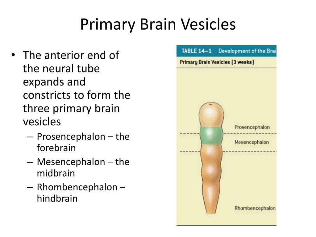
The most anterior of these embryonic brain vesicles is called the “prosencephalon” which is the embryonic precursor of the forebrain. The middle vesicle is the “mesencephalon” which is the precursor of midbrain structures, and the most posterior is the “rhombencephalon” which will become the hindbrain.
How many brain vesicles are there?
Initially there are three primary brain vesicles: prosencephalon, mesencephalon, and rhombencephalon. These develop into five secondary brain vesicles – the prosencephalon is subdivided into the telencephalon and diencephalon, and the rhombencephalon into the metencephalon and myelencephalon.
What are the primary brain vesicles?
It is widely held that three primary brain vesicles (forebrain, midbrain, and hindbrain vesicles) develop into five secondary brain vesicles in all vertebrates (von Baer's scheme).
What are the 3 brain vesicles?
In the textbooks these vesicles are called prosencephalon, mesencephalon, and rhombencephalon.
What is the function of brain vesicles?
In summary, the primary vesicles help to establish the basic regions of the nervous system: forebrain, midbrain, and hindbrain. These divisions are useful in certain situations, but they are not equivalent regions. The midbrain is small compared with the hindbrain and particularly the forebrain.
What are the secondary vesicles?
in neural development, one of the five vesicles that are formed after the prosencephalon and rhombencephalon subdivide. The secondary vesicles include the: telencephalon, diencephalon, mesencephalon, metencephalon, and myelencephalon.
How can I remember the vesicles of my brain?
Three primary vesicles: Forebrain is a pro. Prosencephalon - future forebrain. M for midbrain, mesencephalon. Mesencephalon - future midbrain.
What is the primary brain vesicle of the cerebellum?
The neural tube forms three primary brain vesicles....Development of Brain.Primary vesiclesSecondary vesiclesAdult structuresHindbrain vesicle (rhombencephalon)MetencephalonPons and cerebellumMyelencephalonMedulla3 more rows•Aug 14, 2006
How many primary brains vesicles are formed from the nervous tube?
The cerebrum and brainstem arise from the rostral neural tube. These regions expand and constrict to form the three primary brain vesicles: Forebrain/Prosencephalon, Midbrain/Mesencephalon, and Hindbrain/Rhombencephalon.
Which brain vesicle gives rise to the midbrain?
mesencephalonThe mesencephalon gives rise to the midbrain structures, and the metencephalon the pons and cerebellum.
What are the three primary brain vesicles that form from the neural tube quizlet?
The neural tube becomes 3 "primary" brain vesicles in a 4-week embryo:Prosencephalon ("forebrain")Mesencephalon ("midbrain")Rhombencephalon ("hindbrain")
Which of the primary brain vesicles keeps its original name in the mature brain?
B. The term mesencephalon means "midbrain." As it develops from a primary brain vesicle to a secondary brain vesicle and finally an adult brain structure, it retains its name--the midbrain.
What are the 7 structures of the diencephalon?
There are several structures between the brainstem and the cerebral cortex that make up the diencephalon. These include the epithalamus, thalamus, subthalamus, metathalamus, hypothalamus, hypophysis cerebri and the third ventricle as its cavity.
What is the primary brain vesicle of the cerebellum?
The neural tube forms three primary brain vesicles....Development of Brain.Primary vesiclesSecondary vesiclesAdult structuresHindbrain vesicle (rhombencephalon)MetencephalonPons and cerebellumMyelencephalonMedulla3 more rows•Aug 14, 2006
What are the three primary brain vesicles that form from the neural tube quizlet?
The neural tube becomes 3 "primary" brain vesicles in a 4-week embryo:Prosencephalon ("forebrain")Mesencephalon ("midbrain")Rhombencephalon ("hindbrain")
Which of the primary brain vesicles keeps its original name in the mature brain?
B. The term mesencephalon means "midbrain." As it develops from a primary brain vesicle to a secondary brain vesicle and finally an adult brain structure, it retains its name--the midbrain.
Which primary and secondary brain vesicles ultimately become the structure that contains the 3rd ventricle?
The various brain vesicles give rise to the following adult structures: The telencephalon develops into the cerebrum and lateral ventricles. The diencephalon forms the thalamus, hypothalamus, epithalamus, and third ventricle.
Where does the lens vesiclea vesicle form?
lens vesiclea vesicle formed from the lens pit of the embryo, developing into the crystalline lens.
Where are optic vesicles located?
optic vesicle's Hollow, spherical neuroectodermal protrusions, one on each side of the forebrain. They are derived from the optic pits after closure of the embryonic neural tube. They subsequently invaginate to form the optic cup. The surface ectoderm overlying the optic vesicles invaginates to form the lens vesicle and eventually the crystalline lens. Syn.primary optic vesicle. See anophthalmia; optic cup; ectoderm; optic pit.
What are the three subdivisions of the embryonic neural tube?
brain v's, primarythe three earlier subdivisions of the embryonic neural tube, including the forebrain, midbrain, and hindbrain.
What is the ovoid sac formed by the closure of the auditory pit in the early embryo?
auditory vesiclea det ached ovoid sac formed by closure of the auditory pit in the early embryo, from which the percipient parts of the inner ear develop.
What are the two views of the brain?
Two views of the human brain. a. Lateral view (rostral end is left, caudal is right) shows an apparently uniform surface marked by gyri and sulcal folds (Right hemisphere of J. Piłsudski’s brain, lateral view, image in the public domain). b. Coronal cross-section (cut at approximately the level of the dotted line in A) stained for cell bodies that mark neurons. The neocortex is the thin mantel layer (dark purple) on the surface of the brain. The white areas are connecting fiber pathways. Image reproduced with permission from http://www.brains.rad.msu.eduwhich is supported by the U.S. National Science Foundation. Images obtained with permission from Wiki Commons, http://commons.wikimedia.org/wiki
How many neurons are in the human brain?
The human brain is arguably the most complex of all biological systems. The mature brain is composed of more than 100 billion neurons (Pakkenberg and Gundersen 1997). Neuronsare the information processing cells in the brain (see Fig. 2).
What is the thick layer of the brain?
The neocortex is a 2–5 mm thick layer of cells that lies on the surface of the brain (the word cortex comes from the Latin term meaning bark, as in the bark of a tree). In the cross-section of the brain shown in Fig. 3bthe neocortex is the thin, dark gray strip that follows the brain surface.
What is the enfolding of the brain?
The enfolding of the mature brain is thought to be an adaptation to the dramatic growth in the size of the brain during the course of evolution. The folding of brain tissue allowed large brains to fit in comparatively small cranial vaults that had to remain small to accommodate the birth process (see Fig. 3a). The largest and most important brain information processing networks involve the neocortexand the subcortical nucleithat relay information to and from the neocortex. The neocortex is a 2–5 mm thick layer of cells that lies on the surface of the brain (the word cortex comes from the Latin term meaning bark, as in the bark of a tree). In the cross-section of the brain shown in Fig. 3bthe neocortex is the thin, dark gray strip that follows the brain surface. The subcortical nuclei are clusters of neurons that serve as both signal relay centers communicating between the neocortex and the rest of the body, and as relays among different areas of the cortex. They are located deep in the brain below the cortex and are thus referred to as “subcortical” nuclei. Because both the neocortex and the subcortical nuclei contain the cell bodies of neurons they are gray in appearance, thus giving rise to the term “gray matter”.
What is the ventricular system?
The ventricular system is filled with a fluid called cerebral spinal fluid that is completely recycled several times per day. The ventricular system has a number of important functions including cushioning and protection of the brain, removal of waste material, and transport of hormones and other substances (Brodal 2010). During brain development the walls of the ventricles are the site of most neuron production.
How does the brain develop?
Human brain development is a protracted process that begins in the third gestational week (GW) with the differentiation of the neural progenitor cells and extends at least through late adolescence, arguably throughout the lifespan. The processes that contribute to brain development range from the molecular events of gene expression to environmental input. Critically, these very different levels and kinds of processes interact to support the ongoing series of events that define brain development. Both gene expression and environmental input are essential for normal brain development, and disruption of either can fundamentally alter neural outcomes. But neither genes nor input is prescriptive or determinative of outcome. Rather brain development is aptly characterized as a complex series of dynamic and adaptive processes that operate throughout the course of development to promote the emergence and differentiation of new neural structures and functions. These processes operate within highly constrained and genetically organized, but constantly changing contexts that, over time, support the emergence of the complex and dynamic structure of the human brain (Waddington 1939; Morange 2001; Stiles 2008).
What is the second outcome of brain development?
The second is the outcome of brain development, the mature brain : what are the major structures and what are the basic principles of brain organization. The chapter then considers some of the major milestones of brain development with the aim of illustrating the dynamic, interactive nature of brain development.
How many secondary brain vesicles are there?
List the five secondary brain vesicles, and describe which adult brain structures are formed from each.
What are the three primary vesicles?
The three primary vesicles are the prosencephalon, mesencephalon, and rhombencephalon
What are the four dural septa?
The four cranial dural septa are the falx cerebri, tentorium cerebelli, falx cerebelli , and diaphragma sellae. The falx cerebri projects into the longitudinal fissure between the left and right cerebral hemispheres. Anteriorly, its inferior portion attaches to the crista galli of the ethmoid bone; posteriorly, its inferior portion attaches to the internal occipital crest and the tentorium cerebelli. The tentorium cerebelli separates the occipital and temporal lobes of the cerebrum from the cerebellum. It lies over the posterior cranial fossa and is shaped like a crescent. The falx cerebelli extends into the midsagittal line inferior to the tentorium cerebelli and divides the left and right cerebellar hemispheres. It is located in the posterior cranial fossa. The diaphragma sellae is a continuation of the dural sheet that forms a "roof" over the sella turcica of the sphenoid bone.
Where is the third ventricle located?
The third ventricle is located in the midline of the diencephalon
What is the thick tract that connects the cerebellum and the brainstem?
The cerebellar peduncles are thick tracts linking the cerebellum and the brainstem
Which area of the brain is connected to adjacent association areas?
Association areas integrate new sensory inputs with memories of past experiences. The motor and sensor y regions of the cortex are connected to adjacent association areas that process and interpret incoming data or coordinate a motor response.
Which lobe of the brain is damaged by the athlete?
The athlete damaged an area of his right frontal lobe —specifically, the right primary motor cortex on the medial side adjacent to the longitudinal fissure
Which hemispheres does the forebrain branch into?
The forebrain (prosencephalon) branches into the telencephalon (which turns into the Cerebral hemispheres) and the diencephalon (which turns into the thalamus, hypothalamus, & epithalamus). The Midbrain (mesencephalon) turns into the midbrain. The Hindbrain (Rhombencephalon) branches into the metencephalon (which turns in to the pons and cerebellum) and the myelencephalon (which turns into the medulla oblongata)
What are the Alpha and Beta waves?
Alpha waves (awake and resting), Beta waves (mental activity and sensory stimulation), Theta waves (normal in children and in adults, drowsy or sleeping. can suggest emotional stress or brain disorders) and Delta waves ( normal in infants, in adults deep sleep or serious brain damage)