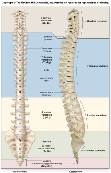
From top to bottom, the vertebrae are:
- Cervical spine: 7 vertebrae (C1–C7)
- Thoracic spine: 12 vertebrae (T1–T12)
- Lumbar spine: 5 vertebrae (L1–L5)
- Sacrum: 5 (fused) vertebrae (S1–S5)
- Coccyx: 4 (3–5) (fused) vertebrae (Tailbone)
What are the 5 parts of the axial skeleton?
The five parts of your axial skeleton include the bones in your skull, ossicles (small bones) of your middle ear, hyoid bone of your neck, vertebra (bones of your spine) and thoracic cage (ribcage). Which bones belong to the axial skeleton?
What are the three regions of the vertebral column?
The vertebrae are divided into three regions: cervical C1–C7 vertebrae, thoracic T1–T12 vertebrae, and lumbar L1–L5 vertebrae. The vertebral column is curved, with two primary curvatures (thoracic and sacrococcygeal curves) and two secondary curvatures (cervical and lumbar curves).
How many vertebrae are in the axial column?
The Vertebral Column - Axial Skeleton The vertebral column (see Figures 3.18–3.20) consists of 26 bones—24 vertebrae, 1 sacrum, and 1 coccyx. Together, they protect the spinal cord, maintain an upright body position, and provide support to the
How many vertebrae are in the thoracic vertebral column?
These vertebrae are larger and denser than the preceding vertebrae, allowing them to support the weight of the upper body. When you add 5 lumbar vertebrae to the 7 cervical vertebrae, you get 12, which is the number of the thoracic vertebrae.

How many vertebrae are in the axial skeleton?
In the human skeleton, it consists of 80 bones and is composed of six parts; the skull (22 bones), also the ossicles of the middle ear, the hyoid bone, the rib cage, sternum and the vertebral column....Axial skeletonTA98A02.0.00.009TA2353Anatomical terminology4 more rows
What are the 4 parts of the axial skeleton?
The axial skeleton includes the bones that form the skull, laryngeal skeleton, vertebral column, and thoracic cage.
What makes up the axial skeleton region?
The axial skeleton consists of the braincase (cranium) and the backbone and ribs, and it serves primarily to protect the central nervous system. The limbs and their girdles constitute the appendicular skeleton.
What are the 3 major regions of the axial skeleton?
The axial skeleton forms the central axis of the human body and consists of the skull, vertebral column, and thoracic cage.
What makes up the axial skeleton quizlet?
The axial skeleton consist of: the skull, hyoid bones, vertebral column, ribs, and sternum. the framework that consist of extremities, shoulders, pelvic and gridle.
Which is not a part of the axial skeleton?
Which of the following bones is not part of the axial skeleton? D. Sternum.
Which is not part of the axial skeleton quizlet?
Which of the following is NOT part of the axial skeleton? Upper limbs are part of the appendicular skeleton, which is composed of the humerus, radius, and ulna. The humerus attaches to the pectoral girdle, which is composed of the scapula and clavicle.
What are the three components of the axial skeleton and their functions?
Answer and Explanation: The axial skeleton contains the core skeletal structure without the limbs. It has three main parts: the skull, the vertebral column, and the thoracic cage. The skull holds and protects the brain as well as forms the underlying structures of the face.
Which of the following is the part of the axial skeleton?
The axial skeleton supports the head, neck, back, and chest and thus forms the vertical axis of the body. It consists of the skull, vertebral column (including the sacrum and coccyx), and the thoracic cage, formed by the ribs and sternum.
What are the regions of the skeletal system?
These are (1) the axial, comprising the vertebral column—the spine—and much of the skull, and (2) the appendicular, to which the pelvic (hip) and pectoral (shoulder) girdles and the bones and cartilages of the limbs belong.
What are the functions of the axial skeleton?
The function of the axial skeleton is to provide support and protection for the brain, the spinal cord, and the organs in the ventral body cavity. It provides a surface for the attachment of muscles that move the head, neck, and trunk, performs respiratory movements, and stabilizes parts of the appendicular skeleton.
What are the three components of the axial skeleton and their functions?
Answer and Explanation: The axial skeleton contains the core skeletal structure without the limbs. It has three main parts: the skull, the vertebral column, and the thoracic cage. The skull holds and protects the brain as well as forms the underlying structures of the face.
What are the main parts of the appendicular skeleton?
The appendicular skeleton is comprised of the upper and lower extremities, which include the shoulder girdle and pelvis. The shoulder girdle and pelvis provide connection points between the appendicular skeleton and the axial skeleton to where mechanical loads transfer.
What are the major parts of the appendicular skeleton?
1: Appendicular skeleton: The appendicular skeleton is composed of the bones of the pectoral limbs (arm, forearm, hand), the pelvic limbs (thigh, leg, foot), the pectoral girdle, and the pelvic girdle.
Which is not part of the axial skeleton quizlet?
Which of the following is NOT part of the axial skeleton? Upper limbs are part of the appendicular skeleton, which is composed of the humerus, radius, and ulna. The humerus attaches to the pectoral girdle, which is composed of the scapula and clavicle.
How many bones are in the vertebral column?
Vertebral Column Anatomy. The vertebral column is part of the axial skeleton, and it is made of 33 individual bones during youth, which anatomists classify as irregular bones. Approximately nine of the bones at the terminal end of the spine later fuse in adulthood to form two larger bones: the sacrum and the coccyx.
What are the four curvatures of the vertebral column?
When viewed from the side, the vertebral column features four curvatures: two are called primary curvatures, and two are called secondary curvatures. Primary curvatures, also called kyphotic curves, are curves that were present during fetal development. These curves are convex, curving outwardly toward the backside.
How many intervertebral discs are there?
Intervertebral Discs Anatomy. Twenty-three inter vertebral discs separate, anchor, and cushion each vertebra. However, there is no intervertebral disc between C1 (atlas) and C2 (axis), or between the sacrum and coccyx bones. As you move down the spine, these shock-absorbing pads of fibrocartilage progressively thicken in size, ...
What is the tailbone of the coccyx?
1 coccyx, which consists of 3-5 fused coccygeal vertebrae (Co1-Co4) – This bone is also called the tailbone, and it represents the terminal end of the vertebral column. It articulates with the sacrum bone above.
How many vertebrae are there in the cervical column?
Anatomists also call this region the cervical spine. Seven and cervical both start with the same “s” sound, so that can help you remember that there are seven cervical vertebrae.
How many thoracic vertebrae are there?
It’s easy to remember that there are 12 thoracic vertebrae, because they articulate with the 12 pairs of ribs to form part of the thoracic cage, and twelve and thoracic both start with the letter “t.”. 5 lumbar vertebrae are inferior (below) the thoracic vertebrae, abbreviated as L1-L5.
What are the two parts of the fibrocartilage?
As you move down the spine, these shock-absorbing pads of fibrocartilage progressively thicken in size, and they consist of two main parts: the nucleus pulposus and the annulus fibrosus.
How many bones are in the vertebral column?
The vertebral column (see Figures 3.18–3.20) consists of 26 bones—24 vertebrae, 1 sacrum, and 1 coccyx. Together, they protect the spinal cord, maintain an upright body position, and provide support to the
What are the parts of the vertebrae?
Each vertebra consists of three parts: the body,vertebral arch, and articular processes (see Figure3.18). The body is thick, disk-shaped, and located an-teriorly. The bodies are interconnected by ligaments. Interspersed between each vertebra are fibrocartilage pads called intervertebral disks . Two vertebral arches lead off posterolaterally from thebody. The two, short, thick processes leading off from the body are known as pedicles. The pedicles have depressions on the superior and inferior surfaces (vertebral notches). The pedicles join the laminae, the flat, posterior part of the arch. These arches meet posteriorly to enclose an opening called thevertebralforamen. Because the vertebrae lie on top of eachother, the successive vertebral foramen form a verte-bral canal. The spinal cord lies in the vertebral canal.Laterally, a small foramen is formed where the notches on the pedicles of successive vertebrae align. This is the intervertebral foramen. The spinal nerves exit from the vertebral canal through these foramen. Posteriorly, each vertebra has a projection called the spinous process. This forms the bumps that are seen in the middle of the back in a lean indi-vidual. The most prominent of these bumps at the base of the neck indicates the location of the C7 spin-ous process. C7 is, therefore, called thevertebraprominens (also see Fig. M 3).
How many vertebrae are there in the cervical column?
The vertebral column is subdivided into the cervical (7 vertebrae), thoracic (12 vertebrae), lumbar (5 ver-tebrae), sacral (1 vertebrae), and coccygeal (1 verte-bra)regions. Although the cervical, thoracic, and lumbar consist of individual vertebrae, the sacrum is formed by the fusion of 5 individual vertebrae and the coccyx is formed by the fusion of 3–5 vertebrae. For ease, the vertebrae are labeled according to the position in individual regions (e.g., the 7th cervical vertebra is labeled C7; 2nd thoracic vertebra as T2; and so on.
Why do vertebrae have a large foramen?
For example, the cervical vertebrae have a large vertebral foramen because all the nerves ascending and descending from the brain form the spinal cord here. As the lower regions are approached, the vertebral foramen become smaller. This is because of the exit of spinal nerves according to the region they supply. As the spinal cord descends, more and more nerves leave; hence, the tapering appearance of the spinal cord. The cervical vertebrae are also smaller as they only must bear the weight of the head. The vertebrae in the other regions become sturdier, the lumbar being the largest (Figure 3.18D) because they have to bear more weight. The thoracic vertebrae have extra facets on the transverse processes and the body that articu-late with the ribs (Figure 3.18C). The transverse processes of the cervical vertebrae have a foramen (transverse foramen), through which the vertebral artery, vein, and nerve pass. The spinous processes of the cervical vertebrae C2–C6 are often bifid.
How are vertebrae separated?
From the axis to the sacrum, the vertebral bodies are separated from each other by fibrocartilage called in-tervertebral disks. Each disk-shaped structure is made of a tough outer layer, the annulus fibrosus. The collagen fibers of this layer attach adjacent bod-ies of the vertebrae. The annulus fibrosus encloses a gelatinous, elastic and soft core called the nucleuspulposus. Seventy-five percent of this core is water,with scattered strands of elastic and reticular fibers. The disks serve as shock absorbers. They also allow the vertebrae to glide over each other slightly, with-out loosing alignment. Because the disks contribute to one-fourth of the length of the vertebral column, the height of the individual diminishes as the disks loose water and become narrower with age.
What is the first cervical vertebra?
The first cervical vertebra is the atlas (Figure 3.18A) as it bears the weight of the head. It articulates with the occipital condyles of the skull. This joint—the atlanto-occipital joint—permits the nodding of the head. The atlas does not have a body and spinous process, instead it has anterior and posterior arches and a thick, lateral mass. The second vertebra is called the axis (Figure 3.18B). It has a projection (dens, or odontoid process) that projects superiorly from theregion of the body of the vertebra. This process is held in place against the inner surface of the atlas by a transverse ligament. This joint allows the head to ro-tate and pivot on the neck. The dens is actually the fu-sion of the body of the atlas with that of the axis.
Which vertebrae are aligned to form four spinal curves?
The individual vertebra of the vertebral column are aligned to form four spinal curves (see Figure 3.17)—the cervical, thoracic, lumbar, and sacralcurvature. The thoracic and sacral curvatures havethe concavity of the curve facing forward.
How many bones are there in the vertebral column?
Definition: The 24 individual bones of the vertebral column
How many vertebrae are in a bone?
Definition: Bone formed from five vertebrae fused together near the base of the spinal column
What is the roughened area between the auricular surface and the lateral sacral crest?
Definition: Roughened area between the auricular surface and the lateral sacral crest; serves as an attachment point for ligaments that stabilize the joint between the sacrum and the pelvic girdle
What is the term for the material that acts as shock absorbers between the vertebrae and allows the back to move?
Definition: Cartilage that act as shock absorbers between the vertebrae and allow the back to move;
How many fused vertebrae form the tailbone?
Definition: 3-5 fused vertebrae that form the tailbone
How many pairs are there in a thoracic vertebra?
Definition: 12 pairs; Attached to the thoracic vertebra
Where is the round central portion of the vertebrae located?
Definition: Round central portion of the vertebrae located on the anterior side of the bone; filled with spongey bone; adjacent bodies are separated and cushioned by intervertebral discs
What are the five regions of the vertebral column?
The vertebral column is grouped into five regions: the cervical spine (C01-C07), the thoracic spine (T01- T-12), the lumbar spine (L01-L05), the sacral spine, and the coccygeal spine. 4. The Bones of the Thoracic Cage Protect Internal Organs.
What is the axial skeleton?
The axial skeleton includes all the bones along the body’s long axis. Let’s work our way down this axis to learn about these structures and the bones that form them. The axial skeleton includes the bones that form the skull, laryngeal skeleton, vertebral column, and thoracic cage. The bones of the appendicular skeleton (the limbs and girdles) ...
What are the ribs in the sternum?
The sternum consists of the manubrium, body of the sternum, and xiphoid process. Ribs 1-7 are called true ribs because they articulate directly to the sternum, and ribs 8-12 are known as false ribs. Download Axial Skeleton Lab Manual. See more from our free eBook library.
How many bones are there in the facial skeleton?
The 14 bones of the facial skeleton form the entrances to the respiratory and digestive tracts. The facial skeleton is formed by the mandible, maxillae (r,l), zygomatics (r,l), and the bones that give shape to the nasal cavity: lacrimals (r,l), nasals (r,l), vomer, palatines (r,l), and the nasal conchae (r,l). Cranial Bones.
What are the cranial bones of a baby?
In fetuses and newborn infants, cranial bones are connected by flexible fibrous sutures, including large regions of fibrous membranes called fontanelles. These regions allow the skull to enlarge to accommodate the growing brain.
What is the tailbone of the human body?
The coccyx, or tailbone, is formed by a fusion of four vertebrae. The bones of the human skeleton are divided into two groups. The appendicular skeleton includes all the bones that form the upper and lower limbs, and the shoulder and pelvic girdles. The axial skeleton includes all the bones along the body’s long axis.
How many vertebrae are in the spinal column?
The vertebral column is a flexible column formed by a series of 24 vertebrae, plus the sacrum and coccyx. Commonly referred to as the spine, the vertebral column extends from the base of the skull to the pelvis. The spinal cord passes from the foramen magnum of the skull through the vertebral canal within the vertebral column. The vertebral column is grouped into five regions: the cervical spine (C01-C07), the thoracic spine (T01- T-12), the lumbar spine (L01-L05), the sacral spine, and the coccygeal spine.
How many vertebrae are there in the cervical spine?
In the neck, there are seven cervical vertebrae, each designated with the letter “C” followed by its number. Superiorly, the C1 vertebra articulates (forms a joint) with the occipital condyles of the skull. Inferiorly, C1 articulates with the C2 vertebra, and so on. Below these are the 12 thoracic vertebrae, designated T1–T12. The lower back contains the L1–L5 lumbar vertebrae. The single sacrum, which is also part of the pelvis, is formed by the fusion of five sacral vertebrae. Similarly, the coccyx, or tailbone, results from the fusion of four small coccygeal vertebrae. However, the sacral and coccygeal fusions do not start until age 20 and are not completed until middle age.
How many curvatures are there in the vertebral column?
The adult vertebral column does not form a straight line, but instead has four curvatures along its length (see Figure 7.20 ). These curves increase the vertebral column’s strength, flexibility, and ability to absorb shock. When the load on the spine is increased, by carrying a heavy backpack for example, the curvatures increase in depth (become more curved) to accommodate the extra weight. They then spring back when the weight is removed. The four adult curvatures are classified as either primary or secondary curvatures. Primary curves are retained from the original fetal curvature, while secondary curvatures develop after birth.
What is the spinal column?
It consists of a sequence of vertebrae (singular = vertebra), each of which is separated and united by an intervertebral disc. Together, the vertebrae and intervertebral discs form the vertebral column. It is a flexible column that supports the head, neck, and body and allows for their movements. It also protects the spinal cord, which passes down the back through openings in the vertebrae.
Why are cervical vertebrae smaller than lumbar vertebrae?
Thus, cervical vertebrae are smaller than lumbar vertebrae due to differences in the proportion of body weight that each supports. Thoracic vertebrae have sites for rib attachment, and the vertebrae that give rise to the sacrum and coccyx have fused together into single bones.
How many cervical vertebrae are there in a giraffe?
An interesting anatomical fact is that almost all mammals have seven cervical vertebrae, regardless of body size. This means that there are large variations in the size of cervical vertebrae, ranging from the very small cervical vertebrae of a shrew to the greatly elongated vertebrae in the neck of a giraffe. In a full-grown giraffe, each cervical vertebra is 11 inches tall.
What are the disorders associated with the curvature of the spine?
Disorders associated with the curvature of the spine include kyphosis (an excessive posterior curvature of the thoracic region), lordosis (an excessive anterior curvature of the lumbar region), and scoliosis (an abnormal, lateral curvature, accompanied by twisting of the vertebral column).
Which vertebrae are larger than cervical vertebrae?
The bodies of the thoracic vertebrae are larger than those of cervical vertebrae ( Figure 7.26 ). The characteristic feature for a typical midthoracic vertebra is the spinous process, which is long and has a pronounced downward angle that causes it to overlap the next inferior vertebra. The superior articular processes of thoracic vertebrae face anteriorly and the inferior processes face posteriorly. These orientations are important determinants for the type and range of movements available to the thoracic region of the vertebral column.
Which vertebrae are axis?
Axis - the second cervical vertebra of mammals; rotary movements of the head occur between the atlas and axis
How many regions are there in the postcranial skeleton?
The postcranial skeleton can be divided into two regions:
What is the body of the vertebra that replaces the notochord?
Centrum - body of the vertebra which replaces the notochord; shape depends on the vertebrate class (Fig. 8.4, p. 277):
What is the term for all vertebral elements fused into a single piece?
holospondyly - all vertebral elements are fused into a single piece
Which vertebrae articulate with the skull?
Cervical - vertebrae of neck (not found in fishes) Atlas - the first cervical vertebra of terrestrial vertebrates which articulates with the skull; nodding movements of the head occur between the atlas and the skull. Axis - the second cervical vertebra of mammals; rotary movements of the head occur between the atlas and axis.
What are the two anatomical relationships between the centra and neural arches?
In tetrapods, two general anatomical relationships occur between centra and neural arches: aspidospondyly - all arch elements (intercentrum, pleurocentrum, and neural arch) remain as separate ossified elements. holospondyly - all vertebral elements are fused into a single piece.
What is the Caudal vertebrae?
Caudal - vertebrae of tail (pygostyle in birds forms attachment for tail feathers)
How many degrees of motion does the anterior thoracic vertebrae have?
The anterior thoracic vertebrae are very mobile (up to 15 degrees of motion) in axial rotation, due to the unique configuration of horizontally oriented ‘prediaphragmatic’-type zygapophyses found at these joints that allow vertebrae to twist (PreZA, Fig. 1a, top panel).
Which outgroup of therapsids has low functional distances?
The outgroup, Ambystoma (tiger salamander), the tegu ( Salvator ), ‘pelycosaurs’ and most non-mammalian therapsids, all have low functional distances with a tight distribution, reflecting functional uniformity of vertebral regions.
Is the vertebral column a metameric structure?
The mammal vertebral column is a classic example of a metameric structure that is both modular, with well-defined morphological regions, and functionally differentiated. How the evolution of regions is related to their functional differentiation in the forerunners of mammals remains unclear.