Key facts about liver ligamentsTable quiz
| Coronary ligament | Coronal ligament Attaches the liver to t ... |
| Triangular ligament | Asymmetrical Right and left components C ... |
| Falciform ligament | Sickle-shaped Anchors the liver the ante ... |
| Ligamentum teres (hepatis) | Round ligament of the liver A remnant of ... |
| Ligamentum venosum | A remnant of the ductus venosum Occupies ... |
What are ligaments suspend the liver from the abdominal wall?
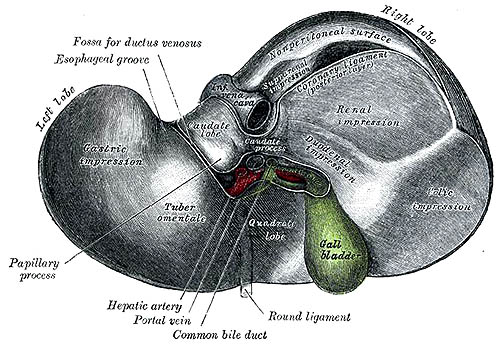
—The liver is connected to the under surface of the diaphragm and to the anterior wall of the abdomen by five ligaments; four of these—the falciform, the coronary, and the two lateral—are peritoneal folds; the fifth, the round ligament, is a fibrous cord, the obliterated umbilical vein.
What is the best ligament supplement?
- Taurine: A potent anti-inflammatory that supports the immune system to promote healing
- Vitamin D: Can promote healing in ligaments and tendons.
- Magnesium: Promotes tissue health and boosts healing in joint cartilage, tendons, and ligaments.
- Silicone: Promotes bone and tissue strength.
- Trace minerals: These include copper, zinc, and manganese. ...
Is a liver lobe the same as a liver lobule?
The lobules of liver, or hepatic lobules, are small divisions of the liver defined at the microscopic scale. The hepatic lobule is a building block of the liver tissue, consisting of a portal triad, hepatocytes arranged in linear cords between a capillary network, and a central vein. Lobules are different from the lobes of the liver: they are the smaller divisions of the lobes. The two-dimensional microarchitecture of the liver can be viewed from different perspectives: Name Shape Model classica
Is the stomach to the right of the liver?
The stomach is located in the upper-left area of the abdomen below the liver and next to the spleen. Its main function is to store and break down the foods and liquids that we consume before those...
See more

What ligament holds the liver in place?
Falciform ligament - this sickle-shaped ligament attaches the anterior surface of the liver to the anterior abdominal wall. Its free edge contains the ligamentum teres, a remnant of the umbilical vein.
Which ligament of liver is derived from a vessel?
Development. The round ligament of the liver is the remnant of the umbilical vein during embryonic development.
What is the round ligament of the liver?
The free, inferior border of the falciform ligament contains the paraumbilical veins and the round ligament of the liver (aka, ligamentum teres hepatis) which courses along a fissure situated between the inferior surface of the right and left lobes.
How many ligaments are present in liver?
It consists of four lobes and two major surfaces. Because it is such a large organ, it requires seven ligaments to keep it relatively immobile in its anatomical location. The largest of the seven hepatic ligaments is the coronary (coronal) ligament.
What is ligamentum teres?
However, the ligamentum teres is a strong intraarticular ligament that is anatomically and biochemically similar to the anterior cruciate ligament of the knee. It is composed of two bands that originate from the acetabular transverse ligament and the pubic and ischial margins of the acetabular notch.
What are ligaments?
A ligament is a fibrous connective tissue that attaches bone to bone, and usually serves to hold structures together and keep them stable.
What is triangular ligament?
The left triangular ligament is a large peritoneal fold. It connects the posterior part of the upper surface of the left lobe of the liver to the thoracic diaphragm.
What is Treitz ligament?
The ligament of Treitz is a thin band of tissue (peritoneum) that connects and supports the end of the duodenum and beginning of the jejunum in the small intestine. It's also called the suspensory muscle of duodenum.
Which ligament attaches the liver to the diaphragm?
The right triangular ligament is formed in a similar fashion adjacent to the bare area and attaches the right lobe of the liver to the diaphragm. Lesser omentum – Attaches the liver to the lesser curvature of the stomach and first part of the duodenum.
Which ligament attaches the anterior surface of the liver to the anterior abdominal wall?
Falciform ligament – this sickle-shaped ligament attaches the anterior surface of the liver to the anterior abdominal wall and forms a natural anatomical division between the left and right lobes of the liver. The free edge of this ligament contains the ligamentum teres, a remnant of the umbilical vein.
What are the two surfaces of the liver?
There are two liver surfaces – the diaphragmatic and visceral: Diaphragmatic surface – the anterosuperior surface of the liver. It is smooth and convex, fitting snugly beneath the curvature of the diaphragm.
What is the liver covered by?
Macroscopic. The liver is covered by a fibrous layer, known as Glisson's capsule. It is divided into a right lobe and left lobe by the attachment of the falciform ligament. There are two further 'accessory' lobes that arise from the right lobe, and are located on the visceral surface of liver:
What is the lobe of the liver?
Macroscopic. The liver is covered by a fibrous layer, known as Glisson’s capsule. It is divided into a right lobe and left lobe by the attachment of the falciform ligament. There are two further ‘accessory’ lobes that arise from the right lobe, and are located on the visceral surface of liver:
Which plexus innervates the parenchyma of the liver?
The parenchyma of the liver is innervated by the hepatic plexus, which contains sympathetic (coeliac plexus) and parasympathetic (vagus nerve) nerve fibres. These fibres enter the liver at the porta hepatis and follow the course of branches of the hepatic artery and portal vein.
Which nerves enter the liver?
Nerve Supply. The parenchyma of the liver is innervated by the hepatic plexus, which contains sympathetic (coeliac plexus) and parasympathetic (vagus nerve) nerve fibres. These fibres enter the liver at the porta hepatis and follow the course of branches of the hepatic artery and portal vein.
Which ligament is continuous with the liver capsule?
The posterior and superior extension of the hepatogastric ligament (lesser omentum) branches, reflects on itself and becomes continuous with the liver capsule. As it branches to the right it reflects on itself, becomes continuous with the liver capsule and becomes the right inferior coronary ligament.
What is the union of the right and left triangular ligaments?
On the right the union is called the right triangular ligament. On the left the left triangular ligament extends toward and attaches to the diaphragm to end in a large fold and a strong fibrous band called the a ppendix fibrosa hepatis.
What is the ligamentum teres?
The ligamentum teres. The ligamentum teres (round ligament) represents the obliterated left umbilical vein. It originates from the umbilicus, courses up the anterior abdominal wall within the the peritoneum and then enters the falciform ligament coursing toward the left portal vein and ligamentum venosum.
What is the falciform ligament?
The falciform ligament is a fold of peritoneum that encloses the round ligament anteriorly. On the superior aspect of the liver it departs from the round ligament, branches, reflects on itself, and becomes continuous with the liver capsule.
What is the bare area of the liver?
The bare area is thus an area on the posterior aspect of the liver bounded by the coronary ligaments. The inferior vena cava traverses the liver through and posterior to the bare area. The right adrenal forms an impression on the liver in the region of the bare area. The ligamentum teres (round ligament) represents the obliterated left umbilical ...
Where does the omentum go in the liver?
Within the liver it courses anteriorly toward the porta hepatis and posteriorly toward the posterior aspects of the liver. The anterior and inferior extension of the lesser omentum divides and becomes continuous with Glissons’ capsule around the porta hepatis.
Which ligament forms the posterior border of the foramen?
The posterior border of the foramen is formed by the ivc. The lesser omentum extends into the ligamentum venosum, and then extends to the porta hepatis and the posterior and inferior components of the coronary ligament. Ligament of the inferior vena cava; Ligaments of the liver.
Development
The round ligament of the liver is the remnant of the umbilical vein during embryonic development. It only exists in placental mammals. After the child is born, the umbilical vein degenerates to fibrous tissue.
Portal hypertension
In adulthood, small paraumbilical veins remain in the substance of the ligament. These act as an important portacaval anastomosis in severe portal hypertension, resulting in a caput medusae.
Abscess
Very rarely, the round ligament of the liver may develop an abscess. This usually requires liver surgery to treat.
Landmark
The umbilical vein/round ligament inserts around the umbilicus, and is an important landmark of the inner surface of the anterior abdominal wall.
Which ligaments are directly related to the liver?
There are five ligaments that are directly related to the liver and they are called: Coronary ligament - formed by the peritoneal reflection from the diaphragm to the liver which has two layers that meet on the right. Left triangular ligament - is a mix of the falciform ligament and the lesser omentum.
Which ligament divides the liver and the diaphragm?
The subphrenic recess which is split by the falciform ligament of the liver, is the division between the liver and the diaphragm. The hepatorenal recess is on the inferior right aspect of the liver and separates it from the kidney anterior inferiorly and the suprarenal gland posterior inferiorly.
What is the function of portal vein?
Vascularization. Functional: portal vein (metabolic processing of the matters absorbed in intestines) Nutritive: hepatic artery (supplying the tissue of the liver with oxygen and nutrients) Drainage: hepatic vein -> inferior vena cava -> right atrium. Innervation.
Why is the liver important?
The liver is a special organ in the sense that it receives more venous blood than arterial blood and this is due to the fact that the liver helps clean the blood via detoxification. The majority of the vascular supply is brought into the organ by the portal vein which carries the blood filled with metabolytes absorbed in the intestines, whereas the rest of the blood comes from the common hepatic artery which originates from the celiac trunk and carries the oxygenated blood to the liver.
What is the left triangular ligament?
Left triangular ligament - is a mix of the falciform ligament and the lesser omentum. Falciform ligament - is not of embryological origin, but a peritoneal reflection of the upper abdominal wall from the umbilicus to the liver and has the round ligament of the liver on its free edge.
What is the fibrous remnant of the umbilical vein?
It is a fibrous remnant of the umbilical vein which still extends from the internal aspect of the umbilicus up to the liver. Ligamentum venosum - is also an embryonic remnant of the ductus venosus. In utero it extended between the umbilical vein and the inferior vena cava.
Where does lymphatic drainage go?
The deep system consists of hepatic lymph vessels which follow the hepatic portal veins, therefore most of the lymph will flow towards the hepatic nodes at the hilum of the liver, which drain to the celiac nodes.
What is the liver?
The liver is reddish-brown and shaped approximately like a cone or a wedge, with the small end above the spleen and stomach and the large end above the small intestine. The entire organ is located below the lungs in the right upper abdomen. It weighs between 3 and 3.5 pounds.
Which ligament separates the two lobes of the liver and connects it to the abdominal wall?
Falciform Ligament: A thin, fibrous ligament that separates the two lobes of the liver and connects it to the abdominal wall. Glisson’s Capsule: A layer of loose connective tissue that surrounds the liver and its related arteries and ducts.
How many lobes are there in the liver?
The liver consists of four lobes: the larger right lobe and left lobe, and the smaller caudate lobe and quadrate lobe. The left and right lobe are divided by the falciform (“sickle-shaped” in Latin) ligament, which connects the liver to the abdominal wall. The liver’s lobes can be further divided into eight segments, which are made up of thousands of lobules (small lobes). Each of these lobules has a duct flowing toward the common hepatic duct, which drains bile from the liver.
What are the functions of the liver?
The liver is an essential organ of the body that performs over 500 vital functions. These include removing waste products and foreign substances from the bloodstream, regulating blood sugar levels, and creating essential nutrients. Here are some of its most important functions: 1 Albumin Production: Albumin is a protein that keeps fluids in the bloodstream from leaking into surrounding tissue. It also carries hormones, vitamins, and enzymes through the body. 2 Bile Production: Bile is a fluid that is critical to the digestion and absorption of fats in the small intestine. 3 Filters Blood: All the blood leaving the stomach and intestines passes through the liver, which removes toxins, byproducts, and other harmful substances. 4 Regulates Amino Acids: The production of proteins depend on amino acids. The liver makes sure amino acid levels in the bloodstream remain healthy. 5 Regulates Blood Clotting: Blood clotting coagulants are created using vitamin K, which can only be absorbed with the help of bile, a fluid the liver produces. 6 Resists Infections: As part of the filtering process, the liver also removes bacteria from the bloodstream. 7 Stores Vitamins and Minerals: The liver stores significant amounts of vitamins A, D, E, K, and B12, as well as iron and copper. 8 Processes Glucose: The liver removes excess glucose (sugar) from the bloodstream and stores it as glycogen. As needed, it can convert glycogen back into glucose.
What does the liver store?
Stores Vitamins and Minerals: The liver stores significant amounts of vitamins A, D, E, K, and B12, as well as iron and copper. Processes Glucose: The liver removes excess glucose (sugar) from the bloodstream and stores it as glycogen. As needed, it can convert glycogen back into glucose.
How to avoid liver disease?
The best way to avoid liver disease is to take active steps toward a healthy life. The following are some recommendations that will help keep the liver functioning as it should: Avoid Illicit Drugs: Illicit drugs are toxins that the liver must filter out. Taking these drugs can cause long-term damage.
How to treat fatty liver?
While the liver can moderate amounts, excessive alcohol use can cause damage. Exercise Regularly: A regular exercise routine will help promote general health for every organ, including the liver. Eat Healthy Foods: Eating excessive fats can make it difficult for the liver to function and lead to fatty liver disease.
What is the liver?
Shaped like a cone, the liver is a dark reddish-brown organ that weighs about 3 pounds. There are 2 distinct sources that supply blood to the liver, including the following: Oxygenated blood flows in from the hepatic artery. Nutrient-rich blood flows in from the hepatic portal vein.
Which part of the liver transports bile?
The common hepatic duct transports the bile made by the liver cells to the gallbladder and duodenum (the first part of the small intestine) via the common bile duct.
What happens when the liver breaks down harmful substances?
When the liver has broken down harmful substances, its by-products are excreted into the bile or blood. Bile by-products enter the intestine and leave the body in the form of feces. Blood by-products are filtered out by the kidneys, and leave the body in the form of urine.
How does the liver help the body?
This helps carry away waste products from the liver. All the blood leaving the stomach and intestines passes through the liver. The liver processes this blood and breaks down, balances, and creates the nutrients and also metabolizes drugs into forms that are easier to use for the rest of the body or that are nontoxic.
How many lobes are there in the liver?
The liver consists of 2 main lobes. Both are made up of 8 segments that consist of 1,000 lobules (small lobes). These lobules are connected to small ducts (tubes) that connect with larger ducts to form the common hepatic duct. The common hepatic duct transports the bile made by the liver cells to the gallbladder and duodenum ...
What is the function of hemoglobin in the liver?
Processing of hemoglobin for use of its iron content (the liver stores iron) Conversion of poisonous ammonia to urea (urea is an end product of protein metabolism and is excreted in the urine) Clearing the blood of drugs and other poisonous substances. Regulating blood clotting.
How does the liver resist infections?
Resisting infections by making immune factors and removing bacteria from the bloodstream. Clearance of bilirubin, also from red blood cells. If there is an accumulation of bilirubin, the skin and eyes turn yellow. When the liver has broken down harmful substances, its by-products are excreted into the bile or blood.
Which ligament is the largest of the liver?
The coronary ligament is the largest of these, having an anterior (frontal) and posterior (back) layers.
What is the ligament that holds the liver to the diaphragm?
Anatomical terminology. The coronary ligament of the liver refers to parts of the peritoneal reflections that hold the liver to the inferior surface of the diaphragm .
Which ligament is reflected from the lower margin of the bare area?
The posterior layer of the coronary ligament is reflected from the lower margin of the bare area and is continuous with the right layer of the lesser omentum . The anterior and posterior layers converge on the right and left sides of the liver to form the right triangular ligament and the left triangular ligament, respectively.
Which surface of the liver has no peritoneal covering?
The diaphragmatic surface of the liver that is in direct contact with the diaphragm (just beyond the peritoneal reflections) has no peritoneal covering, and is termed the bare area of the liver . The anterior layer of the coronary ligament is formed by the reflection of the peritoneum from the upper margin of the bare area ...

Overview
External links
• Anatomy photo:38:12-0106 at the SUNY Downstate Medical Center - "Stomach, Spleen and Liver: The Visceral Surface of the Liver"
• Anatomy image:7819 at the SUNY Downstate Medical Center
• Overview at ucc.edu
Structure
The round ligament connects the liver to the umbilicus. It divides the left part of the liver into medial and lateral sections.
The round ligament of the liver is the remnant of the umbilical vein during embryonic development. It only exists in placental mammals. After the child is born, the umbilical vein degenerates to fibrous tissue.
Clinical significance
In adulthood, small paraumbilical veins remain in the substance of the ligament. These act as an important portacaval anastomosis in severe portal hypertension, resulting in a caput medusae.
Very rarely, the round ligament of the liver may develop an abscess. This usually requires liver surgery to treat.
The umbilical vein/round ligament inserts around the umbilicus, and is an important landmark of …
Additional Images
• Round ligament of liver.Superior surface of liver.