How do you calculate the magnification of a microscope?
On-Screen Magnification
- Firstly, you need to know the magnification of the microscope's objective lens. It is generally printed on the side of the lens. ...
- Now you have to locate the number written on the c-mount adapter. ...
- You have then to measure the diagonal of the screen of the monitor. ...
- Finally, you need to detect the size of the sensor camera present. ...
What are the types of objective lenses on a microscope?
- Scanning Objective lens that has a magnification power of 4x
- A small objective lens that has a magnification power of 10x
- A large objective lens having up to 100x magnification
- Oil-immersion lens having magnification higher than 100x
What is the Typical magnification of a light microscope?
The magnification level of an objective lens on a light microscope will typically range from 5x magnification all the way up to 100x magnification. Some extremely high-performing ocular microscopes require matching magnification levels on the eyepiece to deliver the best performance.
What is the magnification of an objective lens?
What Are the Different Magnifications of Objective Lenses?
- Scanning Objective Lens (4x)
- Low Power Objective (10x)
- High Power Objective Lens (40x)
- Oil Immersion Objective Lens (100x)
- Specialty Objective Lenses (2x, 50x Oil, 60x and 100x Dry)
What is the objective magnification of a microscope?
Most compound microscopes come with interchangeable lenses known as objective lenses. Objective lenses come in various magnification powers, with the most common being 4x, 10x, 40x, and 100x, also known as scanning, low power, high power, and (typically) oil immersion objectives, respectively.
What are the 3 magnifications on a microscope?
The compound microscope typically has three or four magnifications - 40x, 100x, 400x, and sometimes 1000x. At 40x magnification you will be able to see 5mm. At 100x magnification you will be able to see 2mm. At 400x magnification you will be able to see 0.45mm, or 450 microns.
What is the magnification of a microscope with a 10x ocular and a 40x objective?
A microscope's total magnification is a combination of the eyepieces and the objective lens. For example, a biological microscope with 10x eyepieces and a 40x objective has 400x magnification.
What are the 4 types of objective and their magnification?
But most commonly, when talking about types of objective lenses we are referring to the different magnifications and purposes of the four most common types of microscope objective lenses on compound light microscopes. Those four are: The scanning lens (4x)...High Power Lens (40x)Mould.Cells.Germs.Tardigrades.
What is the total magnification of 4x 10x and 40x?
400xGrades 1-8 typically will buy a monocular compound microscope with 3 objective lenses: 4x, 10x, 40x for maximum total magnification of 400x.
What is the magnification of the 3 objective lens?
Objective Lenses: Usually you will find 3 or 4 objective lenses on a microscope. They almost always consist of 4x, 10x, 40x and 100x powers. When coupled with a 10x (most common) eyepiece lens, total magnification is 40x (4x times 10x), 100x , 400x and 1000x.
What is the total magnification of 10x ocular and 100X objective?
1000xThe total magnification is the magnification of the ocular lens (10) multiplied by the objective lens (100). If we multiply 100 x 10 = 1,000. This is noted as the total magnification is 1000x, or 1000 times the size of the specimen.
What is the total magnification of 10x eyepiece and 100X the objectives?
The total magnification is multiplication, so the total is ocular multiplied by objective. So here The ocular is 10 and the objective is 100. So it's 10 times 100 which is 1000. So the total magnification Is 1000 times.
What is the total magnification if you use the 10x objective 40x 100X?
Your total magnification is objective magnification times ocular lens, so the 100x oil immersion objective with the 10x ocular delivers 1000x magnification in total.
Which objective has a magnification of 4x?
Your microscope has 4 objective lenses: Scanning (4x), Low (10x), High (40x), and Oil Immersion (100x).
What is the 10x objective lens called?
Understanding Objective Lens Many microscopes will be equipped with a scanning objective (4x), a low power objective (10x), a high power objective (40x), and perhaps even an oil immersion objective lens.
What are the 4 microscope objectives?
Most microscope objective specifications are listed on the body of the objective itself: the objective design/standard, magnification, numerical aperture, working distance, lens to image distance, and cover slip thickness correction.
What is bigger 10x or 40x?
It's easy to understand. A 40x objective makes things appear 40 times larger than they actually are. Comparing objective magnification is relative—a 40x objective makes things twice as big as a 20x objective while a 60x objective makes them six times larger than a 10x objective.
What does x3 magnification mean?
WHAT DOES IT MEAN WHEN IT SAYS 2x, 3x, 6x ETC ALONGSIDE A MAGNIFIER? This refers to the number of times a magnifier enlarges the subject matter: hence '2x' enlarges it to twice its size, '6x' enlarges to six times the subject size.
What is the difference between 4x 10x and 40x on a microscope?
For example, optical (light) microscopes are usually equipped with four objectives: 4x and 10x are low power objectives; 40x and 100õ are powerful ones. The total magnification (received with 10x eyepiece) of less than 400x characterizes the microscope as a low-powered model; more than 400x as a powerful one.
What is 10x magnification called?
Terms in this set (13) 10x magnification and is the lens closest to your eye. Also called ocular lens.
How to calculate magnification of a microscope?
To calculate the magnification on a microscope multiply the magnification power of the eyepiece you are using by the objective currently in position.
What does microscope magnification mean?
If you’ve used a microscope before you have probably see “100X” or “400X” or heard people talk about magnification, but what does that actually mean in the context of a microscope? Microscope magnification is the microscope’s ability to enlarge an image of an object through a series of lenses to ...
What is the purpose of a compound microscope?
Modern compound microscopes contain an eyepiece, an objective, and a condenser lens and together these lenses work to refract the light that enters our eye and serves to enlarge the specimen under inspection. In fact, the objective lens has within it, several compounding lenses that contribute to higher and higher magnification powers. If you are not familiar with these terms please take a look at my article called Parts of a Compound Microscope: Diagrams and Video to familiarize yourself with the anatomy of a compound microscope.
How to find maximum magnification of an objective lens?
To find the maximum useful magnification for an objective lens multiply 1000 by the numerical aperture.
What happens if you increase the magnification of an objective lens?
If you increase the magnification beyond what the objective lens can resolve you will end up with “Empty Magnification”. You can perform a simple calculation that can tell you before hand what the highest magnification levels will be so you can avoid empty magnification.
What is the objective lens?
The objective lens then gathers the light that has been passed through the specimen and projects an image in the body tube. The eyepiece, being further away from image the objective lenses has projected, is able to further magnify the image and the eye of the person using the microscope sees this secondarily magnified image.
How much magnification does an electron microscope have?
Typically, the standard light microscope will max out at about 1,500X magnification and the electron microscope will be able to achieve 200,000X magnification. To put that into perspective the human eye can see things down to single strand of hair, the thickness of which is about 0.065 millimeters.
What is the total magnification of a microscope?
The total magnification of a microscope = magnification power of the ocular lens x magnification power of the objective lens.
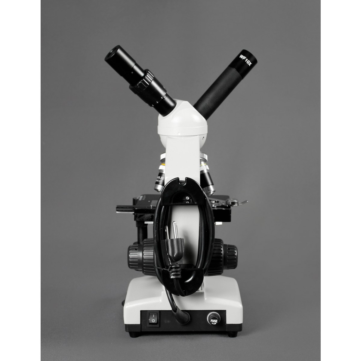
How does a compound light microscope work?
Compound light microscopes magnify objects by using a system of lenses and a light source. They are commonly employed to research bacteria, single cells, and various cell components. You will need to know the power of the ocular and objective lenses to figure out how much your microscope can magnify. The ocular lens is set up in the eyepiece, and ...
How to find the magnification of an ocular lens?
Check for the magnification power of the ocular lens. You can find it marked on the outside of the eyepiece, otherwise, you can look in the manual. The ocular lens usually magnifies 10 times. Next, look for the magnification capacity of your objective lens. You can find this on the side of the lens, or the manual can help. Traditionally the value can vary among 4x, 10x, 40x, 100x.
How many times does an ocular lens magnify?
The ocular lens usually magnifies 10 times. Next, look for the magnification capacity of your objective lens. You can find this on the side of the lens, or the manual can help. Traditionally the value can vary among 4x, 10x, 40x, 100x. As the objective lens is the first one to magnify, it is located on a rotating wheel just above ...
Why is the illumination of a microscope important?
Lighting plays an important role to make the microscope portable for field research. It must concentrate on the specimen to study the details appropriately. There are both top and bottom illumination available for some microscopes but in general, the light is located at the bottom.
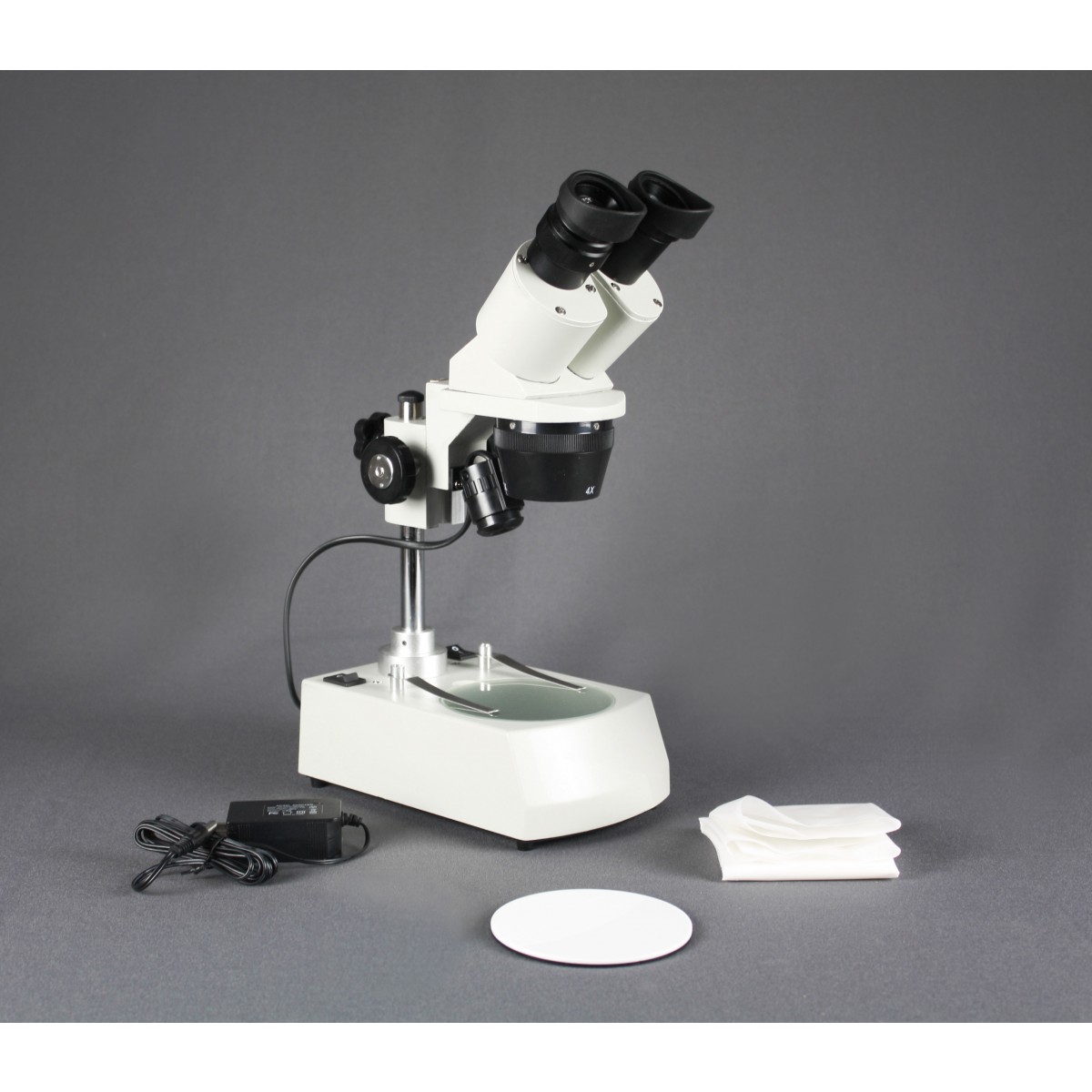
What is a stereo microscope used for?
Stereo Microscope. These are used to study objects that have a decent size such as leaves, flowers, insects, gems, rocks, etc. Stereo microscopes allow moderate magnification and are easy to handle. Production facilities often pick up these kinds of microscopes and even manufacturing plants that need to go through immediate inspection.
What is field diameter?
The field diameter is the viewing area of the lens of the microscope. It is the only number of millimeters or micrometers of the area that you can see. By measuring the field diameter, you can calculate the real size of the objects that are too small to measure.
What is the highest level of correction in a microscope?
The highest level of correction (and expense) is found in apochromatic objectives, illustrated in Figures 2 and 3. Apochromats represent the most highly corrected microscope lenses currently available, and their high price reflects the sophisticated design and careful assembly required in their manufacture. In Figure 3, we compare lens elements in a series of apochromatic objectives ranging from 10x to 100x in magnification. The lower power apochromat objectives (10x and 20x) have a longer working distance and the overall objective length is shorter than in higher power (40x and 100x) apochromat objectives. Traditionally, apochromats are corrected chromatically for three colors (red, green, and blue), almost eliminating chromatic aberration, and are corrected spherically for either two or three wavelengths (see Table 1). Apochromatic objectives are the best choice for color photomicrography in white light. Because of their high level of correction, apochromat objectives usually have, for a given magnification, higher numerical apertures than do achromats or fluorites. Many of the newer high-performance fluorite and apochromat objectives are corrected for four (dark blue, blue, green, and red) or more colors chromatically and four colors spherically.
How to correct spherical aberration?
To remedy this, many high-performance apochromat dry objectives are fitted with correction collars, which allow adjustment to correct for spherical aberration by correcting for variations in cover glass thickness (see Figure 5). Optical correction for spherical aberration is produced by rotating the collar, which causes two of the lens element groups in the objective to move either closer together or farther apart. The objective on the left in Figure 5 has had the correction collar adjusted for a cover glass thickness of 0.20 mm by bringing the adjustable lens elements very close together. In contrast, the objective on the right in Figure 5 has the adjustable lens elements separated by a rather large distance to compensate for very thin cover glasses (0.13 mm). A majority of the correction collar objectives designed for upright transmitted light microscopy have an adjustment range for cover glass thickness variations between 0.10 and 0.23 millimeters. Many of the specialized phase contrast objectives designed for observing tissue culture specimens with an inverted microscope have an even broader compensation range of 0 to 2 millimeters. This allows specimens to be viewed through the bottom of most culture vessels, which often have dramatic thickness fluctuations in this size range. Uncovered specimens, such as blood smears, can also be observed with correction collar objectives when the adjustment is set to 0 to account for the lack of a cover glass.
How to find the field diameter of an optical microscope?
The field diameter in an optical microscope is expressed by the field-of-view number or simply field number, which is the diameter of the viewfield expressed in millimeters and measured at the intermediate image plane. The field diameter in the object (specimen) plane becomes the field number divided by the magnification of the objective. Although the field number is often limited by the magnification and diameter of the ocular (eyepiece) field diaphragm, there is clearly a limit that is also imposed by the design of the objective. In early microscope objectives, the maximum usable field diameter was limited to about 18 millimeters (or considerably less for high magnification eyepieces), but modern planapochromats and other specialized flat-field objectives often have a usable field that can range between 22 and 28 millimeters or more when combined with wide-field eyepieces. Unfortunately, the maximum useful field number is not generally engraved on the objective barrel and is also not commonly listed in microscope catalogs.
What is an objective microscope?
Major microscope manufacturers offer a wide range of objective designs, which feature excellent optical characteristics under a wide spectrum of illumination conditions and provide various degrees of correction for the primary optical aberrations. The objective illustrated in Figure 1 is a 250x long working distance apochromat, which contains 14 optical elements that are cemented together into three groups of lens doublets, a lens triplet group, and three individual internal single-element lenses. The objective also has a hemispherical front lens and a meniscus second lens, which work synchronously to assist in capturing light rays at high numerical aperture with a minimum of spherical aberration. Although not present on this objective, many high magnification objectives of similar design are equipped with a spring-loaded retractable nosecone assembly that protects the front lens elements and the specimen from collision damage. Internal lens elements are carefully oriented and tightly packed into a tubular brass housing that is encapsulated by the objective barrel. Specific objective parameters such as numerical aperture, magnification, optical tube length, degree of aberration correction, and other important characteristics are imprinted or engraved on the external portion of the barrel. Although the objective featured in Figure 1 is designed to operate utilizing air as the imaging medium between the objective front lens and specimen, other objectives have front lens elements that allow them to be immersed in water, glycerin, or a specialized hydrocarbon-based oil.
What is the lens group in an oil immersion objective?
The general design of a practical oil immersion objective includes a hemispherical front len s element, followed by a positive meniscus lens and a doublet lens group. Presented in Figure 6 are the aplanatic refractions that occur at the first two lens elements in a typical apochromatic oil immersion objective. The specimen is sandwiched between the microscope slide and cover glass at point P, the aplanatic point of the hemispherical lens element. Light rays refracted at the rear of the hemispherical lens appear to proceed from point P (1), which is also the center of curvature for the first surface of the meniscus lens. The refracted light rays enter the meniscus lens along the radius of its first surface and experience no refraction at that surface. At the rear surface of the meniscus lens, light rays are refracted aplanatically, so they appear to diverge from point P (2). Refraction of the light rays at the surfaces of subsequent lens groups in the objective complete the convergence of light rays originating from point P, thus forming the intermediate image.
How to determine the brightness of an image?
Just as the brightness of illumination in a microscope is governed by the square of the working numerical aperture of the condenser, the brightness of an image produced by the objective is determined by the square of its numerical aperture. In addition, objective magnification also plays a role in determining image brightness, which is inversely proportional to the square of the lateral magnification. The square of the numerical aperture/magnification ratio expresses the light-gathering power of the objective when utilized with transmitted illumination. Because high numerical aperture objectives are often better corrected for aberration, they also collect more light and produce a brighter, more corrected image that is highly resolved. It should be noted that image brightness decreases rapidly as the magnification increases. In cases where the light level is a limiting factor, choose an objective with the highest numerical aperture, but having the lowest magnification factor capable of producing adequate resolution.
Why are objective microscopes important?
Microscope objectives are perhaps the most important components of an optical microscope because they are responsible for primary image formation and play a central role in determining the quality of images that the microscope is capable of producing. Objectives are also instrumental in determining the magnification of a particular specimen and ...
How are objective lenses classified?
Microscope objective lenses can be classified in several ways, including: By magnification By microscopy technique By lens shape By aberration correction But most commonly, when talking about types of objective lenses we are referring to
How to find the total magnification of a microscope?
To identify the total magnification that you will achieve on a microscope, you need to multiply the magnification of the objective lens with the magnification of your eyepiece. Most microscopes come with 10x, 20x, or 25x eyepieces.
What is an achromatic objective?
In otherwise, this is the standard lens type. An achromatic objective normalizes red and blue light so they meet at the one focal point, while also correcting green light for spherical aberrations.
How to tell if you have a reflected darkfield objective?
You can tell you have a reflected darkfield objective because it will have demarcations such as Neo, BF/DF, or BD written on the objective. See herefor more information on Reflected Darkfield Objectives.
What is a dry objective in a compound light microscope?
By default, compound light microscopes have dry objectives, meaning the space between the specimen and the objective is simply filed with air. Any objective with magnification under 100X you can assume is a dry objective unless otherwise marked.
What is the magnification of a scanning objective lens?
The scanning objective lens usually has 4x magnification and can be identified by a red strip band around the perimeter of the lens.
What is scanning objective?
The scanning objective is designed for getting your bearings right before moving onto the low power lens. Its name, the ‘scanning’ lens, derives from the fact you are zoomed-out enough that you can scan around your specimen at this magnification level to prepare to move on to higher magnifications.