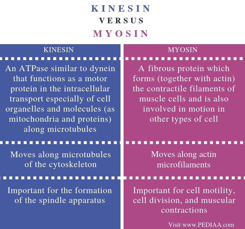
What are the motor proteins that interact with Microfilament?
Actin motors such as myosin move along microfilaments through interaction with actin, and microtubule motors such as dynein and kinesin move along microtubules through interaction with tubulin.
What is the motor protein associated with actin microfilaments?
Myosin motors act upon actin filaments to generate cell surface contractions and other morphological changes, as well as vesicle motility, cytoplasmic streaming and muscle cell contraction.
How do motor proteins move along microfilaments?
Motor proteins, such as myosins and kinesins, move along cytoskeletal filaments via a force-dependent mechanism that is driven by the hydrolysis of ATP molecules (reviewed in [1]).
Do microfilaments have motor proteins?
Like microtubules, microfilaments have associated motor proteins that will actively migrate along the fiber. The most abundant of these is myosin II, which moves toward the plus end of microfilaments, the process being driven by the hydrolysis of ATP.
What are the three types of motor proteins?
In eukaryotes, there are three types of protein fibers in the cytoskeleton: microfilaments, intermediate filaments, and microtubules.
Is dynein a motor protein?
Dyneins are a family of cytoskeletal motor proteins that move along microtubules in cells. They convert the chemical energy stored in ATP to mechanical work. Dynein transports various cellular cargos, provides forces and displacements important in mitosis, and drives the beat of eukaryotic cilia and flagella.
What is dynein and kinesin?
Dyneins and kinesins are microtubule-based molecular motors that play important roles in various cellular processes, including axonal transport, chromosome segregation during mitosis, and flagellar assembly and motility (Vallee and Sheetz, 1996; Hirokawa, 1998; Vale, 2003).
What motor proteins are responsible for movement?
Members of two large families of motor proteins—the kinesins and the dyneins—are responsible for powering the variety of movements in which microtubules participate.
Is actin a motor protein?
Myosin is an actin motor protein, where myosin serves as the engine, the actin filaments provide the tracks that myosin can move along and the energy source that fuels the movement is adenosine triphosphate (ATP).
Which proteins are motor proteins?
There are two motor proteins that seem to be most important; myosin, which with actin filament does the muscle work, and dynein which moves the doublets of tubulin rods (along the axes) in the 9+2 axoneme and makes the flagella whip.
How do microfilaments move?
The microfilaments are often found anchored to proteins in the cell membrane. Sometimes microfilaments are found floating free and connected to other filaments and tubules. Those binding proteins allow the microfilaments to push and pull on the cell membrane to help the cell move.
What are the 4 proteins involved in muscle contraction?
SubstancesActins.Muscle Proteins.Tropomyosin.Myosins.Calcium.
What are the cytoskeleton motor proteins?
Cytoskeleton motors comprise of myosin, kinesin and cytoplasmic dynein. F-actin filaments act as myosin track, while kinesin and cytoplasmic dynein move on microtubules.
Is myosin a motor protein?
Myosins form a superfamily of molecular motor proteins that power muscle contraction, as well as movement on actin filaments in all eukaryotic cells.
What are the 4 proteins involved in muscle contraction?
SubstancesActins.Muscle Proteins.Tropomyosin.Myosins.Calcium.
Are motor proteins part of the cytoskeleton?
Perhaps the most fascinating proteins that associate with the cytoskeleton are the molecular motors called motor proteins. These remarkable proteins bind to a polarized cytoskeletal filament and use the energy derived from repeated cycles of ATP hydrolysis to move steadily along it.
What is the function of microfilaments?
Microfilaments (also called actin filaments) have a double helix-like structure composed of actin protein subunits; microfilaments serve as tracks for the motor protein myosin and are involved in many cellular processes that require motion.
What is the function of motor proteins?
Image credit: OpenStax Biology. Motor proteins use energy in the form of ATP to “walk” along specific cytoskeletal tracks. They are essential for movement of vesicles and other cargoes within cells, as well as for the movement of muscle and cilia/flagella: Myosin is associated with actin microfilaments and is required for movement of muscle.
Why does myosin stay stuck after a power stroke?
Because ATP is required for myosin to be able to release the actin, depletion of ATP due to muscle fatigue will cause muscles to remain locked in a contracted state; this is thought to be one of several sources of cramping after exercise.
How does muscle shortening work?
How does this process work? The motion of muscle shortening occurs as myosin heads bind to actin and pull the actin inwards. This action requires energy, which is provided by ATP. Myosin binds to actin at a binding site on the globular actin protein. Myosin has another binding site for ATP at which enzymatic activity hydrolyzes ATP to ADP, releasing an inorganic phosphate molecule and energy. The process works in a cycle (called the “cross bridge cycle”) like this:
What is a sarcomere?
A sarcomere is the region from one Z line to the next Z line. Many sarcomeres are present in a myofibril, resulting in the striation pattern characteristic of skeletal muscle. Image credit: OpenStax Biology. The protein components of the sarcomere include actin, myosin, tropomyosin, and troponin:
What are the proteins that move the cilia?
ATP, dynein motor proteins, and microtubule tracks are essential for movement of eukaryotic cilia and flagella. Flagella (singular, flagellum) are long, hair-like structures that extend from the cell surface and are used to move an entire cell, such as a sperm. If a cell has any flagella, it usually has one or just a few.
What are the components of a multicellular animal?
In multicellular animals, these components are the skeleton and muscles. In single-celled animals and individual cells, these components are often flagella and/or cilia. All of these structures rely on both motor proteins and components of the cytoskeleton. The cytoskeleton (literally, “cell skeleton”) is a network of filaments ...
Where are ribosomes made?
and the subunits of ribosomes are assembled then they exit through the pores and become full ribosomes. basically ribosomes are made in the nucleoli.
Where are secretory vesicles produced?
secretory are packaged in transport vesicles that are produced in transitional ER , these vesicles take the protein where they need to go likely to the GOLGI!
Where are free ribosomes suspended?
free ribosomes are suspended in cytosol (the cytoplasm) of a cell. their products serve a function within the cytosol such as enzymes. function inside of cell
How does smooth ER increase detox rate?
they are metabolized by the same broad spectrum detox enzymes. they induce proliferation of smooth ER which increases detox rate. this causes tolerance because the body metabolizes it more quickly, requiring higher dose to produce the same effect. if one of these substances the enzyme metabolizes is present it will cause the increase in smooth ER proliferation which means tolerance for any other substance.
Which component is spun first?
the faster it is spun, the smaller components that result. first debris is nucleus then mitochondria, then peices of the membrane, then ribosomes
Which type of cell has a narrow and elongated shape?
nerve ells are narrow and have an elongated shape. intestinal cells have thin surface projections call microvilli that add surface area but very little/negligible amount of volume
What is the name of the protein that spontaneously comes together to form microfilaments?
Related Biology Terms. Actin – The protein that spontaneously comes together to form microfilaments. Cytoskeleton – A network of protein filaments that extends throughout the cytoplasm of the cell. Actomyosin – A complex of the proteins actin and myosin that is responsible for muscle movement. Cytoplasmic streaming – The flow ...
What are the roles of microfilaments in muscle cells?
One of the most important roles of microfilaments is to contract muscles. There is a high concentration of microfilaments in muscle cells, where they form myofibrils, the basic unit of the muscle cell. Actin is an indispensable protein for muscle movement, and microfilaments are often called actin filaments because actin is so prominent in ...
How do microfilaments travel?
It allows nutrients, waste products, and cell organelles to travel from one part of the cell to another. Microfilaments can attach to a cell organelle and then contract, pulling the organelle to a different area of the cell.
What is the structure of a microfilament?
Microfilament Structure. Microfilaments are composed of two strands of subunits of the protein actin (hence the name actin filaments) wound in a spiral. Specifically, the actin subunits that come together to form a microfilament are called globular actin (G-actin), and once they are joined together they are called filamentous actin (F-actin).
Why is actin called a microfilament?
Actin is an indispensable protein for muscle movement, and microfilaments are often called actin filaments because actin is so prominent in the muscular system of the body. In muscle cells, actin works together with the protein myosin to allow the muscles to contract and relax. Here, neither actin nor myosin can work properly without the other, ...
What is the role of microfilaments in the body?
Microfilaments play a role in causing cells to move. This occurs throughout the body and it is also very important for organisms whose entire body consists of one cell, such as amoebae; without microfilaments, they would not be motile. Actomyosin plays a role here just as it does in muscle cells.
What is the role of microfilaments in cytoplasmic streaming?
Cytoplasmic streaming is the flow of cytoplasm (the contents of the cell, including the fluid part called cytosol and cell organelles) throughout the cell. It allows nutrients, waste products, and cell organelles to travel from one part of the cell to another.
What are the two proteins that move along the microtubules?
Kinesin and dynein, the prototypes of microtubulemotor proteins, move along microtubules in opposite directions—kinesintoward the plus end and dynein toward the minus end (Figure 11.45). The first of these microtubule motor proteins to be identified was dynein, which was isolated by Ian Gibbons in 1965. The purification of this form of dynein (called axonemal dynein) was facilitated because it is a highly abundant protein in cilia, just as the abundance of myosinfacilitated its isolation from muscle cells. The identification of other microtubule-based motors, however, was more problematic because the proteins responsible for processes such as chromosome movement and organelle transport are present at comparatively low concentrations in the cytoplasm. Isolation of these proteins therefore depended on the development of new experimental methods to detect the activity of molecular motors in cell-free systems.
Which proteins move in opposite directions?
Microtubule motor proteins. Kinesin and dynein move in opposite directions along microtubules, toward the plus and minus ends, respectively. Kinesin consists of two heavy chains, wound around each other in a coiled-coil structure, and two light chains. (more...)
What are the functions of microtubules?
One of the major roles of microtubules is to transport membrane vesicles and organelles through the cytoplasm of eukaryotic cells . As already discussed, such cytoplasmic organelle transport is particularly evident in nerve cell axons, which may extend more than a meter in length. Ribosomes are present only in the cell body and dendrites, so proteins, membrane vesicles, and organelles (e.g., mitochondria) must be transported from the cell body to the axon. Via video-enhanced microscopy, the transport of membrane vesicles and organelles in both directions can be visualized along axon microtubules, where kinesinand dyneincarry their cargoes to and from the tips of the axons, respectively. For example, secretory vesiclescontaining neurotransmitters are carried from the Golgi apparatusto the terminal branches of the axon by kinesin. In the reverse direction, cytoplasmic dynein transports endocytic vesicles from the axon back to the cell body.
What are microtubules responsible for?
Microtubules are responsible for a variety of cell movements, including the intracellular transport and positioning of membrane vesicles and organelles, the separation of chromosomesat mitosis, and the beating of cilia and flagella. As discussed for actinfilaments earlier in this chapter, movement along microtubules is based on the action of motor proteinsthat utilize energy derived from ATP hydrolysis to produce force and movement. Members of two large families of motor proteins—the kinesinsand the dyneins—are responsible for powering the variety of movements in which microtubules participate.
What is the role of microtubules in mitosis?
As discussed earlier in this chapter, microtubules reorganize at the beginning of mitosisto form the mitotic spindle, which plays a central role in cell division by distributing the duplicated chromosomesto daughter nuclei. This critical distribution of the genetic material takes place during anaphaseof mitosis, when sister chromatids separate and move to opposite poles of the spindle. Chromosome movement proceeds by two distinct mechanisms, referred to as anaphase Aand anaphase B, which involve different types of spindle microtubules and associated motor proteins.
Which family of cells transports vesicles along microtubules?
Transport of vesicles along microtubules. Kinesin and other plus end-directed members of the kinesin family transport vesicles and organelles in the direction of microtubule plus ends, which extend toward the cell periphery. In contrast, dynein and minus (more...)
Which direction does kinesin move?
Further studies demonstrated that kinesintranslocates along microtubules in only a single direction—toward the plus end. Because the plus ends of microtubules in axons are all oriented away from the cell body (see Figure 11.44), the movement of kinesin in this direction transports vesicles and organelles away from the cell body, toward the tip of the axon. Within intact axons, however, vesicles and organelles also had been observed to move back toward the cell body, implying that a different motor protein might be responsible for movement along microtubules in the opposite direction—toward the minus end. Consistent with this prediction, further experiments showed that a protein previously identified as the microtubule-associated protein MAP-1C was in fact a motor protein that moved along microtubules in the minus end direction. Subsequent analysis demonstrated that MAP-1C is related to the dyneinisolated from cilia (axonemal dynein), so MAP-1C is now referred to as cytoplasmic dynein.
What are the functions of motor proteins in the cell?
Motor proteins help in the active transport of proteins in the cell cytoplasm and participate during mitosis and meiosis, triggering cell division. The motor proteins attach with the ATP molecules inside the cells and convert into ADP and P groups, releasing energy. This helps in the conversion of chemical energy derived from mechanical energy. The absence of motor proteins during cell division leads to several diseases. The absence of kinesin causes tooth and some kidney diseases. Lack or deficiency of Dynein may cause respiratory failure due to a lack of cilia movement. Myosin is a vital motor protein involved in muscle contraction, and the deficiency can cause myopathies. Myosin is also used by stereocilia, which aid hearing and the deficiency will lead to non-syndromic deafness.
What is the role of myosin in muscle contraction?
Myosin is an important motor protein found in animals and helps in muscle fiber contraction. Motor proteins help in the active transport of proteins during cellular and metabolic processes. Dyneins and Kinesins form a separate class of motor proteins and help in programming spindle fiber and separation of chromosomes in the nucleus. Certain motor proteins require cytoskeleton for activation and utilize microfilaments or microtubules as substrate. Microtubule motors can be categorized based on their movement on microtubule cable and form plus-end motor and minus-end motors.
What is the function of kinesin?
Kinesin is a major form of motor proteins and uses microtubule fibers to move in a forward direction. Kinesin helps in various cellular processes like mitotic and meiosis. It supports cell division and chromosome differentiation in each cell nucleus. In eukaryotic cells, it helps in the movement of various organelles like Golgi bodies, mitochondria, and other vesicles. Each kinesin molecule is composed of two heavy and light chains. The heavy chain helps in the production of mechanical force by disintegrating ATP molecules. The movement of cargo towards the positive