Explore
What to know about palatine tonsils
- Overview. The palatine tonsils serve as a component of Waldeyer’s ring. ...
- Complications associated with palatine tonsils. The palatine tonsils play an important role in trapping bacteria and viruses as they enter the body.
- Diagnostic tests. ...
- Treatments. ...
- Summary. ...
What is the function of palatine tonsils?
The palatine tonsils are paired lymphoid organs, located in the lateral wall of the pharynx. Smaller and less well-defined lymphoid tissues of the oropharyngeal cavity are the lingual and pharyngeal tonsils. The palatine tonsils consist of follicles populated by B cells and a few CD4+ T cells, and an interfollicular area with T cells.
What are the palatine tonsils?
Tonsils are a pair of lymphoid tissues that are located at the back of the throat. They belong to the lymphatic system and help fight infections. Normally, they are pinkish in color. However, sometimes, tonsils become enlarged, red, and irritated when they get infected. When they are infected, they are covered by a white or yellow film.
What are normal tonsils?
- pharyngeal tonsils (adenoids), which reside behind your nose.
- two palatine tonsils (what people are most commonly referring to when they say the word 'tonsils'), which reside on both sides of the back of the throat.
- lingual tonsils, which are at the back of the tongue.
What are the three sets of tonsils?

What is the role of the palatine tonsils?
The function of the palatine tonsils is thought to be associated with preventing infection in the respiratory and digestive tracts by producing antibodies that help kill infective agents. Frequently, however, the tonsils themselves become the objects of infection, and surgical removal (tonsillectomy) is required.
Where are the palatine tonsils located and what is their function?
The palatine tonsils sit in the back of the throat and are made up of lymphatic tissue. Along with the pharyngeal, tubal, and the lingual tonsils, they act as a defence against possible infections. The palatine tonsils are oval-shaped lymphatic tissue located at both sides of the back of the throat.
How many palatine tonsils are there?
two palatine tonsilsHumans have two palatine tonsils located at the back of the throat, one on either side of the uvula. Each palatine tonsil is composed of 6 to 20 crypts, or pits, which help to increase surface area and trap germs.
Are there 2 palatine tonsils?
The tonsils are a set of lymphoid organs facing into the aerodigestive tract, which is known as Waldeyer's tonsillar ring and consists of the adenoid tonsil, two tubal tonsils, two palatine tonsils, and the lingual tonsils. These organs play an important role in the immune system.
Why is it called palatine tonsils?
…the oral pharynx is a palatine tonsil, so called because of its proximity to the palate. Each palatine tonsil is located between two vertical folds of mucous membrane called the glossopalatine arches.
What causes palatine tonsils to swell?
Tonsillitis is most often caused by common viruses, but bacterial infections also can be the cause. The most common bacterium causing tonsillitis is Streptococcus pyogenes (group A streptococcus), the bacterium that causes strep throat. Other strains of strep and other bacteria also may cause tonsillitis.
Why are palatine tonsils removed?
Tonsillectomy is a surgical procedure in which both palatine tonsils are fully removed from the back of the throat. The procedure is mainly performed for recurrent tonsillitis, throat infections and obstructive sleep apnea (OSA).
Do palatine tonsils get removed?
The palatine tonsils are removed in a tonsillectomy. Palatine tonsils are collections of lymph tissue on the right and left side of the upper throat (also called the oropharynx).
What are the 3 major tonsils?
There are three sets of tonsils in the back of the mouth: the adenoids, the palantine, and the lingual tonsils.
What do swollen palatine tonsils interfere with?
When these tonsils become enlarged they may interfere with breathing and are called adenoids. The palatine tonsils are the ones that are located near the opening of the oral cavity into the pharynx.
What is the definition of Palatine?
Definition of palatine (Entry 1 of 4) 1a : possessing royal privileges. b : of or relating to a palatine or a palatinate. 2a : of or relating to a palace especially of a Roman or Holy Roman emperor.
Can palatine tonsils grow back?
It is possible for tonsils to partially grow back. During a tonsillectomy, most of the tonsils are removed. However, some tissue often remains, so tonsils occasionally can regenerate (regrow) — although they probably won't grow back completely or to their original size.
Where are the palatine tonsils located?
lateral oropharynxThe palatine (or faucial) tonsils, commonly referred to as tonsils, are bundles of lymphatic tissue located in the lateral oropharynx. They sit in the isthmus of the fauces, bordered anteriorly by the palatoglossal arch and posteriorly by the palatopharyngeal arch.
What is the function of the palatine tonsils quizlet?
Functions to prevent infections. These tonsils contain B and T lymphocytes which get activated when harmful bacteria and viruses come in contact with tonsils.
Where are the palatine tonsils located quizlet?
The palatine tonsils are found in which of the following regions? -oropharynx. The palatine tonsils are found in the part of the pharynx that is shared with the oral cavity and is known as the oropharynx.
What are the 3 tonsils and where are they located?
There are three sets of tonsils in the back of the mouth: the adenoids, the palantine, and the lingual tonsils. 1 These tonsils are made up of lymphatic tissue and are typically small in size.
What is the difference between tonsils and lymph nodes?
In contrast to lymph nodes, tonsils are not truly encapsulated and they lack afferent lymph, whereas the reticular crypt epithelium contains many DCs which can transport exogenous antigens to the extrafollicular T-cell areas and to the B-cell follicles. DCs function as APCs in the extrafollicular primary immune responses and occur abundantly around HEVs, often in clusters with T cells that are mainly of the CD4 + phenotype. These lymphocytes consist of both naïve (CD45RA +) and memory (CD45R0 +) subsets ( Figure 5 (a) ), and some express the high-affinity IL-2 receptor (CD25) as a sign of recent activation ( Figure 5 (b) ). Altogether, the tonsils possess the biological armamentarium that should enable them to mount both primary and secondary T-cell responses ( Brandtzaeg and Halstensen, 1992 ).
What is the lymphoid tissue on the oropharynx?
The palatine tonsils are lymphoid tissue on either side of the oropharynx between the palatopharyngeal and palatoglossal arch (Figure 6 ). One of the causes of airway obstruction, especially in children, is tonsillar hypertrophy which has been associated with increase in the respiratory disturbance index.
Why are sIgD and sIgM expressed?
It is still unclear why both sIgD and sIgM must be expressed to render B cells antigen reactive. Likewise, the nature of FDCs remains obscure but their origin is probably mesenchymal. The existence of these cells and their accumulation in the primary follicles depend on the presence of B cells.
What is tonsillar cancer?
Cancer of the palatine tonsil, in the lymphoid region called the Waldeyer’s ring, is usually referred to as tonsillar cancer. Tonsillar cancer is the most common of the oropharyngeal malignancies, and 75% of all tonsillar carcinomas are squamous cell carcinomas (Genden et al., 2003 ).
Where are the palatine tonsils located?
They are housed in the tonsillar fossa, bordered by the palatoglossus anteriorly, palatopharyngeus posteriorly, superior constrictor laterally, and the base of the tongue inferiorly.
How does GC work?
The GC reaction is initiated by stimulation of naïve B cells immediately outside the lymphoid follicles through cognate help from activated CD4 + T cells ( Figure 3 (b) ). These helper T (Th) cells have received processed foreign antigen from DCs in the context of class II molecules of the major histocompatibility complex—in humans also called HLA class II, such as the classical HLA-DR, -DQ, and -DP molecules. The activated B cells can pick up antigen and, after processing, further present it as peptides to the cognate T cells in an interaction that provides mutual support. Some of these B cells (sIgD + IgM + CD38 + CD77 +) will then colonize the follicles and act as founder cells for GCs ( Figure 6 ).
What tissue is in the lower center of Waldeyer's ring?
At lower center is the lymphoid tissue (red). Apart from the pair of palatine tonsils, there is a pair of lingual tonsils and a pair of pharyngeal tonsils (adenoids) which contribute to the Waldeyer's ring of lymphoid tissue. (With permission from Photo Science Library.) View chapter Purchase book.
What is the lymphoid tissue on the oropharynx?
The palatine tonsils are lymphoid tissue on either side of the oropharynx between the palatopharyngeal and palatoglossal arch (Figure 6 ). One of the causes of airway obstruction, especially in children, is tonsillar hypertrophy which has been associated with increase in the respiratory disturbance index.
What is the palatine tonsil?
The palatine tonsils, in addition to the adenoids and lingual tonsils, are classified as mucosa-associated lymphoid tissue. They are housed in the tonsillar fossa, bordered by the palatoglossus anteriorly, palatopharyngeus posteriorly, superior constrictor laterally, and the base of the tongue inferiorly. The fossa and tonsil are lined by nonkeratinizing stratified squamous epithelium that on the tonsillar surface forms multiple crypts. A connective tissue “capsule,” consisting of loose areolar tissue, separates the tonsil from the pharyngobasilar fascia and the superior constrictor muscle. Nerves and vessels pass through the fascia. The palatine tonsils are supplied by branches of the external carotid artery: the inferior tonsillar artery via the facial artery, the superior tonsillar artery via the greater palatine branch of the maxillary artery, the posterior tonsillar artery from the ascending pharyngeal arteries and the facial artery, and the anterior tonsillar artery from the dorsal lingual artery. The venous drainage is via the peritonsillar plexus, which drain to the pharyngeal plexus or facial vein and into the internal jugular vein. Sensation is provided via the tonsillar branches of the glossopharyngeal nerve and the lesser palatine branch of the maxillary division of the trigeminal nerve. Some taste is provided by the tonsillar pillars and that is supplied by the glossopharyngeal nerve.3,8–11,13
What is tonsillar cancer?
Cancer of the palatine tonsil, in the lymphoid region called the Waldeyer’s ring, is usually referred to as tonsillar cancer. Tonsillar cancer is the most common of the oropharyngeal malignancies, and 75% of all tonsillar carcinomas are squamous cell carcinomas (Genden et al., 2003 ).
How to aspirate palatine tonsils?
12.38) Place cotton wool soaked in hydrogen superoxide into the aperture of the aspirator tube. Position the tip of the tube over the palatine tonsil and ‘milk’ the tonsil by pressing the rubber bulb. The pressure on the rubber bulb must not be so great that you have to forcibly detach the tip of the aspirator tube from the tonsil. Afterwards the drainage products from the tonsil can be inspected on the cotton wool (greasy, possibly yellowish, or granular).
How does GC work?
The GC reaction is initiated by stimulation of naïve B cells immediately outside the lymphoid follicles through cognate help from activated CD4 + T cells ( Figure 3 (b) ). These helper T (Th) cells have received processed foreign antigen from DCs in the context of class II molecules of the major histocompatibility complex—in humans also called HLA class II, such as the classical HLA-DR, -DQ, and -DP molecules. The activated B cells can pick up antigen and, after processing, further present it as peptides to the cognate T cells in an interaction that provides mutual support. Some of these B cells (sIgD + IgM + CD38 + CD77 +) will then colonize the follicles and act as founder cells for GCs ( Figure 6 ).
What is a crypt in anatomy?
Crypts represent extension of surface mucosa into underlying lymphoid stroma; most are simple, but may be branched, and they extend to connective tissue pseudocapsule
Where are the palatine tonsils located?
The palatine tonsils are paired lymphoid organs, located in the lateral wall of the pharynx. Smaller and less well-defined lymphoid tissues of the oropharyngeal cavity are the lingual and pharyngeal tonsils. The palatine tonsils consist of follicles populated by B cells and a few CD4+ T cells, and an interfollicular area with T cells. Squamous cell epithelium overlies the tonsils and forms crypts that extend deeply into the lymphoid tissue. The arterial blood supply comes from the tonsilar artery, a branch of the lingual artery. Numerous efferent lymphatics merge into two or three lymph vessels that drain into the medial retropharyngeal lymph node ( Belz and Heath, 1995b ).
What is the palatine tonsil?
The palatine tonsil is one of the mucosa-associated lymphoid tissues (MALT), located at the entrance to the upper respiratory and gastrointestinal tracts to protect the body from the entry of exogenous material through mucosal sites. In consequence it is a site of, and potential focus for, infections, and is one of the chief immunocompetent tissues in the oropharynx. It forms part of the Waldeyer's ring, which comprises the adenoid, the paired tubal tonsils, the paired palatine tonsils and the lingual tonsils. From the pharyngeal side, they are covered with a stratified squamous epithelium, whereas a fibrous capsule links them to the wall of the pharynx. Through the capsule pass trabecules that contain small blood vessels, nerves and lymphatic vessels. These trabecules divide the tonsil into lobules.
How many episodes of tonsillitis are there in a year?
Recurrent infection has been variably defined as from four to seven episodes of acute tonsillitis in one year, five episodes for two consecutive years or three episodes per year for 3 consecutive years.
How many crypts are in a palatine tonsil?
Palatine tonsils consist of approximately 15 crypts, which result in a large internal surface.
What type of cells produce antibodies?
Tonsillar (relating to palatine tonsil) B cells can mature to produce all the five major Immunoglobulin (Ig, aka antibody) classes. Furthermore, when incubated in vitro with either mitogens or specific antigens, they produce specific antibodies against diphtheria toxoid, poliovirus, Streptococcus pneumoniae, Haemophilus influenzae, Staphylococcus aureus, and the lipopolysaccharide of E. coli. Most Immunoglobulin A produced by tonsillar B cells in vitro appears to be 7S monomers, although a significant proportion may be l0S dimeric IgA.
Where does the blood supply to the palatine tonsils come from?
Blood supply and innervation. The nerves supplying the palatine tonsils come from the maxillary division of the trigeminal nerve via the lesser palatine nerves, and from the tonsillar branches of the glossopharyngeal nerve. The glossopharyngeal nerve continues past the palatine tonsil and innervates the posterior 1/3 of ...
What is the role of tonsils in infectious disease?
The pathogenesis of infectious/inflammatory disease in the tonsils most likely has its basis in their anatomic location and their inherent function as organ of immunity, processing infectious material, and other antigens and then becoming , paradoxically, a focus of infection/inflammation.
Which artery supplies blood?
Blood supply is provided by tonsillar branches of five arteries: the dorsal lingual artery (of the lingual artery ), ascending palatine artery (of the facial artery ), tonsillar branch (of the facial artery), ascending pharyngeal artery (of the external carotid artery ), and the lesser palatine artery (a branch of the descending palatine artery, itself a branch of the maxillary artery ). The tonsils venous drainage is by the peritonsillar plexus, which drain into the lingual and pharyngeal veins, which in turn drain into the internal jugular vein .
What is the palatine tonsil?
The palatine tonsils, (also known as the faucial tonsils or simply " the tonsils ") are a bilateral collection of lymphoid tissue in the pharyngeal mucosa. They form part of Waldeyer's ring.
How many borders does the tongue have?
It is often described to have two borders, two poles and two surfaces: anterior and posterior borders (described in relations below) upper and lower poles: extending to the soft palate and dorsum of the tongue respectively.
Which artery supplies the arterial supply?
arterial supply is primarily from the tonsillar branch of facial artery, with contributions from the ascending pharyngeal , lingual and ascending palatine arteries.
What is strep throat?
Strep throat happens when the tonsils are infected by bacteria called Streptococcus, usually class ified by two different strains, A and B. Strep is usually a problem that affects children, though adults can get strep throat, too.
What are tonsils called?
Technically, there are three sets of tonsils in the body: the pharyngeal tonsils, commonly known as adenoids, the palatine tonsils and the lingual tonsils, which are lymphatic tissue on the surface tissue of the base of the tongue, according to Encyclopedia Britannica. When people refer to tonsils, though, they are usually talking about the palatine tonsils. These tonsils are oval, pea-sized clusters of lymph cells in the pharynx at the opening of either side of the throat. Though they may seem large in children, the size of the tonsils tends to get smaller when a person becomes an adult.
What happens if you get strep infection untreated?
If strep goes untreated, it can cause scarlet fever impetigo, cellulitis, toxic shock syndrome and necrotizing fasciitis (flesh-eating disease), according to the National Institutes of Health. Rheumatic fever may also occur from an untreated strep infection.
What is the name of the fluid that is a clear and colorless fluid?
For example, tonsils sample bacteria and viruses entering the body through the mouth or nose and flush them using lymph. Lymph is a clear and colorless fluid; the name comes from the Latin word lympha, which means "connected to water," according to the National Lymphadema Network. RECOMMENDED VIDEOS FOR YOU... CLOSE.
Where are tonsils located in the throat?
The tonsils are in the back of the throat. Strep throat and tonsillitis can lead to inflammation. (Image credit: solar22/Shutterstock)
What is the best medicine for tonsil stones?
Typically, doctors will prescribe antibiotics, such as Augmentin, to rid the body of the bacteria. Tonsil stones are a typical affliction of the throat area, as well. This happens when debris gets caught in the groves of the tonsils. Then, white blood cells attack the debris, creating a rock-like stone.
What are the bumps on the back of the throat?
On top of all that, they produce white blood cells and antibodies, according to the Mayo Clinic . According to the American Academy of Otolaryngology, these bumps on the back of the throat are the "first line of defense as part of the immune system.".
How does tonsil cancer form?
Tonsil cancer forms when healthy cells in the tonsils develop changes in their DNA. A cell's DNA contains the instructions that tell a cell what to do. The changes tell the cells to grow out of control and to continue living when healthy cells would normally die. The accumulating cells form a tumor that can grow beyond the tonsils ...
What is the name of the two pads in the back of your mouth?
Tonsils . Your tonsils are two oval-shaped pads in the back of your mouth. Your tonsils are part of your body's germ-fighting immune system. Tonsil cancer is an abnormal growth of cells that forms in a tons il.
What is HPV and throat cancer?
Close. HPV and throat cancer. HPV and throat cancer. Human papillomavirus (HPV) is a common sexually transmitted infection that increases the risk of certain types of throat cancer. HPV has been linked to cancer that affects the soft palate, tonsils, back of the tongue, ...
How to reduce the risk of tonsil cancer?
To reduce your risk of tonsil cancer: Don't use tobacco. If you don't use tobacco, don't start. If you currently use tobacco of any kind, talk with your doctor about strategies to help you quit. Limit alcohol if you choose to drink. If you choose to drink alcohol, do so in moderation.
How do you know if you have tonsil cancer?
Signs and symptoms of tonsil cancer include: Difficulty swallowing. A sensation that something is caught in the back of your throat. Swelling and pain in the neck. Earache. Jaw stiffness.
What does a dentist check for?
Get regular dental care. During your appointment, your dentist will check your mouth for signs of cancer and precancerous changes.
Can tonsil cancer be transmitted?
This common sexually transmitted infection is detected in most tonsil cancers in the United States. Tonsil cancer caused by HPV tends to occur at a younger age and is more likely to respond well to available treatments.
Overview
Palatine tonsils, commonly called the tonsils and occasionally called the faucial tonsils, are tonsils located on the left and right sides at the back of the throat, which can often be seen as flesh-colored, pinkish lumps. Tonsils only present as "white lumps" if they are inflamed or infected with symptoms of exudates (pus drainage) and severe swelling.
Structure
The palatine tonsils are located in the isthmus of the fauces, between the palatoglossal arch and the palatopharyngeal arch of the soft palate.
The palatine tonsil is one of the mucosa-associated lymphoid tissues (MALT), located at the entrance to the upper respiratory and gastrointestinal tracts to protect the body from the entry of exogenous material through mucosal sites. …
Function
Tonsillar (relating to palatine tonsil) B cells can mature to produce all the five major Immunoglobulin (Ig, aka antibody) classes. Furthermore, when incubated in vitro with either mitogens or specific antigens, they produce specific antibodies against diphtheria toxoid, poliovirus, Streptococcus pneumoniae, Haemophilus influenzae, Staphylococcus aureus, and the lipopolysaccharide of E. coli. Most Immunoglobulin A produced by tonsillar B cells in vitro appear…
Clinical significance
The pathogenesis of infectious/inflammatory disease in the tonsils most likely has its basis in their anatomic location and their inherent function as organ of immunity, processing infectious material, and other antigens and then becoming, paradoxically, a focus of infection/inflammation. No single theory of pathogenesis has yet been accepted, however. Viral infection with secondary …
Additional images
• Lymphatic system
• The mouth cavity. The cheeks have been slit transversely and the tongue pulled forward.
• Throat after tonsillectomy
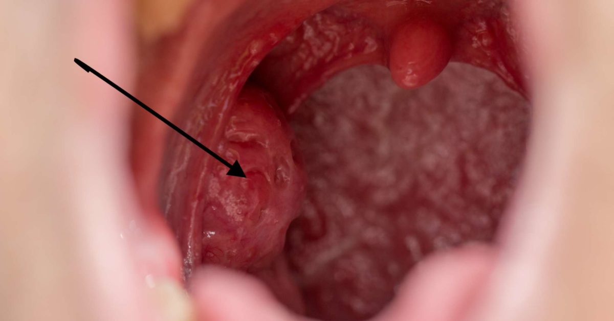
• Anterior photograph of the oral cavity showing palatine tonsils (inflamed) and uvula.
External links
• "Anatomy diagram: 05287.011-1". Roche Lexicon - illustrated navigator. Elsevier. Archived from the original on 2013-04-22.