
Figure 3: Major currents during the cardiac ventricular action potential
| Current ( I ) | α subunit protein | α subunit gene | Phase / role | |
| Na + | INa | Na V 1.5 | SCN5A [31] | 0 |
| Ca 2+ | ICa (L) | Ca V 1.2 | CACNA1C [32] | 0-2 |
| K + | Ito1 | K V 4.2/4.3 | KCND2 / KCND3 | 1, notch |
| K + | IKs | K V 7.1 | KCNQ1 | 2,3 |
What are the three phases of action potential?
Illustrations:
- Types of neurons and synapse (diagram) - Paul Kim
- Action potential curve and phases (diagram) - Jana Vasković
- Ions exchange in action potential (diagram) - Jana Vasković
What are the different phases of action potential?
It provides complete analysis on pipeline drugs detailing Phases ... of action (MoA) targeted by different Gingivitis companies are identified to support decision makers target the most potential ...
What are the steps in action potential?
Ben Simmons is back doing “more shooting” and “more work” as he steps up a potential return to the court with Philadelphia in the coming weeks and months. The Aussie vowed never to play for the team again as he attempted to push through a trade ...
What are the three phases of cardiac arrest?
Cardiac Arrest Management: Part 1
- American Heart Association. 2005 American Heart Association guidelines for cardiopulmonary resuscitation and emergency cardiovascular care. ...
- Weisfeldt M, Becker L. Resuscitation after cardiac arrest: A 3-phase time-sensitive model. ...
- Ewy G. Cardiocerebral resuscitation: The new cardiopulmonary resuscitation. ...
- Kellum W, Dennedy K, Ewy G. ...
What are the 4 phases of cardiac action potential?
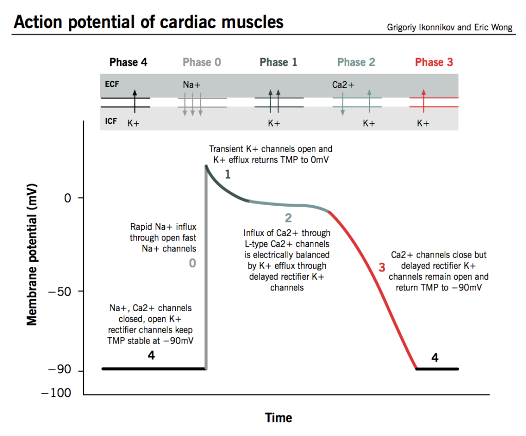
Action potentials and impulse conductionPhase 4: The resting phase.Phase 0: Depolarization.Phase 1: Early repolarization.Phase 2: The plateau phase.Phase 3: Repolarization.
What are the phases of action potential in cardiac muscle cell?
Phases of the Cardiac Action Potential. The cardiac transmembrane action potential consists of five phases:phase 0, upstroke or rapid depolarization;phase 1, early rapid repolarization;phase 2, plateau;phase 3, final rapid repolarization; andphase 4, resting membrane potential and diastolic depolarization (Fig.
What is phase 3 of cardiac action potential?
Repolarization (phase 3 of the action potential) occurs because of an increase in potassium permeability. At the SA node, potassium permeability can be further enhanced by vagal stimulation. This has the effect of hyperpolarizing the cell and reducing the rate of firing. Sympathetic stimulation has the opposite effect.
What are the three phases of action potential?
The action potential has three main stages: depolarization, repolarization, and hyperpolarization. Depolarization is caused when positively charged sodium ions rush into a neuron with the opening of voltage-gated sodium channels.
How many phases are in the action potential cycle?
Phases of the Cardiac Action Potential. The cardiac transmembrane action potential consists of five phases:phase 0, upstroke or rapid depolarization;phase 1, early rapid repolarization;phase 2, plateau;phase 3, final rapid repolarization; andphase 4, resting membrane potential and diastolic depolarization (Fig.
How do you remember cardiac action potential?
Let's first discuss the action potentials of non-pacemaker cardiac myocytes - the contractile cardiac muscle cells that contract the atria and ventricles. All you need to memorize is the following phrase: “summit, plummet, continue, plummet”.
What is cardiac depolarization and repolarization?
Depolarization with corresponding contraction of myocardial muscle moves as a wave through the heart. 7. Repolarization is the return of the ions to their previous resting state, which corresponds with relaxation of the myocardial muscle. 8.
Why is the plateau phase important?
This phase is known as phase 2 and is commonly referred to as the plateau phase. This plateau phase allows for a longer muscle contraction and gives time for the nearby cardiac muscle cells to depolarize. This is important in allowing the heart to contract in a steady, uniform and forceful manner.
What are the steps of action potential quizlet?
Terms in this set (4)Step 1 - Resting Potential. Sodium and potassium channels are closed. ... Step 2 - Depolarization. Sodium channels open in response to a stimulus. ... Step 3 - Repolarization. Na+ channels close and K+ channels open. ... Step 4 - Resting Conditions. Na+ and K+ channels are closed.
What are the 5 steps of an action potential quizlet?
Terms in this set (5)Threshold (-55mV) ... Depolarization (inside less negative) ... Resting. ... Repolarization. ... Refractory (hyper-polarization)
What is the order of events in an action potential quizlet?
Terms in this set (14)depolarization of cell membrane.generation of action potential.action potential moves down axon.repolarization occurs.action potential arrives at axon terminal.calcium channels open and calcium ions move into the neuron.neuron makes and stores neurotransmitters in the vesicles.More items...
What is cardiac action potential?
Cardiac action potential. Typically described cardiac action potential is that of the myocardial cell. Action potential of tissues like sinus node will be different and characterized by diastolic depolarization which contributes to the automaticity.
What is the action potential of myocardial cells?
It may be noted that the cardiac action potential is different from the surface electrocardiogram which represent the sum total of all electrical activity of the heart as recorded from the body surface. Myocardial action potential is recorded with intracellular electrode under experimental conditions.
What is the action potential of a heart cell?
The cardiac action potential is a brief change in voltage ( membrane potential) across the cell membrane of heart cells. This is caused by the movement of charged atoms (called ions) between the inside and outside of the cell, through proteins called ion channels. The cardiac action potential differs from action potentials found in other types of electrically excitable cells, such as nerves. Action potentials also vary within the heart; this is due to the presence of different ion channels in different cells (see below).
How many action potentials does a San have?
They produce roughly 60-100 action potentials every minute. This action potential passes along the cell membrane causing the cell to contract, therefore the activity of the SAN results in a resting heart rate of roughly 60-100 beats per minute.
Why does the membrane voltage increase in a pacemaker cell?
In pacemaker cells (e.g. sinoatrial node cells ), however, the increase in membrane voltage is mainly due to activation of L-type calcium channels. These channels are also activated by an increase in voltage, however this time it is either due to the pacemaker potential (phase 4) or an oncoming action potential.
What is phase 4 in a cell?
In these cells, phase 4 is also known as the pacemaker potential. During this phase, the membrane potential slowly becomes more positive, until it reaches a set value (around -40 mV; known as the threshold potential) or until it is depolarized by another action potential, coming from a neighboring cell.
How does sodium channel work?
These sodium channels are voltage-dependent, opening rapidly due to depolarization of the membrane, which usually occurs from neighboring cells, through gap junctions. They allow for a rapid flow of sodium into the cell, depolarizing the membrane completely and initiating an action potential. As the membrane potential increases, these channels then close and lock (become inactive). Due to the rapid influx sodium ions (steep phase 0 in action potential waveform) activation and inactivation of these channels happens almost at exactly the same time. During the inactivation state, Na + cannot pass through (absolute refractory period). However they begin to recover from inactivation as the membrane potential becomes more negative (relative refractory period).
What is the voltage of a ventricular cell?
Similar to skeletal muscle, the resting membrane potential (voltage when the cell is not electrically excited) of ventricular cells, is around -90 millivolts (mV; 1 mV = 0.001 V) i.e. the inside of the membrane is more negative than the outside.
How are cardiac muscle cells linked?
All cardiac muscle cells are electrically linked to one another, by structures known as gap junctions (see below) which allow the action potential to pass from one cell to the next. This means that all atrial cells can contract together, and then all ventricular cells.
What are the phases of cardiac action potential?
Cardiac action potential consists of four distinct phases (Figure 2a ). In phase 0, upstroke occurs due to rapid transient influx of Na +. Later, Na + channels are inactivated, combined with a transient efflux of K +. In phase 2, also known as the plateau phase, the efflux of K + and the influx of Ca 2+ are counterbalanced. At the end of the plateau, sustained repolarization occurs due to K+ efflux via the delayed rectifier K + channels exceeding Ca 2+ influx; this constitutes phase 3 of the action potential. Finally, as part of phase 4, resting potential in myocytes is maintained.
Where does the action potential originate?
The cardiac action potential originates in specialized cells at the right atrium called the sinoatrial (SA) node, the natural pacemaker of the heart. The cells in the SA node are enriched in hyperpolarization-activated cyclic nucleotide-gated (HCN) channels. HCN channels slowly activate at hyperpolarizing voltages when most voltage-gated channels are closed and pass both Na+ and K +. The opening of HCN channels gradually raises the membrane potential. Because HCN channels open more readily with increasing cyclic adenosine monophosphate (cAMP), sympathetic stimulation (which increases cAMP) induces an increase in heart rate. Conversely, parasympathetic stimulation reduces cAMP and decreases heart rate. Pacemaking cells lack NaV channels and depolarization is caused by opening of Ca V channels, resulting in a much slower upstroke when compared to action potentials in axons. Ca V inactivation and activation of K V channels terminate the action potential.
What is the AP of fish heart?
The cardiac action potential (AP) and the ion channels that open and close to excite the cardiac myocyte are discussed in DESIGN AND PHYSIOLOGY OF THE HEART | Action Potential of the Fish Heart; this article focuses on the cellular Ca fluxes that result from the cardiac AP and lead to myocyte contraction. Specifically, this article explores the sources and routes of Ca movement during each contraction–relaxation cycle. The contraction–relaxation cycle is the cellular equivalent of the heartbeat. Video Clip 1 shows the contraction and relaxation cycle of a rainbow trout (Oncorhynchus mykiss) ventricular myocyte.Routes of cellular Ca flux associated with the contraction–relaxation cycle are very important for understanding heart contractility because the rate (how fast) and magnitude (how much) Ca movement directly determines the rate and strength heart contraction. How Ca actually causes contraction the myofilaments in fish cardiac cells is explained in DESIGN AND PHYSIOLOGY OF THE HEART | Cardiac Excitation–Contraction Coupling: Calcium and the Contractile Element.
How does temperature affect cardiac AP?
The cardiac AP is strongly changed by temperature ( Fig. 4). Lowering of temperature increases the duration and reduces the rate of upstroke of cardiac AP . As a consequence, heart rate and velocity of impulse conduction over the heart are depressed, and the duration of contraction of each cardiac myocyte lasts longer. The longer duration of AP allows more time for calcium influx through the sarcolemma and at least partly compensates for the temperature-dependent decrease in calcium influx via ICa. Prolonged depolarization also allows more time for the myofilaments to generate force.
Which cell has the most excitable cells in the heart?
We consider here the action potential of SA nodal cells and ventricular muscle cells. The SA node contains the most excitable cells in the heart, and so it sets the pace of the heart. The SA nodal cells have an unstable resting membrane potential that spontaneously depolarizes due to a pacemaker potential.
What is the refractory period after an AP?
After an AP is triggered, the cardiac myocyte is unable to initiate another AP for some duration of time (which is slightly shorter than the duration of action potential itself). This period of time is referred to as the refractory period.
What is the cardiac action potential?
The cardiac action potential is a measurement of the membrane potential waveform of the cardiac myocytes signifying the electrical activity of the cell during the contraction and relaxation of the heart.
What is the AP of fish heart?
The cardiac action potential (AP) and the ion channels that open and close to excite the cardiac myocyte are discussed in DESIGN AND PHYSIOLOGY OF THE HEART | Action Potential of the Fish Heart; this article focuses on the cellular Ca fluxes that result from the cardiac AP and lead to myocyte contraction. Specifically, this article explores the sources and routes of Ca movement during each contraction–relaxation cycle. The contraction–relaxation cycle is the cellular equivalent of the heartbeat. Video Clip 1 shows the contraction and relaxation cycle of a rainbow trout ( Oncorhynchus mykiss) ventricular myocyte.Routes of cellular Ca flux associated with the contraction–relaxation cycle are very important for understanding heart contractility because the rate (how fast) and magnitude (how much) Ca movement directly determines the rate and strength heart contraction. How Ca actually causes contraction the myofilaments in fish cardiac cells is explained in DESIGN AND PHYSIOLOGY OF THE HEART | Cardiac Excitation–Contraction Coupling: Calcium and the Contractile Element.
Which cell has the most excitable cells in the heart?
We consider here the action potential of SA nodal cells and ventricular muscle cells. The SA node contains the most excitable cells in the heart, and so it sets the pace of the heart. The SA nodal cells have an unstable resting membrane potential that spontaneously depolarizes due to a pacemaker potential.
How long is cardiac action potential?
Typical neural AP duration is around 1ms and those of skeletal muscle are roughly 2-5ms, whereas cardiac action potentials range from 200-400ms.
What are the forces that facilitate the transfer of ions across the cell membrane?
The main forces responsible for facilitating the transfer of ions across the cell membrane are: Chemical potential. causes an ion to move down its concentration gradient. Electrical potential. causes an ion to move away from similarly-charged particles.
What is the transmembrane potential?
The Transmembrane Potential is the voltage difference between the intra and extracellular environments. A net movement of positive ions out of the cell, causes the TMP to become more negative, and vice versa. Cardiac ion channels have various properties that enable them to carry out their function: Selective.
Which cell uses Ca ions in depolarization?
Nervous and muscle cells (as well as non-pacemaker cardiac cells) use the opening of Na channels to facilitate the depolarisation phase, whereas cardiac pacemaker cells use Ca ions in depolarisation. The transfer of ions from the intracellular environment to the extracellular environment, and vice versa, is what allows for ...
Summary
Phases
The standard model used to understand the cardiac action potential is that of the ventricular myocyte. Outlined below are the five phases of the ventricular myocyte action potential, with reference also to the SAN action potential.
In the ventricular myocyte, phase 4 occurs when the cell is at rest, in a period known as diastole. In the standard non-pacemaker cell the voltage during this …
Overview
Similar to skeletal muscle, the resting membrane potential (voltage when the cell is not electrically excited) of ventricular cells is around -90 millivolts (mV; 1 mV = 0.001 V), i.e. the inside of the membrane is more negative than the outside. The main ions found outside the cell at rest are sodium (Na ), and chloride (Cl ), whereas inside the cell it is mainly potassium (K ).
The action potential begins with the voltage becoming more positive; this is known as depolariza…
Refractory period
Cardiac cells have two refractory periods, the first from the beginning of phase 0 until part way through phase 3; this is known as the absolute refractory period during which it is impossible for the cell to produce another action potential. This is immediately followed, until the end of phase 3, by a relative refractory period, during which a stronger-than-usual stimulus is required to produce another action potential.
Gap junctions
Gap junctions allow the action potential to be transferred from one cell to the next (they are said to electrically couple neighbouring cardiac cells). They are made from the connexin family of proteins, that form a pore through which ions (including Na , Ca and K ) can pass. As potassium is highest within the cell, it is mainly potassium that passes through. This increased potassium in the neighbour cell causes the membrane potential to increase slightly, activating the sodium cha…
Channels
Ion channels are proteins that change shape in response to different stimuli to either allow or prevent the movement of specific ions across a membrane. They are said to be selectively permeable. Stimuli, which can either come from outside the cell or from within the cell, can include the binding of a specific molecule to a receptor on the channel (also known as ligand-gated ion channels) or a change in membrane potential around the channel, detected by a sensor (als…
Autorhythmicity
Electrical activity that originates from the sinoatrial node is propagated via the His-Purkinje network, the fastest conduction pathway within the heart. The electrical signal travels from the sinoatrial node (SAN), which stimulates the atria to contract, to the atrioventricular node (AVN) which slows down conduction of the action potential, from the atria to the ventricles. This delay allows the ventri…
See also
• Electrical conduction system of the heart
• Excitation–contraction coupling
• Cardiac excitation-contraction coupling
• Action potential