A brain herniation is considered a serious emergency. Signs and symptoms may include: dilated pupils. headache. drowsiness. difficulty concentrating. high blood pressure. loss of reflexes.
- High blood pressure.
- Irregular or slow pulse.
- Severe headache.
- Weakness.
- Cardiac arrest (no pulse)
- Loss of consciousness, coma.
- Loss of all brainstem reflexes (blinking, gagging, and pupils reacting to light)
- Respiratory arrest (no breathing)
What are the symptoms of a brain herniation?
- Collection of pus and other material in the brain, usually from a bacterial or fungal infection ( abscess)
- Bleeding in the brain (hemorrhage)
- Buildup of fluid inside the skull that leads to brain swelling ( hydrocephalus)
- Strokes that cause brain swelling
- Swelling after radiation therapy
Which sign indicates impending herniation?
Which sign indicates impending herniation? Asymmetric pupillary response, unilateral or bilateral pupillary dilation, and abnormal motor posturing are signs of impending herniation from an uncontrolled increase in intracranial pressure
Do you know the warning signs of a brain injury?
Traumatic brain injury can have wide-ranging physical and psychological effects. Some signs or symptoms may appear immediately after the traumatic event, while others may appear days or weeks later. Mild traumatic brain injury. The signs and symptoms of mild traumatic brain injury may include: Physical symptoms. Headache; Nausea or vomiting
What are the warning signs of brain tumors?
October 22, 2021
- Headaches. Everyone has a headache on occasion. ...
- Seizures. A tumor can irritate parts of the brain, leading to a seizure. ...
- Changes in motor function. This could include trouble speaking, understanding, hearing, seeing, swallowing or remembering. ...
- Mood changes. ...
- Weakness or numbness in the face, arms or legs. ...
- Ringing in the ears. ...
- Loss of smell. ...

How is cerebral herniation diagnosed?
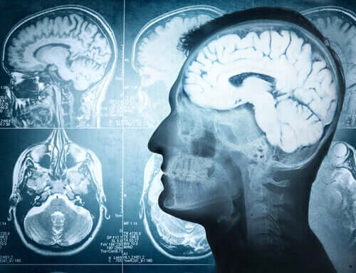
Computed tomography or magnetic resonance imaging is done to diagnose brain herniation. Doctors treat causes if possible and take measures to support breathing (such as mechanical ventilation) and to reduce the increased pressure within the skull.
What is the most common type of brain herniation?
Subfalcine hernia, also known as midline shift or cingulate hernia, is the most common type of cerebral hernia. It is generally caused by unilateral frontal, parietal, or temporal lobe disease that creates a mass effect with medial direction, pushing the ipsilateral cingulate gyrus down and under the falx cerebri.
What are the stages of brain herniation?
Brain herniation can progress from a subtle finding of pupillary asymmetry (uncal herniation) to an altered level of consciousness (compression of the reticular activating system), then progressing to the moribund stage of abnormal posturing (compression of the diencephalon and the brainstem), and finally death ...
Can a person survive brain herniation?
The outlook varies, depending on where in the brain the herniation occurs. Without treatment, death is likely. There can be damage to parts of the brain that control breathing and blood flow. This can rapidly lead to death or brain death.
Where does your brain go when herniated?
During a central herniation, the temporal lobe gets pushed down to the tentorial notch, which is the closest part of the brain to the spine. Tonsillar herniations. These occur in the infratentorial area of the brain. The herniation pushes back brain tissue into the area that connects the skull to the spine.
What causes the brain to herniate?
Bleeding or swelling in the brain can increase pressure within the skull. The pressure may force the brain sideways and downward in the skull through small openings in the relatively rigid sheets of tissue that separate the brain into compartments. The result is brain herniation.
Which brain herniation is the most life threatening?
Central herniation Downward herniation can stretch branches of the basilar artery (pontine arteries), causing them to tear and bleed, known as a Duret hemorrhage. The result is usually fatal.
What are 3 symptoms of a brain injury?
Physical symptomsLoss of consciousness from several minutes to hours.Persistent headache or headache that worsens.Repeated vomiting or nausea.Convulsions or seizures.Dilation of one or both pupils of the eyes.Clear fluids draining from the nose or ears.Inability to awaken from sleep.More items...•
What patient presentation is indicative of the first stage of brain herniation?
A patient with impending uncal herniation will initially present with symptoms of increased intracranial pressure. These symptoms include headache, nausea, vomiting, and altered mental status.
Can brain herniation cause sudden death?
Understanding brain herniation The condition is usually caused by swelling from a head injury, stroke, bleeding, or brain tumor. A brain herniation is a medical emergency and requires immediate medical attention. It's often fatal if not treated right away.
What is a clinical symptom of central herniation?
The clinical syndrome of central herniation classically manifests as a rostral to caudal progression of deficits attributed to brainstem dysfunction, including cranial nerve III (oculomotor nerve) palsy, diminished level of consciousness, decerebrate or decorticate posturing, rigidity or paralysis, abnormal respiratory ...
What happens when your brain shifts?
Diffuse axonal injury is the shearing (tearing) of the brain's long connecting nerve fibers (axons) that happens when the brain is injured as it shifts and rotates inside the bony skull. DAI usually causes coma and injury to many different parts of the brain.
Which brain herniation is the most life threatening?
Central herniation Downward herniation can stretch branches of the basilar artery (pontine arteries), causing them to tear and bleed, known as a Duret hemorrhage. The result is usually fatal.
What is the most common type of head injury?
Head injuries include: Concussion, in which the brain is shaken, is the most common type of traumatic brain injury.
What are the two most common brain injuries?
Depending on the cause, there are two types of brain injuries: Traumatic Brain Injuries (TBI) and Non-Traumatic Acquired Brain Injuries (ABI).
What is the most serious type of brain injury?
Diffuse Axonal Injury (DAI) Diffuse axonal injuries are one of the most severe types of traumatic brain injury. They occur when the brain is shaken or twisted inside the skull.
How long does it take for ICP to show signs?
The genesis for this approach is the changes in ICP being evident 6 hours before clinical signs and symptoms are observable in patients following herniation syndrome.[3] However, studies from ICP guided rescue therapy have produced mixed results.
What causes tonsillar herniation?
Central involves herniation of both temporal lobes through the tentorial notch. A tonsillar herniation is caused by an infratentorial mass, forcing the cerebellar tonsils through the foramen magnum. Upward herniation occurs when an infratentorial mass compressed the brainstem. Etiology.
Why is brain herniation labeled as a brain code?
Brain herniation can be labeled as “brain code” to connate the emergent need to timely counteract such disastrous brain processes. [1]
Why is brain herniation important?
Brain herniation, also called brain code, requires early diagnosis and prompt management in order to prevent irreversible pathological cascades that eventually lead to respiratory arrest and subsequent death. This activity reviews the evaluation and management of brain herniation and highlights the role of the interprofessional team in evaluating and improving care for patients with this condition.
What causes a duret hemorrhage?
As the pathological descent of the brainstem through the incisura progresses, venous congestion along with stretching and tearing of small perforators create Duret hemorrhage s. Clinically there is a progression from abnormal flexor posturing to abnormal extensor response through the involvement of rubrospinal and the vestibulospinal tracts. Soon following this stage comes either a vegetative state or impending death due to damage to respiratory and cardiac centers in medulla by the herniated cerebellar tonsils through the foramen magnum. Patients may exhibit characteristic triple components of Cushing triad constituting of hypertension, bradycardia, and irregular respirations.
What is the difference between anterior and posterior transalar herniation?
In the posterior (descending) variant of transalar herniation, there may be infarction within the middle cerebral artery territory resulting from its compression within the sphenoid ridge. In anterior (ascending) transalar herniation, compression of the supra-clinoid segment of the internal carotid artery against the anterior clinoid process leads to infarction within the territory of anterior and middle cerebral arteries.
Why do you need to be paralyzed when you have mechanical ventilation?
Most patients require mechanical ventilation and need to be paralyzed to avoid straining or agitation. Sedatives should be used to calm the patient.
What is an upward transtentorial herniation?
Upward transtentorial herniation can occur when an infratentorial mass (eg, tumor in the posterior fossa, cerebellar hemorrhage) compresses the brain stem, kinking it and causing patchy brain stem ischemia. The posterior 3rd ventricle becomes compressed.
Why does the skull have a cushing reflex?
Because the skull is rigid after infancy, intracranial masses or swelling may increase intracranial pressure, sometimes causing protrusion (herniation) of brain tissue through one of the rigid intracranial barriers (tentorial notch, falx cerebri, foramen magnum). When intracranial pressure is increased sufficiently, regardless of the cause, Cushing reflex and other autonomic abnormalities can occur. Cushing reflex includes systolic hypertension with increased pulse pressure, irregular respirations, and bradycardia.
What is the cingulate gyrus pushed under?
The cingulate gyrus is pushed under the falx cerebri by an expanding mass high in a cerebral hemisphere. In this process, one or both anterior cerebral arteries become trapped, causing infarction of the paramedian cortex. As the infarcted area expands, patients are at risk of transtentorial herniation, central herniation, or both.
Which structure is herniated in the brain?
Brain herniation is classified based on the structure through which tissue is herniated: Transtentorial (uncal) herniation: The medial temporal lobe is squeezed by a unilateral mass across and under the tentlike tentorium that supports the temporal lobe.
What nerves are involved in herniation?
As herniation progresses, the ipsilateral cerebral peduncle. In about 5% of patients, the contralateral 3rd cranial nerve and cerebral peduncle. Eventually, the upper brain stem and the area in or around the thalamus.
Why do temporal lobes herniate?
Central herniation: Both temporal lobes herniate through the tentorial notch because of bilateral mass effects or diffuse brain edema.
How much serum osmolality should be maintained?
Diuretics: Serum osmolality should be kept at 295 to 320 mOsm/kg. Osmotic diuretics (eg, mannitol) may be given IV to lower ICP and maintain serum osmolality. These drugs do not cross the blood-brain barrier. They pull water from brain tissue across an osmotic gradient into plasma, eventually leading to equilibrium. Fluid and electrolyte balance should be monitored closely while osmotic diuretics are used. A 3% saline solution is another potential osmotic agent to control ICP.
What causes a herniated brain?
Herniation of the brain can also be caused by other factors that lead to increased pressure inside the skull, including: 1 Collection of pus and other material in the brain, usually from a bacterial or fungal infection ( abscess) 2 Bleeding in the brain (hemorrhage) 3 Buildup of fluid inside the skull that leads to brain swelling ( hydrocephalus) 4 Strokes that cause brain swelling 5 Swelling after radiation therapy 6 Defect in brain structure, such as a condition called Arnold-Chiari malformation
Why does my brain bleed?
This is most often the result of brain swelling or bleeding from a head injury, stroke, or brain tumor. Brain herniation can be a side effect of tumors in the brain, including: Metastatic brain tumor. Primary brain tumor. Herniation of the brain can also be caused by other factors that lead to increased pressure inside the skull, including:
What causes swelling in the brain after radiation?
Bleeding in the brain (hemorrhage) Buildup of fluid inside the skull that leads to brain swelling ( hydrocephalus) Strokes that cause brain swelling. Swelling after radiation therapy. Defect in brain structure, such as a condition called Arnold-Chiari malformation.
What is the best way to reduce brain swelling?
Medicines that decrease brain swelling, such as mannitol, saline, or other diuretics. Placing a tube in the airway ( endotracheal intubation) and increasing the breathing rate to reduce the levels of carbon dioxide (CO 2) in the blood.
What to do if you have decreased alertness?
Call 911 or the local emergency number or take the person to a hospital emergency room if they develop decreased alertness or other symptoms, especially if there has been a head injury or if the person has a brain tumor or blood vessel problem. Prevention. Expand Section.
Where does a brain herniation occur?
Brain herniation can occur: From side to side or down, under, or across rigid membrane like the tentorium or falx. Through a natural bony opening at the base of the skull called the foramen magnum. Through openings created during brain surgery.
What is the loss of all brainstem reflexes?
Loss of all brainstem reflexes (blinking, gagging, and pupils reacting to light) Respiratory arrest (no breathing) Wide (dilated) pupils and no movement in one or both eyes. Exams and Tests. Expand Section. A brain and nervous system exam shows changes in alertness.
What is the term for the extension of the dura mater that separates the cerebellum from the cereb?
The tentorium is an extension of the dura mater that separates the cerebellum from the cerebrum. There are two major classes of herniation: supratentorial and infratentorial. Supratentorial refers to herniation of structures normally found above the tentorial notch, and infratentorial refers to structures normally found below it.
Why is herniation fatal?
Because herniation puts extreme pressure on parts of the brain and thereby cuts off the blood supply to various parts of the brain, it is often fatal. Therefore, extreme measures are taken in hospital settings to prevent the condition by reducing intracranial pressure, or decompressing (draining) an hematoma which is putting local pressure on ...
What is the term for the pressure in the skull that causes the brain to shift?
Brain herniation. Brain herniation is a potentially deadly side effect of very high pressure within the skull that occurs when a part of the brain is squeezed across structures within the skull. The brain can shift across such structures as the falx cerebri, the tentorium cerebelli, and even through the foramen magnum ...
What is Kernohan's notch?
Another important finding is a false localizing sign, the so-called Kernohan's notch, which results from compression of the contralateral cerebral crus containing descending corticospinal and some corticobulbar tract fibers. This leads to Ipsilateral hemiparesis in reference to the herniation and contralateral hemiparesis with reference to the cerebral crus.
What are the symptoms of a brain herniation?
One or both pupils may be dilated and fail to constrict in response to light. Vomiting can also occur due to compression of the vomiting center in the medulla oblongata. Severe headaches and seizures as a result of increased intracranial pressure are not uncommon . Cardiovascular and pulmonary symptoms may also be present as the brain loses function, but might also be associated with bleeding. They can include: hypertension, respiratory depression, arrhythmia and in severe cases cardiac arrest.
Which part of the temporal lobe is squeezed so much that it moves towards the tentorium?
In uncal herniation, a common subtype of transtentorial herniation, the innermost part of the temporal lobe, the uncus, can be squeezed so much that it moves towards the tentorium and puts pressure on the brainstem, most notably the midbrain.
What happens if the brain stem is disrupted?
The disrupted brainstem can lead to decorticate posture, respiratory center depression and death. Other possibilities resulting from brain stem distortion include lethargy, slow heart rate, and pupil dilation.
What happens if a herniated primary lesion is large enough?
If the primary lesion becomes large enough, uncal or central herniation may develop. As the lesion grows and herniation becomes prominent, symptoms progress and patients may develop anisocoria, a decreased level of consciousness, changes in respiratory pattern, changes in muscle tone and posturing.
What is the most common brain herniation?
Subfalcine herniation or cingulate herniation, the most common brain herniation pattern, is characterized by displacement of the brain (typically the cingulate gyrus) beneath the free edge of the falx cerebri due to raised intracranial pressure.
Why is uncal herniation bad?
Uncal herniation carries a bad prognosis due to the direct compression of the vital midbrain centers. They often require emergency neurosurgical decompression.
Why is my subfalcine hernia missed?
Subfalcine herniation may present with very subtle clinical symptoms. The hernia may be missed because the symptoms don’t warrant neural imaging.
What is the term for the extension of the dura mater that separates the cerebellum from the cereb?
The tentorium is an extension of the dura mater that separates the cerebellum from the cerebrum. There are two major classes of herniation: supratentorial and infratentorial. Supratentorial refers to herniation of structures normally found above the tentorial notch, and infratentorial refers to structures normally found below it.
What is the name of the condition where the brain is displaced?
Brain herniation is also called cerebral herniation, acquired intracranial herniation or brain herniation syndrome, is a condition in which a portion of the brain, cerebrospinal fluid (CSF) and blood vessels is displaced because of increased pressure inside the skull. Increase in pressure results in progressive damage to brain tissue that may include life-threatening damage to the brainstem.
What happens when the fontanels close in infancy?
Once the fontanels close in infancy, the cra nium becomes a rigid structure with a fixed volume containing three components the brain, the cerebrospinal fluid (CSF), and blood. The Monroe-Kellie Doctrine states that intracranial volume is constant and an increase in the volume of one component will cause a decrease in the volume of one or both of the others 1). Within a rigid structure, this change can have significant effects, such as decreased cerebral blood flow or herniation of brain tissue. When brain herniation occurs, the location of the herniation affects the presenting symptoms and the clinical outcomes.