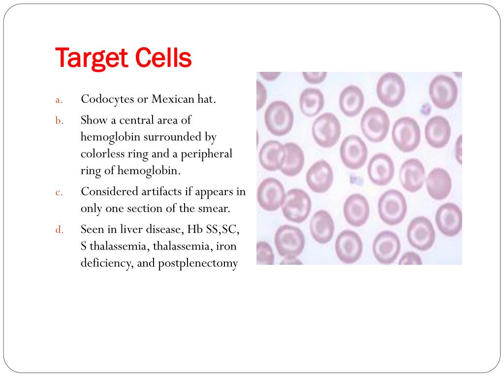
What is the significance of target cells?
Significance. Target cells are abnormal, but are not associated with a single specific disease. Therefore, always look for the context: where target cells are the dominant abnormal form look for macrocytosis that may imply liver disease, or if MCV is normal or low consider a haemoglobinopathy (HbC, D or E). If the target cell is part of a ...
What causes target cells?
- Causes of Target Cells
- Liver disease (obstructive or intrahepatic)
- Haemoglobinopathies
- Beta thalassaemia
- Iron deficiency anaemia
- Sideroblastic anaemia
- Sickle cell anaemia
- Post splenectomy
- Lead poisoning
What is the definition of target cells?
target cell. n. 1. A cell selectively affected by a particular agent, such as a virus, drug, or hormone. 2. An abnormal red blood cell having a dark center surrounded by a light band that is itself encircled by a darker ring, seen in certain anemias and after splenectomy. American Heritage® Dictionary of the English Language, Fifth Edition.
What two cellular structures would you target?
Well upon my research of what antibacterial & parasites medications do is target the outside of the capsule working the way to the inner side of the prokaryotic cell to destroying the DNA to where it can not reproduce, there fore the bacterial infection is being eliminated. The two cellular structures I have chosen to target is the capsule & DNA .
What is an example of a target cell?
An erythrocyte in target cell anaemia, with a dark centre surrounded by a light band that again is encircled by a darker ring; it thus resembles a shooting target; such cell's also appear after splenectomy.
Where are the target cells?
Macrocytic target cells can be seen in liver disease, and microcytic target cells may be seen in thalassemia. Target cells may also be seen in hemoglobin C and E disease, and following splenectomy; they can also occur as an artifact of slide preparation.
What are target cells or organs?
Target cells are cells that are receptive to a secreted hormone. dependent on three factors; the hormone levels in the blood, the receptor levels on the target cell, and hormone–receptor affinity.
How many target cells are there?
16 Target Cells (Codocytes)
Why are there target cells?
Presence of cells called target cells may be due to: Abnormal hemoglobin, the protein in RBCs that carries oxygen (hemoglobinopathies) Deficiency of an enzyme called lecithin cholesterol acyltransferase. Iron deficiency.
Why target cells are formed?
Target cells appear in conditions that cause the surface of the red cell to increase disproportionately to its volume. This may result from a decrease in hemoglobin, as in iron deficiency anemia, or an increase in cell membrane.
What are target cells quizlet?
A target cell is a cell that possesses specific receptors for a particular hormone.
What is a target cell RBC?
Codocytes, also known as target cells, are red blood cells that have the appearance of a shooting target with a bullseye.
What are target tissues?
Target tissue refers to the intended site that a hormone will affect such as muscle. Receptor site. Receptor sites are special sites located on every target tissue and only communicate with the specific hormone intended for the target tissue.
What does few target cells mean?
There are four major circumstances in which target cells appear as the major morphologic abnormality: thalassemia, hepatic disease with jaundice, hemoglobin C disorders, and the postsplenectomy state. Lesser numbers of target cells are found in sickle cell anemia, iron deficiency, and lead intoxication.
What are target cells in sickle cell anemia?
A fraction of erythrocytes appear as target cells in stained blood smears in sickle cell disease, due to a inheritance of the hemoglobin variant Hb S, polymerizing upon deoxygenation. These cells appear in a three dimension as thin cups.
What is pencil cells in blood?
Cigar cells (also referred to as pencil cells) are red blood cells that are cigar- or pencil-shaped on peripheral blood smear. Cigar cells are commonly associated with hereditary elliptocytosis.
What are target cells quizlet?
A target cell is a cell that possesses specific receptors for a particular hormone.
What are target cells on peripheral smear?
Target cells (codocytes) have a centrally located disk of hemoglobin surrounded by an area of pallor with an outer rim of hemoglobin adjacent to the cell membrane giving the cell the appearance of a target. Leptocytes (or wafer cells) are thin, flat cells with the hemoglobin at the periphery of the cell.
What are target cells in sickle cell anemia?
A fraction of erythrocytes appear as target cells in stained blood smears in sickle cell disease, due to a inheritance of the hemoglobin variant Hb S, polymerizing upon deoxygenation. These cells appear in a three dimension as thin cups.
Are target cells seen in alpha thalassemia?
Alpha thalassemia trait The peripheral blood smear typically shows hypochromia, microcytosis, and target cells.
What is the target cell?from imagebank.hematology.org
Target Cells. Target cells are erythryoctes with an increased cell membrane-to-volume ratio, due either to gain of membrane lipids or to a reduction in cell volume.
How are target cells formed?from laboratoryinfo.com
Codocytes or popularly known as target cells are formed if the red blood cell’s surface is increased disproportionately to its volume. Target cells are actually red blood cells, which are extremely thin and have an excessive cell membrane. Hence, the red blood cells assume the shape ...
What is the shape of a codo cell?from laboratoryinfo.com
On the other hand, if you are going to examine the target cells using an electron microscope, you will notice that they look very thin and look like the shape of a bell. That is why target cells are called codocytes as codo means bell.
Why are codocytes associated with diseases?from laboratoryinfo.com
The formation of diseases can be linked to two reasons – direct increase in the surface area by having a direct impact on the concentration of lipids in the red blood cell membrane and decreased concentration of hemoglobin within the red blood cells.
Why are target cells different from normal red blood cells?from laboratoryinfo.com
Target cells differ from the usual red blood cells because they are more resistant to hypertonic solutions than normal red blood cells. Hemolysis may less likely to take place until the saline concentration reaches 1.5% to 2%. Their envelope is too large for hemoglobin content.
What does it mean when target cells are elevated?from laboratoryinfo.com
Hence, if there is an elevation in target cells, it could indicate a shift in the equilibrium exchanges between the red blood cells and the cholesterol. In other words, target cells have an abnormally high surface area for their volume. (2, 3, 4, 5, and 6)
Why do blood cells look thinner?from laboratoryinfo.com
In a film of blood, target cells look thinner than usual because of their pale appearance. In target cells, there is a disproportional increase in the ratio of surface membrane area to volume.
What is the role of target cell metabolism?
Target cell metabolism has been shown to play an important role in several steroid hormone systems [116 ]. In mineralocorticoid-responsive tissues, the receptor that mediates the action of aldosterone binds glucocorticoids with equal affinity. Selectivity is achieved in these tissues by efficient degradation of glucocorticoids, rendering them unavailable to the receptor. Alternatively, cells can convert hormones to more active metabolites as in the conversion of T 4 to T 3 or estrone (E 1) to estradiol (E 2 ).
How do target cells regenerate tissue?
Tissue regeneration is a complex process where healing occurs through various synergistic effects of secreted cytokines, inflammatory cells and signaling molecules. Inflammations have a direct relation with healing process. Immune cells attracted to the wound/implant produces certain cytokines and chemokines which cause chemotaxis of epithelial and endothelial cells at the wound site promoting regeneration/vascularization. Generally, monocytes in circulation polarize into two main phenotypes: M1 and M2, which have distinct effect on recruiting and performance of cells [66,67]. Directed migration and oriented differentiation of stem cells could be achieved by cultivating immune cells on the material surface. M1 macrophages promote long distance rostral migration of neural stem cells in CXCR4 dependent pathway [68]. Macrophages are found to capture mesenchymal stem cells (MSCs) and direct their own phenotype transition from M1 to M2 as determined by their M2/ M1 ratio marker (CD163/CCR7) positive cells [69].
What is the target effect of LC cells?
Target cells could stimulate the LC cells and this target effect was shown to be mediated by interneuronal contact and also by glial-soluble factors. A target stimulatory effect was also reported in other aminergic systems (Prochiantz et al., 1979; Azmitia and Whitaker-Azmitia, 1987 ). Stimulation of dopaminergic and serotonergic af-ferents by target areas was also of neuronal origin ( Di Porzio et al, 1980 ); however, the existence of a glial stimulatory soluble factor is suggested in the serotonergic system ( Whitaker-Azmitia and Azmitia, 1989 ). In addition to the stimulatory effect, target cells can also exert an inhibitory effect on the development of LC cells. Our results suggest that the inhibitory effect is mediated by target glial membranal molecules which are expressed in, or interact with, serum-containing medium. The role of cell contact in target afferent interaction has been demonstrated in the dopaminergic system ( Prochiantz et al, 1981 ). Inhibitory target influence in vitro was also found in the dopaminergic system and the morphological aspect of this phenomenon has been explored ( Denis-Donini et al, 1983 ). The existence of inhibitory mechanisms in the CNS may be of great importance in regard to the specificity of neuronal connections. In a previous section we mentioned that although the NA innervation is very extensive in the CNS, certain areas receive only sparse NA innervation. Indeed, an inhibitory interaction mediated by contact between LC and septal transplants was shown to restrict the fiber growth into area dentata transplants in oculo ( Goldowitz et al, 1984 ). It is possible that target areas may provide signals that can stimulate neunte outgrowth on the one hand and on the other hand transmit signals that limit further outgrowth. A balance between both types of signals can be crucial both for regulating neurite expansion and for creating specific innervation patterns.
What are the chemokines in NK cells?
Target cell recognition by freshly isolated human NK cells induces a set of chemokines, including CCL3, CCL4, CCL5, as well as the cytokines TNF-α and IFN-γ (Fauriat et al., 2010 ). Chemokines are induced within one hour of stimulation, whereas secretion occurs several hours after activation. Importantly, experiments varying the signaling input for NK cell activation have revealed a hierarchy in requirements for the induction of chemokines and cytokines, with chemokines induced by weakly activating signals, degranulation induced by intermediate levels of activating stimuli, and cytokines requiring the strongest activation. This hierarchy is reflected in the requirements for induction of different effector responses. PLC-γ is required for all responses ( Tassi et al., 2005; Caraux et al., 2006 ). A deficiency in SOCE, as seen in NK cells from humans with autosomal recessive STIM1 and ORAI1 mutations, results in defective degranulation and cytokine production induced upon target cell recognition, but only partially impairs chemokine production ( Maul-Pavicic et al., 2011 ). Notably, NK cells from PI3K p110δ-deficient mice displayed selectively impaired cytokine production, whereas knockout of both p110δ and p110γ was required to impair cytotoxicity ( Kim et al., 2007; Tassi et al., 2007 ). Moreover, PKCθ-deficient mice had defects in IFN-γ transcription and secretion due to impaired JNK, AP-1, and NFAT activation ( Tassi et al., 2008 ). Yet, NK cell-mediated cytotoxicity was not impaired in PKCθ-deficient mice. Curiously, a Rap1b deficiency in mice selectively deters NK cell chemokine and cytokine production, but not cytotoxicity ( Awasthi et al., 2010 ). Thus, a few proteins, including PI3K p110δ and PKCθ may be specifically required for transcription of cytokine genes. Further studies are required to understand how these proteins are integrated in the signaling pathways for NK cell activation and how the engagement of different activating receptor controls their function.
What is the target cell specificity of polyplexes?
Target cell specificity of polyplexes has been achieved by the incorporation of cell-binding ligands into the surfaces of nanoparticulate vectors , most notable for polymer–DNA complexes. A variety of targeting ligands have been attached covalently or noncovalently to polycations such as Tf, epithelial growth factor (EGF), folate, galactose-containing ligands, low-density lipoprotein (LDL), basic fibroblast growth factor (bFGF), and antibodies.224,244–251
How does sensitivity to hormones affect the target cell?
Target cell sensitivity to a certain hormone is partly based on the amount of receptors for that hormone that the target cell has. The more receptors, the higher the target cell’s sensitivity. Hormone receptors are continuously broken down and replaced. This ensures that all parts of the cell are new and functioning correctly, and also allows numbers of receptors to be altered as needed. When new receptor synthesis occurs faster than old receptor degradation, the target cell becomes more sensitive to a hormone. This is often referred to as up-regulation since the number of receptors increases (see Fig. 1.7 ). When the opposite occurs, it is called down-regulation.
What are the two death-inducing factors in exocytosis?
Granule exocytosis can provide additional death-inducing factors. One factor is perforin. Perforin can form polymeric channels in cell membranes. Affected membranes lose their structural integrity, and lysis of the cell soon follows. The second factor is granzyme B. Granzymes may gain access to the target cell, either directly by fusion of the granule vesicles or dense core with the membrane of the target cell or because of a sub-lytic quantity of perforin that allows granzymes to gain access to the cytosol of the target cell. When in the cytosol, granzyme B can activate apoptotic proteolysis.
What is the target cell?
Target Cells. Target cells are erythryoctes with an increased cell membrane-to-volume ratio, due either to gain of membrane lipids or to a reduction in cell volume.
What is the target cell of erythrocytes?
Target cells are erythryoctes with an increased cell membrane-to-volume ratio, due either to gain of membrane lipids or to a reduction in cell volume. The characteristic bull’s-eye appearance results from clustering of intensely-staining hemoglobin around the periphery and in the center of the cell.
What is the target cell morphology?
Target cells adopt a “ bullseye” morphology where hemoglobin is concentrated in the center and on the periphery with a colourless zone in between the two areas. Other target cells may also look folded or bell shaped. 1-3
What is the artifact of target cell formation?
Artifact: Target cell formation occurs when blood smears are made when humidity is high. 1. Hemoglobinopathies: There is a uneven distribution of hemoglobin within the cell, and an increased surface area to volume ratio. 1. Note: Target cells have an increased surface area to volume ratio and decreased osmotic fragility. 1,3.
Content
A target cell or white cell (from English target cell) is any cell in which a hormone recognizes its receptor. In other words, a target cell has specific receptors where hormones can bind and exert their effect.
Definition of target cells
In the branch of endocrinology, a target cell is defined as any cell type that has specific receptors to recognize and interpret the message of hormones.
Interaction characteristics
The molecule that is acting as a chemical messenger attaches itself to its receptor in the same way that an enzyme does to its substrate, following the pattern of the key and the lock.
Cell signaling
Target cells are a key element in cell signaling processes, since they are in charge of detecting the messenger molecule. This process was elucidated by Earl Sutherland, and his research was awarded the Nobel Prize in 1971.
Reception
During the first stage, the detection of the target cell of the signal molecule occurs, which comes from outside the cell. Thus, the chemical signal is detected when the binding of the chemical messenger to the receptor protein occurs, either on the surface of the cell or inside it.
Transduction
The union of the messenger and the receptor protein alters the configuration of the latter, initiating the transduction process. At this stage, the signal is converted into a form that is capable of eliciting a response.
Reply
The last stage of cell signaling consists of the origin of the response, thanks to the transduced signal. The answer can be of any kind, including enzymatic catalysis, organization of the cytoskeleton, or activation of certain genes.
What Are The Target Cells Of Insulin And Glucagon?
These processes activate adenal cyclase, which raises cyclic adenosine monophosphate in target cells. When affected by insulin, liver cells are stimulated to conduct glucose uptake. Insulin binds to the target cells and allows the cell to pull glucose in through its membranes via signal transduction. The glucose is then used as an energy source for the cell. Learn more about Human Anatomy Continue reading >>
What hormone regulates the level of sugar in the blood?
Insulin, hormone that regulates the level of sugar (glucose) in the blood and that is produced by the beta cells of the islets of Langerhans in the pancreas. Insulin is secreted when the level of blood glucose rises—as after a meal. When the level of blood glucose falls, secretion of insulin stops, and the liver releases glucose into the blood. Insulin was first reported in pancreatic extracts in 1921, having been identified by Canadian scientists Frederick G. Banting and Charles H. Best and by Romanian physiologist Nicolas C. Paulescu, who was working independently and called the substance “pancrein.” After Banting and Best isolated insulin, they began work to obtain a purified extract, which they accomplished with the help of Scottish physiologist J.J.R. Macleod and Canadian chemist James B. Collip. Banting and Macleod shared the 1923 Nobel Prize for Physiology or Medicine for their work. Insulin is a protein composed of two chains, an A chain (with 21 amino acids) and a B chain (with 30 amino acids), which are linked together by sulfur atoms. Insulin is derived from a 74-amino-acid prohormone molecule called proinsulin. Proinsulin is relatively inactive, and under normal conditions only a small amount of it is secreted. In the endoplasmic reticulum of beta cells the proinsulin molecule is cleaved in two places, yielding the A and B chains of insulin and an intervening, biologically inactive C peptide. The A and B chains become linked together by two sulfur-sulfur (disulfide) bonds. Proinsulin, insulin, and C peptide are stored in granules in the beta cells, from which they are released into the capillaries of the islets in response to appropriate stimuli. These capillaries empty into the portal vein, which carries blood from the stomach, intestines, and pancrea Continue reading >>