There are three types of cone cells:
- Red-sensing cones (60 percent)
- Green-sensing cones (30 percent) and
- Blue-sensing cones (10 percent)
What are the three different types of cones cells?
What are the three cone cells? We have three types of cones: blue, green, and red. The human eye only has about 6 million cones. Many of these are packed into the fovea, a small pit in the back of the eye that helps with the sharpness or detail of images. Other animals have different numbers of each cell type. Click to see full answer.
How are cone cells adapted to their function?
What are the properties of cone?
- One circular face.
- One vertex.
- A circular base and one continuous curve.
- Apex is a point above the centre of the base.
- Funnels are cone-shaped.
- You can get ice cream in cones.
- Birthday hats are cone-shaped.
What type of cells contain a true nucleus?
The nucleus is a membrane-bound organelle that contains cellular DNA. Some cells, such as yours, contain a nucleus. Other cells, such as bacteria, do not. The nucleus-containing cells are called eukaryotic cells. Eukaryote means having membrane-bound organelles.
What structure contains cells called rods and cones?
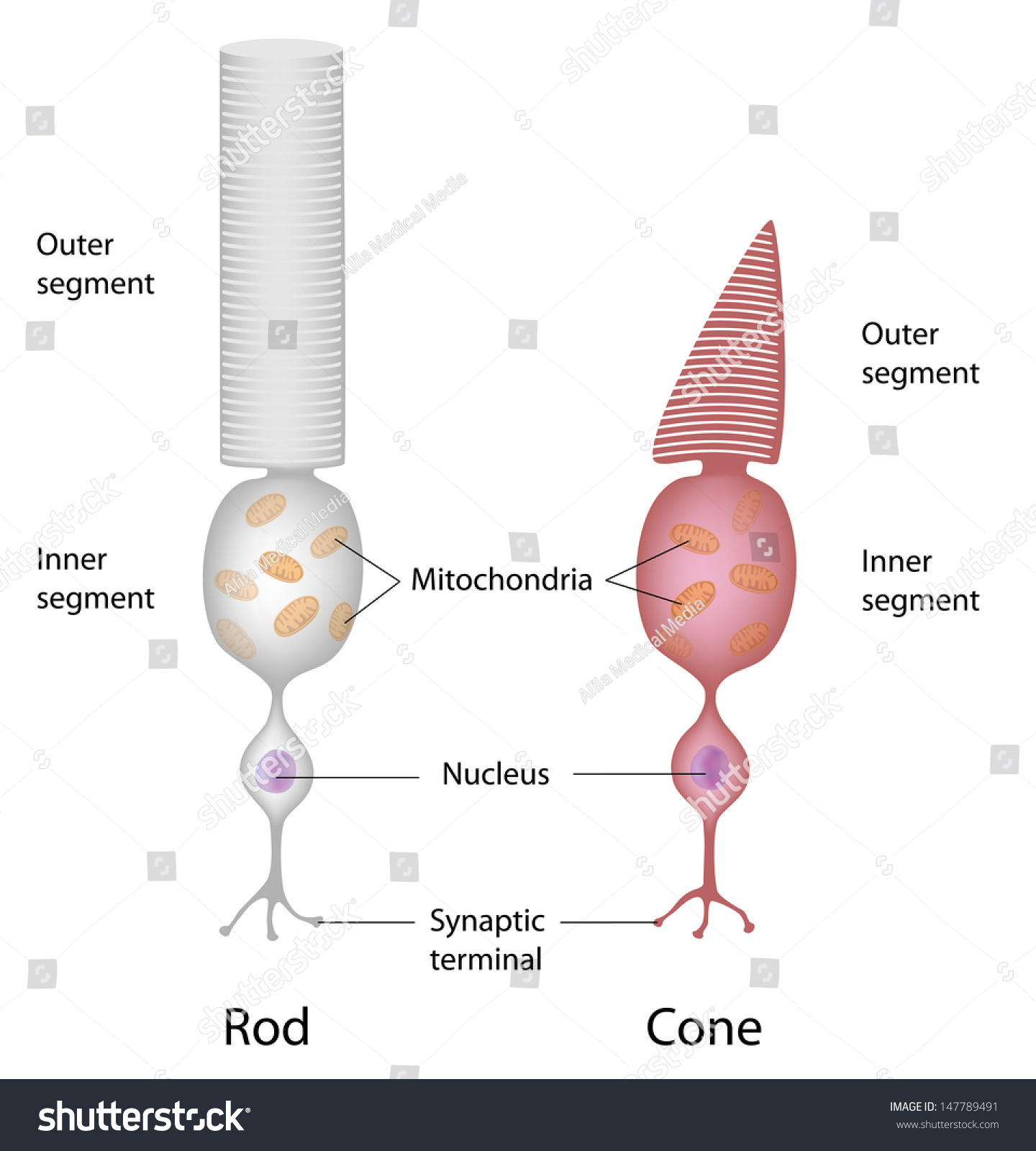
They consist of five principal regions:
- outer segment
- connecting cilium
- inner segment
- nuclear region
- synaptic region
What are the 3 types of cone cells?
There are three types of cone cells:Red-sensing cones (60 percent)Green-sensing cones (30 percent) and.Blue-sensing cones (10 percent)
What are the three types of cone photoreceptors?
There are 3 types of cones which we will refer to as the short-wavelength sensitive cones, the middle-wavelength sensitive cones and the long-wavelength sensitive cones or S-cone, M-cones, and L-cones for short. The light levels where both are operational are called mesopic.
What are the 3 cone pigments?
For example, human rhodopsin, the rod pigment that mediates dim-light vision, absorbs at 498 nm [2,3], while the three cone pigments contained in human cone cells are responsible for trichromatic (color) vision peak at 425 (blue), 530 (green), and 560 nm (red).
What are the 3 types of cones and how do they differ?
Humans normally have three types of cones, usually designated L, M and S for long, medium and short wavelengths respectively. The first responds the most to light of the longer red wavelengths, peaking at about 560 nm. The majority of the human cones are of the long type.
How many types of cone cells are there?
three typesCones require a lot more light and they are used to see color. We have three types of cones: blue, green, and red. The human eye only has about 6 million cones. Many of these are packed into the fovea, a small pit in the back of the eye that helps with the sharpness or detail of images.
What are the 3 color receptors?
3 Different Cone ReceptorsShort-wavelength cone receptors.Middle-wavelength cone receptors.Long-wavelength cone receptors.
What are rod cells and cone cells?
The human retina has two types of photoreceptors to gather light namely rods and cones. While rods are responsible for vision at low light levels, cones are responsible for vision at higher light levels. The light levels where both are functional are known as mesopic.
How many cone cells are in the retina?
Despite the fact that perception in typical daytime light levels is dominated by cone-mediated vision, the total number of rods in the human retina (91 million) far exceeds the number of cones (roughly 4.5 million).
What are the types of photoreceptors?
Photoreceptor types. There are four photoreceptor types in the human retina. Short-wavelength cones (blue), medium-wavelength cones (green), long-wavelength cones (red) and rods.. Three different cone mechanisms can be detected in behavioral, psychophysical and physiological testing (Fig.
What are cones and rods What are their functions?
What is the function of rods and cones in the eye? Rods are responsible for vision at low light levels or scotopic vision. Whereas, the cones are responsible for vision at higher light levels or photopic vision.
What are the types of photoreceptors and their functions?
There are two types of photoreceptors: cone photoreceptors and rod photoreceptors. These cells function by sensing light and/or color and delivering the message back to the brain through the optic nerve. While cone photoreceptors detect color through bright light, rod photoreceptors are sensitive to low-light levels.
What are the different types of photoreceptors in plants?
Six classes of photoreceptors are known: light-oxygen-voltage (LOV) sensors, xanthopsins, phytochromes, blue-light sensors using flavin adenine dinucleotide (BLUF), cryptochromes, and rhodopsins.
What type of cell is the cone?
Cones are a type of photoreceptor cell in the retina. They give us our color vision. Cones are a type of photoreceptor cell in the retina. They give us our color vision.
How many cones are there in the retina?
The retina has approximately 120 million rodsand 6 million con es. There are three types of cone cells:
What are cone cells?
Anatomical terms of neuroanatomy. Cone cells, or cones, are photoreceptor cells in the retinas of vertebrate eyes including the human eye. They respond differently to light of different wavelengths, and are thus responsible for color vision, and function best in relatively bright light, as opposed to rod cells, which work better in dim light.
Where are cone cells located?
Cone cells are densely packed in the fovea centralis , a 0.3 mm diameter rod-free area with very thin, densely packed cones which quickly reduce in number towards the periphery of the retina. Conversely, they are absent from the optic disc, contributing to the blind spot.
What is the wavelength of light in the S cone?
S Cones are most sensitive to light at wavelengths around 420 nm. However, the lens and cornea of the human eye are increasingly absorptive to shorter wavelengths, and this sets the short wavelength limit of human-visible light to approximately 380 nm, which is therefore called ' ultraviolet ' light.
What is the role of S cones in the circadian system?
It is possible that S cones may play a role in the regulation of the circadian system and the secretion of melatonin but this role is not clear yet. The exact contribution of S cone activation to circadian regulation is unclear but any potential role would be secondary to the better established role of melanopsin.
What are the three types of cones?
Cones are normally one of the three types, each with different pigment, namely: S-cones, M-cones and L-cones. Each cone is therefore sensitive to visible wavelengths of light that correspond to short-wavelength, medium-wavelength and longer-wavelength light. Because humans usually have three kinds of cones with different photopsins, ...
What is the synaptic terminal of a cone cell?
The synaptic terminal forms a synapse with a neuron such as a bipolar cell. The inner and outer segments are connected by a cilium.
How are cone cells different from rods?
Cone cells are somewhat shorter than rods, but wider and tapered, and are much less numerous than rods in most parts of the retina, but greatly outnumber rods in the fovea. Structurally, cone cells have a cone -like shape at one end where a pigment filters incoming light, giving them their different response curves. They are typically 40–50 µm long, and their diameter varies from 0.5 to 4.0 µm, being smallest and most tightly packed at the center of the eye at the fovea. The S cone spacing is slightly larger than the others.
What are the different types of cones?
Located on each of the two retinas are actually three different types of cones: Red cones, which account for 60% of all cones. Green cones, which make up 30% of the cones. Blue cones, limited to just 10% of the cones 1 .
What are the problems with the eye cones?
Problems With Eye Cones. Color Blindness. Seeing the world in all its beautiful glory is made possible by a type of nerve cells on the retina known as cones. There are about 6 million of these cones here that allow us to see the world in all its colorful hues.
What color are rods and cones?
But, when it's not entirely dark, such as around dusk or twilight, both rods and cones are able to work and you see some colors, as well as shades of grey. 5 . The red cones, also known as L-cones, are stimulated by long-wavelength light. The green cones, dubbed M-cones, respond to medium-wavelength light.
What is the term for a group of malfunctioning gene-related, inherited disorders that can affect both cones
There is a group of malfunctioning gene-related, inherited disorders known as dystrophies that can affect both cones and rods. By mid-adulthood these result in legal blindness. Those with these dystrophies may experience the follow symptoms:
How does light bounce off a flower stimulate the cones in the eye?
In bright sunshine it's all about the cones. It works this way. Light bouncing off a yellow flower, for example, would stimulate both the red and green cones in your eyes. This signal would then run from the optic nerve to the brain, which interprets the type of signal coming in based on its strength.
What is the disorder of the blue cone?
One cone-related disorder, blue cone monochromacy, is also inherited. This mainly affects males. With this condition, while the blue cones function perfectly normally, neither the red nor the green cones work properly. Uncontrolled back and forth shaking of the eye known as nystagmus.
How many nanometers can the eye see?
Our eyes can actually perceive light frequencies as short as 380 nanometers and as long as 700 nanometers. 3 Although these cones mainly respond to light in their own color zone, there is overlap between these. Each is actually able to respond to a variety of wavelengths. Getty Images.
Cone cells Definition
A cone cell is a type of photoreceptor cell present in the eye's retina that performs well in very bright light and enable color vision, with larger visual acuity as compared to the rod cells which are more sensitive towards dim light and cannot distinguish the colors.
Overview of Cone Cells
Cones or cone cells are one of the types of photoreceptor cells that are present in the eye's retina and these cells are responsible for eye color sensitivity and color vision. Cone cells perform well in fairly bright light as compared to the rod cells which perform better in dim light.
Types of cone cells
Cone cells are classified into three different types of cone cells which include:
Structure
Cone cells have a light-sensing part which is relatively shorter but it is tapered and wider. Cone cells are present in fewer numbers compared to the rod cells. But they are larger in number on the fovea.
What is the best material to study cone cell machinery?
One of the best materials to study the cone cell machinery is chicken retina, which shows a relatively high ratio of cone to rod cells in comparison to other animals. This chapter describes the methods of preparation and characterization of chicken rod and cone pigments.
What are the functions of ectoplacental cones?
The cone cells secrete a plethora of signaling, immune, and metabolic factors that regulate many functions in maternal tissues and the fate of the cells, which are essential for successful gestation . The strategic localization of this structure in contact with uterine maternal tissues, vessels, and immune cells is also critical in infection control or the vertical transmission of parasites, viruses, and bacteria. We have presented several models in which the ectoplacental cone can be studied in isolation or associated with maternal cells in different circumstances to help our comprehension of its differentiation process and functional participation in the maternal–fetal interaction.
What are CCs in the eye?
The CCs are the first nonneuronal cells to be recruited in the eye imaginal disc, and this occurs immediately after PR specification is complete ( Fig. 5.7 A). In fact, CCs are derived from a common precursor pool of 5 cells, known as the R7 equivalence group, which gives rise to both the R7 PR and the four CCs ( Dickson et al., 1992; Tomlinson et al., 1987 ). Cells within the R7 equivalence group express the Sevenless tyrosine kinase receptor, the EGF receptor, and the Notch receptor ( Cagan and Ready, 1989b; Fortini et al., 1993; Jennings et al., 1994; Rebay et al., 1993; Tomlinson and Struhl, 2001; Tomlinson et al., 1987 ). Each of these cells require EGF and Notch signaling to form. However, only one of these cells differentiates into the R7 neuron due to the fact that only a single cell comes in direct contact with the Sevenless ligand, membrane-bound Boss, which is expressed on the previously specified R8 precursor. Since the Sevenless receptor signals through the same Ras/MAPK pathway as the EGF receptor, this Boss-receiving cell receives higher Ras signaling, and becomes specified as a neuron, while the remaining 4 cells adopt the default fate, that of cone (or Semper) cell ( Cagan et al., 1992; Hart et al., 1990; Kramer et al., 1991; Reinke and Zipursky, 1988; Van Vactor et al., 1991 ). Overactivating Sevenless receptor signaling or overexpressing activated Ras in CC precursors can transform them into ectopic R7 PRs, and removing Sev signaling from the eye causes a failure in R7 differentiation but maintains the normal complement of four CCs ( Basler et al., 1991; Dickson et al., 1992; Tomlinson and Ready, 1986 ). Together, these data led to the model that cells within the R7 equivalence group are all similarly capable of becoming R7 or CCs, and that this fate choice merely requires Sev-activated signaling. While these findings have been critical for defining the components of the Ras signaling pathway, the molecular mechanisms that mediate the dose-dependent neural (R7) versus nonneural (CC) fate decision remain unclear. Interestingly, however, not all cells within the R7 equivalence group respond the same to different mutants affecting R7/CC fate decisions instead, only one to two cells are generally affected ( Basler et al., 1991; Bhattacharya and Baker, 2009; Dickson et al., 1992; Flores et al., 2000; Hayashi et al., 1998; Lai and Rubin, 1992; Matsuo et al., 1997; Tsuda et al., 2002 ). These data suggest that cells within the R7 equivalence group are actually not equivalent and that some bias toward R7 or CC fate exists in among these cells. Consistent with this idea, we have recently found that differential expression of two transcription factors, Pros and dPax2, in different CC precursors are important for establishing this bias ( Fig. 5.7 B), and that concurrent regulation of these factors is necessary to completely convert cells within the R7 equivalence group into R7 or CC fates (Charlton-Perkins and Cook, submitted).
What is the outer nuclear layer?
The outer nuclear layer (ONL) contains the rod and cone cell bodies; the cone cell body and nucleus are larger than those of the rod. Cone outer fibers are very short, and therefore the cone nuclei lie in a single layer close to the external limiting membrane; cell bodies of the rods are arranged in several rows inner to the cone cell bodies. The ONL is 8 to 9 cells thick on the nasal edge of the optic disc and 4 rows thick at the temporal edge and is thickest in the fovea, where it contains approximately 10 layers of cone nuclei.2
What are cone cells?
Description. Cone cells, or cones, are one of the two types of photoreceptor cells that are in the retina of the eye which are responsible for color vision as well as eye color sensitivity; they function best in relatively bright light, as opposed to rod cells that work better in dim light. Cone cells are densely packed in ...
Where are cone cells located in the eye?
There are about six to seven million cones in a human eye and are most concentrated towards the macula.
How big are the cones of the eye?
They are typically 40–50 µm long, and their diameter varies from 0.5 to 4.0 µm, being smallest and most tightly packed at the center of the eye at the fovea. The S cones are a little larger than the others. Photobleaching can be used to determine cone arrangement.
What is the synaptic terminal of a cone cell?
The synaptic terminal forms a synapse with a neuron such as a bipolar cell.
What is the response of cone cells to light?
The response of cone cells to light is also directionally nonuniform, peaking at a direction that receives light from the center of the pupil; this effect is known as the Stiles–Crawford effect. This definition incorporates text from the wikipedia website - Wikipedia: The free encyclopedia. (2004, July 22).
Which type of cell is shorter, rods or cone cells?
Cone cells are somewhat shorter than rods, but wider and tapered, and are much less numerous than rods in most parts of the retina, but greatly outnumber rods in the fovea. Structurally, cone cells have a cone-like shape at one end where a pigment filters incoming light, giving them their different response curves.
Why are cones less sensitive to light than rod cells?
They are also able to perceive finer detail and more rapid changes in images, because their response times to stimuli are faster than those of rods.

Overview
Cone cells, or cones, are photoreceptor cells in the retinas of vertebrate eyes including the human eye. They respond differently to light of different wavelengths, and the combination of their responses is responsible for color vision. Cones function best in relatively bright light, called the photopic region, as opposed to rod cells, which work better in dim light, or the scotopic region. Con…
Structure
Humans normally have three types of cones, usually designated L, M and S for long, medium and short wavelengths respectively. The first responds the most to light of the longer red wavelengths, peaking at about 560 nm. The majority of the human cones are of the long type. The second most common type responds the most to light of yellow to green medium-wavelength, peaking at 530 nm. …
Function
The difference in the signals received from the three cone types allows the brain to perceive a continuous range of colors, through the opponent process of color vision. (Rod cells have a peak sensitivity at 498 nm, roughly halfway between the peak sensitivities of the S and M cones.)
All of the receptors contain the protein photopsin, with variations in its confor…
Clinical significance
One of the diseases related to cone cells present in retina is retinoblastoma. Retinoblastoma is a rare cancer of the retina, caused by the mutation of both copies of retinoblastoma genes (RB1). Most cases of retinoblastoma occur during early childhood. One or both eyes may be affected. The protein encoded by RB1 regulates a signal transduction pathway while controlling the cell cycle progression as normally. Retinoblastoma seems to originate in cone precursor cells prese…
See also
• Cone dystrophy
• Disc shedding
• Double cones
• RG color space
• Tetrachromacy
External links
• Cell Centered Database – Cone cell
• Webvision's Photoreceptors
• NIF Search – Cone Cell via the Neuroscience Information Framework
• Model and image of cone cell
Structure of Eye Cones
Types of Cones
- Located on each of the two retinas are actually three different types of cones: 1. Red cones, which account for 60% of all cones 2. Green cones, which make up 30% of the cones 3. Blue cones, limited to just 10% of the cones1
Function of Cones
- These cones contain photopigments, known as opsin amino acids, that are sensitive to different wavelengths of visible light. Fact is, each of the different colors of the rainbow have a different wavelength. Our cones are able to capture these various frequencies thanks to these color-sensitive photopigments. Our eyes can actually perceive light frequencies as short as 380 nano…
Color Vision
- Color vision brings the world to life. In bright sunshine it's all about the cones. It works this way. Light bouncing off a yellow flower, for example, would stimulate both the red and green cones in your eyes. This signal would then run from the optic nerveto the brain, which interprets the type of signal coming in based on its strength. In this case, it would peg it as yellow. On the other hand, i…
Problems with Eye Cones
- Not everybody necessarily sees colors the same way. Color vision is tested with the Ishihara color palettes—a series of dots of different hues. This test, which identifies color issues, was named for Japanese ophthalmologist Shinobu Ishihara and includes numbers embedded in each of a set of circular images. The idea is to detect if you are unable to see certain colors.7 Unfortunately, eye …
Color Blindness
- If you are being tested with the Ishihara exam and can’t pick out some of the numbers amid the different shades of dots, it means that the color frequency isn’t registering because some of your cones aren’t functioning properly. You likely have some sort of color blindness.7 The term color blindness is a bit of a misnomer, however. In most cases, this does not mean that yo…