See more

What are the layers of the jejunum?
The jejunum has the typical histological pattern as the entire small intestine: mucosa, submucosa, muscularis and serosa. The mucosa is lined by simple columnar epithelium towards the lumen (lamina epithelialis).
What is the outermost layer of the jejunum?
Serosa. The serosa is the outermost layer that covers the small intestine.
What are the 3 subparts of the small intestine?
The small intestine has three parts: the duodenum, jejunum, and ileum. It helps to further digest food coming from the stomach. It absorbs nutrients (vitamins, minerals, carbohydrates, fats, proteins) and water from food so they can be used by the body. The small intestine is part of the digestive system.
What is the jejunum wall?
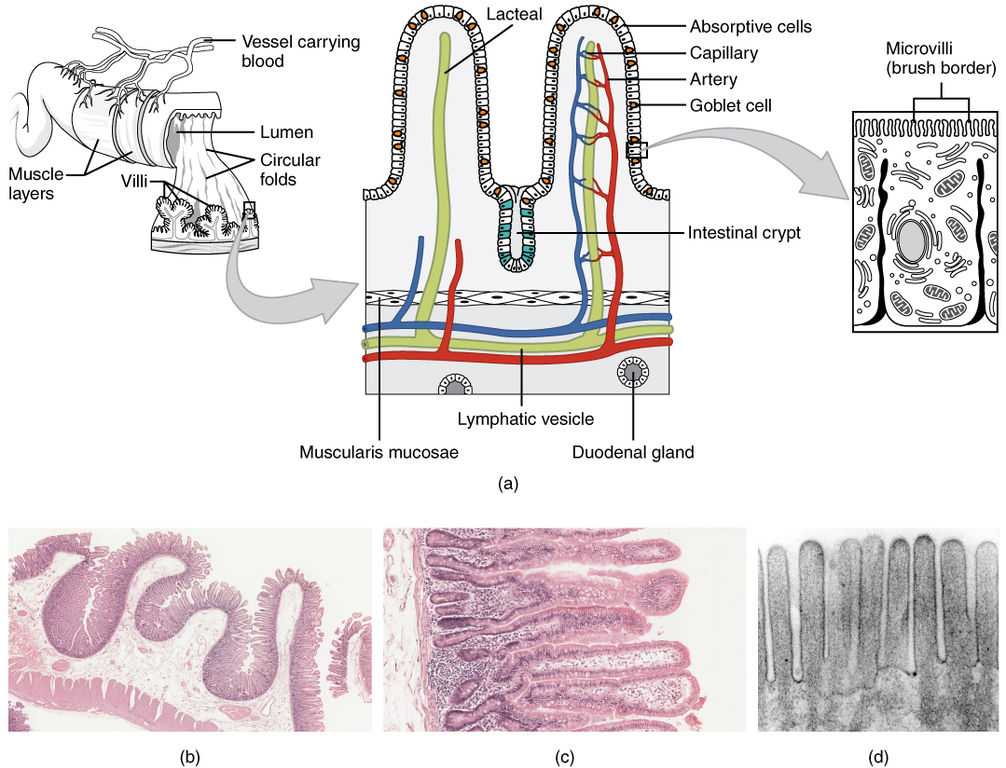
The inner wall of the small intestine is covered by numerous folds of mucous membrane called plicae circulares. The surface of these folds contains tiny projections called villi and microvilli, which further increase the total area for absorption.
Does jejunum have serosa or adventitia?
& E., 50 x. The basic pattern and arrangement of layers in the intestinal wall are seen in both the duodenum (A) and jejunum (B). In each, there is a mucosa, submucosa, muscularis, and an adventitia or a serosa. The mucosa has finger-like projections, the villi, lined by simple columnar epithelium.
What are layers of small intestine?
intestinal wall: The wall of the small intestine is composed of four layers, from the outside to the inside: serosa, muscularis, submucosa, and mucosa.
What are the functions of the 3 parts of the small intestine?
The small intestine is the longest part of the digestive system....Small intestine.DefinitionA part of the alimentary tract which extends from the stomach (pyloric orifice) to the large intestine (ileal orifice)FunctionFinal stages of food digestion Absorption of nutrients and water4 more rows
What are the 3 parts of the large intestine?
The large intestine is one long tube, but slightly different things happen in different parts of it. Its three parts are the colon, the rectum and the anus. The colon can also be divided into parts. The entry point, about six inches long, is called the cecum.
What is the 3rd portion of the duodenum?
The third portion of the duodenum (the transverse portion) crosses midline from right to left, immediately ventral to the anterior wall of the aorta and dorsal to the origin of the superior mesenteric artery (SMA).
Is villi present in jejunum?
The interior surface of the jejunum—which is exposed to ingested food—is covered in finger–like projections of mucosa, called villi, which increase the surface area of tissue available to absorb nutrients from ingested foodstuffs.
Does jejunum have Peyer's patches?
Abstract. In pigs there are two types of Peyer's patches in the small intestine: discrete patches in the jejunum (jejPP) and a continuous patch in the terminal ileum (ilPP).
Is there villi in the jejunum?
A vast surface area is a prerequisite for the optimal absorption of nutrients, so the wall of the jejunum contains the following features that increase its surface area: Circular folds; Villi; Microvilli.
What are the functions of the jejunum?
The main tasks of the jejunum are: cleavage of nutrients (e.g. by amylase, proteinase) absorption of lipophilic nutrients (proteins, fats, cholesterol and the fat-soluble vitamins A, D, E and K) absorption of water (about 90% of the secreted water, 6 to 8 liters/day).
What is the role of the jejunum in digestion?
It plays an important role for digestion as 40% of the whole small intestine is jejunum. Its functions include absorbing water and nutrients. Histologically speaking, its mucosa is lined by simple columnar epithelium and contains the characteristic crypts of Lieberkuhn together with villi. Key facts about the jejunum. Definition.
What is the outer layer of the muscularis?
As usual, the muscularis has an inner circular and outer longitudinal layer of smooth musculature between which the Auerbach’s plexus lies. The entire jejunum is covered by serosa from the outside which consists of simple squamous epithelium and a connective tissue layer underneath (lamina propria serosae).
What is the jejunum in 2021?
Last reviewed: May 31, 2021. Reading time: 5 minutes. The jejunum is the middle of the three parts of the small intestine between the duodenum and ileum. It's arterial supply is provided by the jejunal arteries, while the innervation by the celiac and superior mesenteric plexi together with the vagus nerve.
What are the parallel running circular folds in the mucosa?
Macroscopically noticeable are the many parallel running circular folds in the mucosa ( valves of Kerckring ). Like all intraperitoneal organs both the jejunum and ileum are attached to the posterior wall of the abdomen by the mesentery.
Where does the transition from the extraperitoneal ascending part of the duodenum to the intraperitone
The transition from the extraperitoneal ascending part of the duodenum to the intraperitoneal jejunum occurs at the duodenojejunal flexure ( at the height of L2) . The transition to the ileum is not sharply marked and only visible microscopically.
What is the columnar epithelium?
simple columnar epithelium (enterocytes and goblet cells), crypts of Lieberkuhn and finger-like villi. inner circular and outer longitudinal layer of smooth musculature. absence of Brunner’s glands (-> duodenum) and Peyer’s patch (-> ileum) absorption of water and nutrients.
Where is the jejunum located?
The jejunum is the second segment of the small intestine. It is located between the first part, the duodenum, and the last part, the ileum. Most of the nutrients in food are absorbed in the small intestine. While it is only one part of the small intestine, most of this absorption takes place in the jejunum. ericsphotography / E+ / Getty Images.
What is the name of the tube that is placed through the wall of the abdomen and into the jejunum?
One way is through a tube that is placed through the wall of the abdomen and into the jejunum. This is called a feeding jejunostomy. A feeding jejunostomy is used in select patients for certain conditions and is often a life-saving procedure.
Why is the jejunum so difficult to access?
Because of its location, the jejunum can be difficult to access. There are, however, several tests that might be used to assess any issues that are happening in the middle of the small intestine. Capsule endoscopy: During this test, a small camera that's shaped like a pill is swallowed.
What is the layer of muscle that moves food through the intestine called?
The next layer is connective tissue, called the submucosa, which contains nerves and blood and lymphatic vessels.
What is the second section of the small intestine called?
In most adults, the second section, the jejunum, is about 8 feet (2.5 meters) long. The small intestine contains several layers. The outer layer is called the serosa and contains the mesothelium and epithelium. The next layer is called the muscularis, and it consists of two layers of muscle.
What is the condition that affects the jejunum?
Associated Conditions. Crohn's disease is a form of inflammatory bowel disease that can affect any part of the digestive tract, including the jejunum. When Crohn's disease affects the jejunum, it is called jejunoileitis. This form of Crohn's disease is less common.
Which layer of the small intestine is responsible for the uptake of nutrients?
The many cells of the villi in the mucosal layer of the small intestine facilitate the uptake of nutrients. The jejunum has a specialized role in digestion. In the duodenum, complex proteins called enzymes begin to break down food. Small nutrient molecules are extracted.
What is the protective layer of the jejunum?
Serosa, a layer of tissue which has the protective function. Serosa provides protection to the jejunum from any shock and trauma that could possibly be caused by rubbing with other organs located in the abdomen. This protection is provided through secretion of serous fluids which coat the jejunum in lubricating liquid.
What is the jejunum?
Location. Jejunum is the second of the three segments which, together, make up the small intestine. The beginning of the jejunum is clearly marked by the duodenojejunal flexure, encircled by the Ligament of Treitz. The end of the jejunum is not clearly identified by any anatomic structure but it is recognized through the changes which occur in ...
What is the function of the jejunum?
The jejunum’s main function is to absorb micro and macro nutrients contained within the ingested foods and to move the foods and liquids further through the digestive tube in order to complete the digestion process. The anatomy of the jejunum is best explained with its main function in focus.
What are the symptoms of jejunum obstruction?
Some symptoms that can usually be present is the case of jejunum obstruction are vomiting, nausea, dehydration, absence of flatus and abdominal pain. As it is the case with any other organ, jejunum is also not exempted from a possibility of being affected by a tumor formation although this is not considered to be a frequent pathology since it only makes up approximately 1% of all gastrointestinal neoplasms and 0,3% of all tumors in the body. The neoplastic pathologies in the jejunum are classically diagnosed as a consequence of the malabsorption symptoms presentation or the signs of obstruction appearing.
How is the end of the jejunum identified?
The end of the jejunum is not clearly identified by any anatomic structure but it is recognized through the changes which occur in the jejunum histology and anatomy during its gradual passing into the ileum after taking a long and winding path through the abdomen.
What part of the small intestine absorbs nutrients?
These nutrients, absorbed in the jejunum, include: amino acids, water soluble vitamins and fat while the rest of food passes further to the third and last part of the small intestine known as “ileum”.
Which layer of the body produces mucus?
The mucosa layer produces mucus through its goblet cells in order to coat all the surfaces and provide lubrication to all the food that is moving along the organ. Jejunum receives its blood irrigation by the superior mesenteric artery and receives its innervation by the Vagus nerve (cranial nerve X) .
What is the membrane that covers the outside of the jejunum?
Like the rest of the small intestine, the outside of the jejunum is covered by a thin membrane called the mesentery. In addition to supporting the jejunum, the mesentery also insulates the jejunum, helping to keep it warm. Muscle in the jejunum helps to move food through the digestive system.
Where is the transition between the duodenum and the jejunum?
Anatomy. The transition between the duodenum and jejunum occurs at the suspensory ligament, or Ligament of Treitz, that is typically present in the left upper quadrant of the abdomen and just behind the stomach. 2 There is no clear indication where the duodenum ends and the final segment of the small intestine, or ileum, begins.
What is the middle portion of the small intestine?
Updated on December 04, 2019. The jejunum is the middle portion of the small intestine, connecting the first portion of the small intestine (duodenum) with the last section (ileum).
What is the role of the jejunum in gastric bypass surgery?
Gastric bypass surgery is a technique used to treat several disorders but is most commonly used to facilitate weight reduction in extremely obese people.
What are the disorders of the jejunum?
A few of these include: 3 . Bleeding. Celiac disease. Infections. Intestinal cancer. Intestinal obstruction. Irritable bowel syndrome.
Which part of the small intestine absorbs nutrients?
The jejunum, along with the other areas of the small intestine, is responsible for absorbing nutrients from digested food into the bloodstream. The jejunum is able to absorb these nutrients because it is lined with finger-like projections that are called villi.
Where are nutrients absorbed?
The nutrients are absorbed into the bloodstream where they can be utilized for energy by the entire body. 1 . The jejunum and the rest of the small intestine make it possible to change the food that we eat into energy that we need for daily activities.
Structure
The wall structure of the small intestine includes four layers. From inside to outside, these are:
Mesentery of the Small Intestine
It is a broad fan-shaped fold of the peritoneum, which suspends the small intestine (jejunum and ileum) via the posterior abdominal wall .It has the root (connected margin) as well as free margin (intestinal margin).The root expanding via the duodenojejunal flexure to the ileocaecal junction and is connected to an oblique line throughout the posterior abdominal wall .The duodenojejunal flexure lies to the left side of second lumbar vertebra, whereas ileocaecal junction lies at the right sacroiliac joint..
Arterial Supply
The jejunum and ileum are supplied by the jejunal and ileal branches (12 – 15 in number) of the superior mesenteric artery. They enter the mesentery to reach the intestine and emerge via the left side of the superior mesenteric artery.
Venous Drainage
The veins represent the branches of superior mesenteric artery and drain right into the portal vein, which brings the items of protein and carbohydrates towards the liver.
Lymphatic Drainage
The lymphatics of the small intestine have a rounded route in its wall. The lymph vessels via the small intestine travel through a great deal of mesenteric nodes ( lymph nodes found in the mesentery) and finally drain into superior mesenteric nodes present around the origin of superior mesenteric artery.
Nerve Supply
The sympathetic supply is originated from T10-T11 spinal sections via splanchnic nerves and superior mesenteric plexus. The small intestine is innervated by both sympathetic and parasympathetic nerve fibers.
Which layer of the jejunum supports the other tissue layers?
Deep to the mucosa is the submucosa layer that supports the other tissue layers. Many blood vessels and nerves pass through the submucosa to provide oxygen, nutrients, and nerve signals to tissues of the jejunum.
What is the outermost layer of the jejunum?
Finally, the serosa forms the outermost layer of the jejunum and functions as the skin of the intestine. Serosa is made of simple squamous epithelial tissue and secretes a thin slippery liquid known as serous fluid.
How does chyme enter the jejunum?
As chyme enters the jejunum, it is mixed by segmentations, or localized smooth muscle contractions in the walls of the jejunum. These segmentations help to circulate chyme and increase its contact with the walls of the jejunum.
Where is the jejunum located?
Jejunum. The jejunum is the middle segment of the small intestine found between the duodenum and the ileum. Most of the nutrients present in food are absorbed by the jejunum before being passed on to the ileum for further absorption.
Where does the jejunum begin?
The jejunum is a continuation of the small intestine following the duodenum. It begins at the duodenojejunal flexure, where the small intestine turns sharply toward the anterior direction. « Back Show on Map ». Anatomy Term.
Which structure is optimized for the absorption of nutrients from chyme?
Thus, the entire structure of the jejunum is optimized for the absorption of nutrients from chyme. By the time chyme has passed through the jejunum and enters the ileum, around 90% of all available nutrients have been absorbed into the body.
Which layer of the jejunum surrounds the submucosa?
The muscularis is the next layer of the jejunum that surrounds the submucosa and contains smooth muscle tissue. Contractions of the smooth muscle in the muscularis allow food to be mixed and propelled through the jejunum.
What is the jejunum?
Outline of anatomy. v. t. e. The jejunum is the second part of the small intestine in humans and most higher vertebrates, including mammals, reptiles, and birds. Its lining is specialised for the absorption by enterocytes of small nutrient molecules which have been previously digested by enzymes in the duodenum .
Why is the Jejunum called the Jejunum?
Jejunum is derived from the Latin word jējūnus, meaning " fasting ." It was so called because this part of the small intestine was frequently found to be void of food following death, due to its intensive peristaltic activity relative to the duodenum and ileum .
What is the surface area of the jejunum?
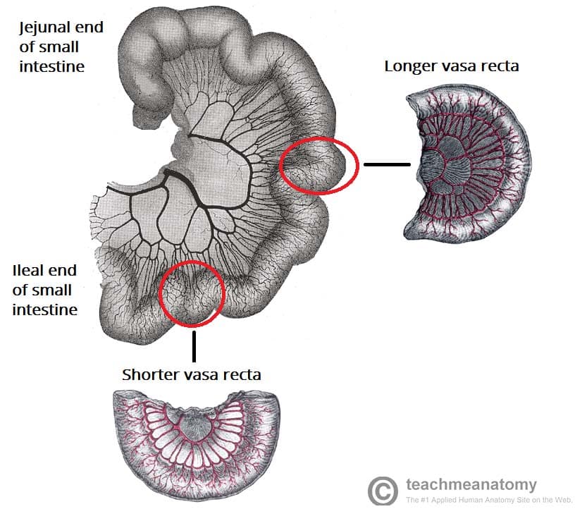
The interior surface of the jejunum—which is exposed to ingested food—is covered in finger–like projections of mucosa, called villi, which increase the surface area of tissue available to absorb nutrients from ingested foodstuffs. The epithelial cells which line these villi have microvilli. The transport of nutrients across epithelial cells through the jejunum and ileum includes the passive transport of sugar fructose and the active transport of amino acids, small peptides, vitamins, and most glucose. The villi in the jejunum are much longer than in the duodenum or ileum.
Which has less fat inside its mesentery?
The jejunum has less fat inside its mesentery than the ileum. The jejunum is typically of larger diameter than the ileum. The villi of the jejunum look like long, finger-like projections, and are a histologically identifiable structure.
Which part of the body absorbs nutrients?
The lining of the jejunum is specialized for the absorption by enterocytes of small nutrient particles which have been previously digested by enzymes in the duodenum. Once absorbed, nutrients (with the exception of fat, which goes to the lymph) pass from the enterocytes into the enterohepatic circulation and enter the liver via the hepatic portal vein, where the blood is processed.
Where is the jejunum attached?
They are attached to the posterior abdominal wall by mesentery (a double layer of peritoneum). The jejunum begins at the duodenojejunal flexure. There is no clear external demarcation between the jejunum and ileum – although the two parts are macroscopically different.
What are the two parts of the small intestine?
Jejunum and Ileum. The jejunum and ileum are the distal two parts of the small intestine. In contrast to the duodenum, they are intraperitoneal. They are attached to the posterior abdominal wall by mesentery (a double layer of peritoneum).
What is the name of the muscle that connects the duodenum to the aorta?
Located at the duodenojejunal junction is a slip of muscle called the suspensory muscle of the duodenum.
What is the first section of the duodenum?
The first section of the duodenum is known as ‘the cap ’. It ascends upwards from the pylorus of the stomach, and is connected to the liver by the hepatoduodenal ligament. This area is most common site of duodenal ulceration.
Where does the ileum end?
The ileum ends at the ileocaecal junction. At this junction, the ileum invaginates into the cecum to form the ileocecal valve. Although it is not developed enough to control movement of material from the ileum to the cecum, it can prevent reflux of material back into the ileum (if patent, see below).
What are the three parts of the small bowel?
Anatomically, the small bowel can be divided into three parts: the duodenum, jejunum, and ileum.
Where is the descending duodenum located?
It lies posteriorly to the transverse colon, and anterior to the right kidney. Internally, the descending duodenum is marked by the major duodenal papilla – the opening at which bile and pancreatic secretions to enter from the ampulla of Vater (hepatopancreatic ampulla).
What Is jejunum?
- The small intestine is a long, hollow tube, with an empty space inside called the lumen. It is located in the digestive system between the stomach and the large intestine. After food is chewed in the mouth and swallowed, it travels down the esophagus, into the stomach, and then on into the lumen inside the small intestine. The small intestine is ma...
Location
Structure of The Jejunum
Function
Clinical Significance