Full Answer
What are the five steps of hemostasis?
What are the five steps of homeostasis?
- First step. Stimulus; a stimulus occurs such as a change in in body temperature.
- Second step. Receptors; the stimulus is acknowledged by the receptors.
- Third step.
- Fourth step.
- Fifth step.
- Final step.
- Negative Feedback.
- Positive Feebback.
What are the 3 mechanisms of hemostasis?
What is Hemostasis | Its Mechanism in 4 Stages
- Vasoconstriction. In these stages, the injured blood vessel’s channel gets narrow so as to minimize the blood flow. ...
- Plug formation by platelets. The sticky platelets adhere to each and release adenosine diphosphate ( ADP ). ...
- Coagulation. In this stage, another layer of the cover is formed on the previous platelet plug formed. ...
- Fibrinolysis. ...
What does hemostatic disorders mean?
hemostasis. that lead to an increased susceptibility to bleeding (also known as. hemorrhagic diathesis. ). They are classified into disorders of primary hemostasis (when caused by a. platelet. abnormality), disorders of secondary hemostasis (when caused by defects in the extrinsic and/or intrinsic pathway of the. coagulation cascade. ), and.
What are hemostatic disorders?
Hemostatic Disorders General Principles Normal hemostasis involves a sequence of interrelated reactions that lead to platelet aggregation (primary hemostasis) and activation of coagulation factors (secondary hemostasis) to produce a durable vascular seal.

What are hemostasis disorders?
Disorders of primary hemostasis: low platelet count or abnormal platelet function test with normal INR and PTT. Disorders of secondary hemostasis: abnormal INR or PTT with normal platelet counts and platelet function.
How many hemostasis disorders are there?
The three most common hereditary bleeding disorders are hemophilia A (factor VIII deficiency), hemophilia B (factor IX deficiency) and von Willebrand disease.
What are two clotting disorders?
Bleeding disorders such as hemophilia and von Willebrand disease result when the blood lacks certain clotting factors.
What are the two opposing categories of hemostasis disorders?
There are two types: Type A, which is a deficiency in factor VIII, and Type B, also known as Christmas disease, which is a deficiency in factor IX.
What are the 4 types of hemostasis?
The mechanism of hemostasis can divide into four stages. 1) Constriction of the blood vessel. 2) Formation of a temporary “platelet plug." 3) Activation of the coagulation cascade. 4) Formation of “fibrin plug” or the final clot.
What are the types of hemostasis?
Though they look like separate processes, these all happen at the same time when your body forms a blood clot.Primary hemostasis (platelet clotting) Primary hemostasis is when your body forms a temporary plug to seal an injury. ... Secondary hemostasis (coagulation cascade) ... Fibrin clot remodeling.
What is the most common blood clotting disorder?
Antiphospholipid syndrome (APS): This is the most common acquired clotting disorder. APS is an Autoimmune condition where the body makes antibodies that mistakenly attack cell molecules called phospholipids. Higher levels of APS antibodies in the blood raise the risk of blood clots.
What is the most common bleeding disorder?
The most common bleeding disorder affecting women is von Willebrand disease (VWD), which results from a deficiency or defect in the body's ability to produce a certain protein that helps blood clot.
What are the three major groups of bleeding disorders?
Hemophilia A, the most common type of hemophilia, which is caused by a lack of clotting factor VIII or low levels of clotting factor VIII. Hemophilia B, which occurs when you are missing clotting factor IX or have low levels of clotting factor IX. Hemophilia C, a rare condition also known as factor XI deficiency.
What are the two most common inherited bleeding disorders?
The following two are the most common:Hemophilia A (Classic Hemophilia) This type is caused by a lack or decrease of clotting factor VIII.Hemophilia B (Christmas Disease) This type is caused by a lack or decrease of clotting factor IX.
Is hemophilia A disorder of hemostasis?
Hemophilia A, the most common hereditary disorder of hemostasis, occurs in one out of 5000 males and accounts for 80% of hemophilia cases. Hemophilia A occurs in more than 400000 males worldwide, many of whom remain undiagnosed in the developing world.
What are the 5 events of hemostasis?
Terms in this set (16) Vessel Spasm. ... Formation of Platelet Plug. ... Blood Coagulation. ... Clot Retraction. ... Clot Dissolution (Lysis)
What is the rarest bleeding disorder?
Glanzmann's Thrombasthenia an ultra-rare bleeding disorder.
What are the three major groups of bleeding disorders?
Hemophilia A, the most common type of hemophilia, which is caused by a lack of clotting factor VIII or low levels of clotting factor VIII. Hemophilia B, which occurs when you are missing clotting factor IX or have low levels of clotting factor IX. Hemophilia C, a rare condition also known as factor XI deficiency.
What is the rarest blood clotting disorder?
CATASTROPHIC ANTIPHOSPHOLIPID SYNDROME (CAPS) Catastrophic antiphospholipid syndrome, also known as CAPS or Asherson's syndrome, is an extremely rare variant of APS in which multiple blood clots affect various organ systems of the body potentially causing life-threatening multi-organ failure.
What is the role of GPVI in platelet adhesion?
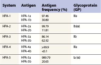
Thus, in summary, platelet adhesion is initiated by GPIbα binding to immobilized VWF and GPVI binding to collagen, which is exposed to the blood due to endothelial injury. These platelets and other local platelets are then activated, and adhesion and aggregation is strengthened and expanded via platelet-platelet connections between αIIbβ3 bound to fibrinogen, VWF, fibronectin or vitronectin as well as between αvβ3 bound to vitronectin or thrombospondin, with α5β1-fibronectin and α6β1-laminin interactions perhaps also playing a role. In addition, adherence to subendothelial collagen is strengthened via interaction of integrin α2β1 and collagen. This platelet plug is also stabilized by deposition of insoluble fibrin generated by the coagulation cascade (see below).
What is the role of platelet activation?
Activation of platelets is critical for aggregation. In particular the integrins, αIIbβ3, α2β1 and αvβ3 are normally present on the platelet surface in an inactive form, but platelet activation induces a conformational transition in these receptors that exposes ligand binding sites (Luo et al., 2006;Xiao et al., 2004). αIIbβ3 is arguably the most important of these receptors as it is present at the highest density on the platelet surface. In addition, αIIbβ3 binds to multiple ligands that promote platelet-platelet aggregation. These include fibrinogen, VWF, collagen, fibronectin and vitronectin (Varga-Szabo et al., 2008). α2β1, αvβ3, α5β1 and α6β1 play smaller roles, binding primarily to collagen, vitronectin, fibronectin or laminin, respectively (Emsley et al., 2000;Lam et al., 1989;Sonnenberg et al., 1988;Varga-Szabo et al., 2008), though other ligands have also been identified for each of these. All of the integrins are maintained in an inactive state on quiescent platelets.
What is the function of TF in hemostasis?
Thrombin performs multiple functions, including fibrin generation, platelet activation, positive feedback activation of the intrinsic pathway, and negative feedback activation of the activated protein C pathway. Procoagulant reactions are shown in blue, and anticoagulant reactions are shown in pink. APC = activated protein C, FV = factor V, PS = protein S and TM = thrombomodulin.
What are the components of hemostasis?
There are two main components of hemostasis. Primary hemostasis refers to platelet aggregation and platelet plug formation . Platelets are activated in a multifaceted process (see below), and as a result they adhere to the site of injury and to each other, plugging the injury. Secondary hemostasis refers to the deposition of insoluble fibrin, which is generated by the proteolytic coagulation cascade. This insoluble fibrin forms a mesh that is incorporated into and around the platelet plug. This mesh serves to strengthen and stabilize the blood clot. These two processes happen simultaneously and are mechanistically intertwined. The fibrinolysis pathway also plays a significant role in hemostasis. Pathological thrombus formation, called thrombosis, or pathological bleeding can occur whenever this process is dis-regulated. The complexity of these systems has been increasingly appreciated in the last few decades.
What is the receptor of GPIb-IX-V?
Receptor GPIb-IX-V binds to immobilized von Willebrand factor (VWF) specifically through an interaction between GPIbα and the A1 domain of VWF. VWF is a large multimeric protein secreted from endothelial cells and megakaryocytes that is always present in the soluble state in the plasma as well as in the immobilized state in subendothelial matrix (Ruggeri, 2007). However, soluble VWF in the circulation does not bind with high affinity to GPIbα (Yago et al., 2008). The high affinity interaction may be dependent upon high sheer stress exerted by flowing blood on immobilized VWF, whether that VWF is immobilized on subendothelial matrix or other activated platelets (Siedlecki et al., 1996).
How does hemostasis stop bleeding?
Blood loss is stopped by formation of a hemostatic plug. The endothelium in blood vessels maintains an anticoagulant surface that serves to maintain blood in its fluid state, but if the blood vessel is damaged components of the subendothelial matrix are exposed to the blood. Several of these components activate the two main processes of hemostasis to initiate formation of a blood clot, composed primarily of platelets and fibrin. This process is tightly regulated such that it is activated within seconds of an injury but must remain localized to the site of injury.
What is the feedback activation of nearby platelets surrounding a new site of injury?
Feedback activation of nearby platelets surrounding a new site of injury is critical for further aggregation and propagation of the platelet plug. This activation is mainly mediated by agonists released by activated platelets themselves acting on G protein-coupled receptors. ADP is released from platelet dense granules and binds to receptors P2Y1and P2Y12(Mills, 1996). Thromboxane A2is synthesized de novo by activated platelets and binds to the thromboxane receptor primarily, and other prostanoid receptors to a lesser degree, locally on platelets (Hanasaki et al., 1988). Serotonin is also secreted from dense granules and contributes to platelet activation.
What are the causes of bleeding disorders?
Bleeding disorders can be caused by#N#platelet disorders#N#(#N#primary hemostasis#N#defects), coagulation defects (#N#secondary hemostasis#N#defects), or increased clot degradation (#N#hyperfibrinol ysis#N# ). Coagulation defects may be general or further divided into either intrinsic or extrinsic defects according to the specific pathway of the#N#coagulation cascade#N#that is affected. Bleeding disorders may be inherited or acquired. Although clinical features may overlap, mucocutaneous bleeding (e.g.,#N#epistaxis#N#,#N#petechiae#N#,#N#gastrointestinal bleeding#N#) is associated more with#N#platelet disorders#N#, while bleeding into potential spaces (e.g.,#N#hemarthrosis#N#, muscular bleeding) is more characteristic of coagulation defects. A basic understanding of physiological processes during#N#hemostasis#N#and#N#fibrinolysis#N#is necessary for properly interpreting laboratory studies and accurately diagnosing bleeding disorders. Treatment depends on the underlying cause and may involve#N#blood transfusion#N#and replacement of#N#coagulation factors#N#.
Why is fibrinolysis necessary?
fibrinolysis. is necessary for properly interpreting laboratory studies and accurately diagnosing bleeding disorders. Treatment depends on the underlying cause and may involve. blood transfusion. and replacement of. coagulation factors. .
What is hemorrhagic diathesis?
Hemorrhagic diathesis is the abnormally increased susceptibility to bleeding.
What is advanced laboratory study?
Advanced laboratory studies help identify specific disorders. The choice of studies depends on the suspected underlying pathology and usually requires specialist consult.
Is coagulation defect inherited?
coagulation cascade. that is affected. Bleeding disorders may be inherited or acquired. Although clinical features may overlap, mucocutaneous bleeding (e.g., epistaxis. ,
Which system inhibits anticoagulant balance?
and the processes that inhibit it occur simultaneously in the circulatory system (procoagulant-anticoagulant balance).
Which factor activates factor X of the common pathway?
and factor VII form a complex that activates factor X of the common pathway.
What is the process of activation and amplification of the coagulation cascade?
This process is called fibrinolysis.
What are the two main phases of hemostasis?
The modern model of hemostasis is divided into 2 principal phases, the first being defined as primary hemostasis which involves the platelet-vessel interplay, while the second, defined as secondary hemostasis, mainly involves coagulation factors and surfaces of activated cells.
What is the balance of hemostasis?
Hemostasis is a complicated biological system, where the balance between procoagulation and anticoagulation processes maintains fluidity of blood through intact blood vessels and creates thrombi when it is needed to prevent bleeding from the impaired vessels. The modern model of hemostasis is divided into 2 principal phases, the first being defined as primary hemostasis which involves the platelet-vessel interplay, while the second, defined as secondary hemostasis, mainly involves coagulation factors and surfaces of activated cells. The activation and amplification of the coagulation cascade is regulated by natural inhibitors of coagulation. The blood clots which arise to prevent loss of blood must subsequently be broken down and the compact blood vessel wall must be restored. This process is called fibrinolysis. Bleeding and thromboses are manifestations of the impaired hemostatic balance. Detection of its cause is important for efficient treatment and prevention of the condition. This requires a combined evaluation of the family and personal history, a clinical anamnesis along with the evaluation of laboratory results. It is not reasonable or economical to perform all the available tests of hemostasis at the same time. It is recommendable to proceed from the global to the screening and then special tests and, through the process of elimination, obtain an explanation of the bleeding or prothrombotic phenotypes. The purpose of this report is to provide a brief overview of the principal causes of the hemostatic disorders and the practices which facilitate their diagnosis. Key words: diagnosis of hemorrhagic manifestations - diagnostics of thrombophilias - DIC diagnostics - hemophilia A - thrombopathy - von Willebrand disease.
Is it reasonable to perform all the hemostasis tests at the same time?
It is not reasonable or economical to perform all the available tests of hemostasis at the same time. It is recommendable to proceed from the global to the screening and then special tests and, through the process of elimination, obtain an explanation of the bleeding or prothrombotic phenotypes.
What are the different types of hemostasis?
In this section, we have split up hemostastic disorders based on physiology, although some disorders, which encompass multiple aspects of hemostasis, are placed as separate categories: 1 Table summary of the expected test results with different disorders that affect hemostasis. 2 Primary hemostasis: Vessel wall defects, platelet number, platelet function, von Willebrand disease ( vWD ). 3 Secondary hemostasis: Inherited factor deficiencies 4 Inhibitors 5 Any or multiple aspects of hemostasis: Vitamin K deficiency or antagonism, disseminated intravascular coagulation ( DIC ), underlying disease (e.g. renal, hepatic, neoplastic), drugs and trauma.
What are the most common hemostatic abnormalities in veterinary practice?
The most common hemostatic abnormalities encountered in private veterinary practice are acquired disorders, in particular immune-mediated thrombocytopenia, anticoagulant rodenticide toxicosis, and metabolic diseases that affect hemostasis (such as liver disease) or induce disseminated intravascular coagulation (DIC; such as severe inflammation, sepsis, cancer).
What is the most common inherited disease?
The most common inherited diseases are von Willebrand disease ( primary hemostasis), which is the most common inherited disorder of hemostasis, and hemophilia A (factor VIII deficiency, secondary hemostasis). Inherited disorders of the blood vessel wall, platelet number, platelet function, and inhibitors are quite rare.
What is secondary hemostasis?
Secondary hemostasis: Inherited factor deficiencies. Inhibitors. Any or multiple aspects of hemostasis: Vitamin K deficiency or antagonism, disseminated intravascular coagulation ( DIC ), underlying disease (e.g. renal, hepatic, neoplastic), drugs and trauma. Hemostatic disorders can also be inherited or acquired.
Can hemostasis be acquired in older animals?
Acquired disorders of hemostasis can occur in an animal of any age, but are more common in older animals due to underlying diseases. Animals presenting with clinical signs of hemorrhage should be thoroughly examined for underlying diseases (e.g. hemogram, biochemistry panel, radiographic and ultrasonographic examination) that may have precipitated the hemorrhage. This is particularly true for older animals. Also since drugs may cause or exacerbate a bleeding tendency (e.g. aspirin), a thorough drug history should be taken from clients with a bleeding animal.
Can an animal have inherited hemostasis?
Inherited disorders of hemostasis should be suspected in an animal presenting with bleeding symptoms at a young age, specially those that are clinically healthy, are of a predisposed breed, demonstrate recurrent bleeding episodes, or have a known family history. Some animals with mild inherited bleeding disorders may only be diagnosed when they are adults as their bleeding symptoms are mild or instigated by trauma or surgical procedures. Therefore, an inherited bleeding disease should not be excluded in adults. It is imperative to obtain a good history from clients of an adult bleeding animal in order to ascertain at what age the hemorrhage was first noticed, if it is recurrent and if it is precipitated by trauma (even the kind of trauma sustained from chewing a bone or rawhide). Recurrent hemorrhage (especially that separated by days, weeks or months or that is precipitated by trauma of some kind) is a good indicator of an inherited bleeding disorder.
Is hemostasis inherited or acquired?
Hemostatic disorders occur in all pathways of hemostasis and can be inherited or acquired. They are usually recognized clinically by excessive hemorrhage. History, signalment and clinical signs can guide a clinician as to the likely underlying disorder. For instance, disorders of primary hemostasis are characterized by mucosal hemorrhage and small bleeds (petechiae) when there is thrombocytopenia or thrombopathia. Disorders of secondary hemostasis result in larger bleeds (hematomas) and intracavity bleeding. Thus, a thorough history (travel, exposure to toxins, drug treatment) and clinical examination (assessment of the skin and all mucosal surfaces for hemorrhage) are mandatory in bleeding animals. Ancillary diagnostic testing (e.g. hemograms, biochemical panels, radiographic and ultrasonographic examination) may be indicated in individual animals, particularly those that are sick, to confirm or rule out underlying disease as a cause for the hemostatic disorder. Acquired disorders are by far the most common, particularly thrombocytopenia. Inherited disorders predisposing animals to thrombosis (deficiencies in anticoagulants, defective fibrinolysis) are rare and it is difficult to recognize thrombosis clinically, leading to under-recognition of acquired prothrombotic diseases.
