What is C1 in vertebrae?
C1 (Atlas) - 1st Cervical Vertebra. The C1 vertebra, known as the atlas, is the superior-most vertebra in the spinal column.
What is the Atlas of the C1?
The C1, or first cervical vertebra, is commonly called the atlas due to its unique position in the spine.
What is C1 bone made up of?
C1 Spine (Atlas)Bony Landmark The atlas is made up of the anterior and posterior arch, 2 transverse processes, 2 prominent lateral masses and the transverse foramen. Lateral mass is the thickest part of the bone. This region supports the weight of the skull.
What is C2 bone in the spine?
Below the atlas bone is the axis bone (C2). Pivot and gliding joints of the atlas & axis bones allow the head to move side-to-side. C1 Spine (Atlas)Bony Landmark The atlas is made up of the anterior and posterior arch, 2 transverse processes, 2 prominent lateral masses and the transverse foramen.
What structure does C1 articulate with?
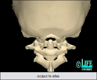
Atlas (C1) and Axis (C2) The first cervical vertebra is unique in its articulation with the occipital condyle of the cranium. This articulation is the basis for significant flexion and extension of the head.
What does C1 attach to?
The cervical vertebrae C1 is attached directly to the skull, which allows for any nodding lateral motions.
What does the superior articular facet of C1 articulate with?
Superior articular facet (Facies articularis superior) is an oval-shaped, concave structure on the superior aspect of the lateral mass. It articulates with an occipital condyle on the underside skull.
How do C1 and C2 articulate?
C1 and C2 form a unique set of articulations that provide a great degree of mobility for the skull. C1 serves as a ring or washer that the skull rests upon and articulates in a pivot joint with the dens or odontoid process of C2.
What are the symptoms of a pinched nerve at C1?
Common Symptoms and Signs Stemming from C1-C2Radiating pain up to the back and/or top of the head.Pain in the temple and/or behind the eyes and/or ears.Neck-tongue syndrome resulting in neck and/or head pain with numbness or abnormal sensation on one side of the tongue.Sensitivity to light.Fatigue.Dizziness.Nausea.
What nerves are affected by C1?
The C1 Nerve Root supplies neurological impulses for blood supply to the head, the pituitary gland, the scalp, bones of the face, the brain itself, inner and middle ear, and the sympathetic nerve system.
What is unique about C1?
The Atlas (the first cervical vertebra - C1) differs from the other cervical vertebrae in that it has no body or spinous process. It is comprised of two bony arches with two bony masses laterally. It articulates with the Occiput above and C2 (the Axis) below.
Is C1 connected to skull?
C1 Vertebra (the atlas). The atlas connects to the occipital bone above to support the base of the skull and form the atlanto-occipital joint. More of the head's forward/backward range of motion occurs at this joint compared to any other spinal joint.
How do C1 and C2 differ from other vertebrae?
C1 and C2 Anatomy The C1 and C2 vertebrae are uniquely shaped and have vertebral foramen (spaces within the bone) which allow the vertebral arteries and veins to reach through to the brain and supply it with blood. This unique formation is only seen in the cervical vertebrae.
What does C1 vertebrae control?
Despite being the smallest vertebrae in the cervical region, they are responsible for the range of motion of the head. The C1 vertebra (the atlas) is the uppermost vertebra. It connects to the base of the skull and forms the atlanto-occipital joint. This joint allows you to nod your head up and down.
What happens if C1 is damaged?
Complications in the management of C1 fractures range from minor discomfort to death. The primary concern with C1 fractures is establishing and maintaining cervical stability. Atlanto-occipital and atlantoaxial instability threatens the brainstem and spinal cord, potentially causing myelopathy and even mortality.
What is a C1 spinal injury?
High-Cervical Nerves (C1 – C4) Most severe of the spinal cord injury levels. Paralysis in arms, hands, trunk and legs. Patient may not be able to breathe on his or her own, cough, or control bowel or bladder movements. Ability to speak is sometimes impaired or reduced.
Where is C1 protein located?
blood plasmaC1 esterase inhibitor (C1-INH) is a protein found in the fluid part of your blood. It controls a protein called C1, which is part of the complement system. The complement system is a group of nearly 60 proteins in blood plasma or on the surface of some cells.
What is C1 pathway?
The C1 complex (complement component 1, C1) is a protein complex involved in the complement system. It is the first component of the classical complement pathway and is composed of the subcomponents C1q, C1r and C1s.
What is required for activation of C1?
Activation of C1 which, in vivo, is controlled by C1 inhibitor, can be achieved by various activators, such as immune complexes; it appears to result from the suppression of a negative control and resides in a positive modulation of the intrinsic autocatalytic potential of C1r inside C1.
Which domain of IgG binds to 1st complement component?
Binding of the complement component C1q to the CH2 domain of antigen-bound immunoglobulin gamma (IgG) activates the classical complement pathway and depends on its close proximity to Fc fragments of neighboring antibodies.
What is the C1 and C2?
The C1 and C2 vertebrae are the first two vertebrae at the top of the cervical spine. Together they form the atlantoaxial joint, which is a pivot joint. The C1 sits atop and rotates around C2 below. More of the head’s rotational range of motion comes from C1-C2 than any other cervical joint. 1. Spinal Motion Segment: C1-C2 (Atlantoaxial Joint) ...
What does C1 C2 pain mean?
Vertebral pain at C1-C2 can range anywhere from a dull a che to a sharp, burning pain in the neck. C1-C2 pain may either last for a short while or become chronic. If a C2 nerve root becomes inflamed or injured, additional symptoms may include: Radiating pain up to the back and/or top of the head.
Which vertebrae are responsible for the spinal cord?
See Cervical Spinal Nerves. The spinal cord is protected by the C1-C2 vertebrae in the upper cervical area. These vertebrae have several small and large foramens. The spinal cord passes through the large, centrally placed vertebral foramen. The smaller foramens facilitate the passage of blood vessels around the spine.
Where does the C2 nerve exit?
At the atlantoaxial joint, the C2 spinal nerve exits the spinal cord through a small bony hole or foramen above the C2, called the intervertebral foramen. This nerve has a sensory root and a motor root. The C2 dermatome is an area of skin that receives sensations through the C2 nerve.
Why is C1-C2 painful?
While C1-C2 is relatively sturdy and resistant to injury, it can become quite painful and problematic due to trauma or degenerative conditions. These problems may also affect the spinal nerves, vertebral artery, and/or the spinal cord at the at the C2 level. See Cervical Spine Anatomy.
What causes a fracture of the C1 vertebrae?
Fractures may result from diving in shallow water, falling, motor vehicle accidents, 1 and/or hitting an obstacle with the forehead or chin. 2 Trauma to C1-C2 may also cause whiplash injury, spondylolisthesis, nerve injury, and/or spinal cord injury.
What is crowned dens syndrome?
Crowned dens syndrome results from the deposition of calcium on the dens of C2 from the surrounding ligaments, causing pain and reduced mobility of the neck. Read Neck Pain from Crowned Dens Syndrome
Which ligament surrounds the articular facets?
The capsular ligaments are thin and loose; they surround the joints between the articular facets and are lined with synovial membrane. Each is strengthened at its posterior and medial part by an accessory ligament, which is attached below to the body of the axis near the base of the odontoid process, and above to the lateral mass of the atlas near the transverse ligament.
What is the capular ligament?
The capsular ligaments are thin and loose; they surround the joints between the articular facets and are lined with synovial membrane. Each is strengthened at its posterior and medial part by an accessory ligament, which is attached below to the body of the axis near the base of the odontoid process, and above to the lateral mass ...
How are the two vertebrae connected?
In front the two vertebra are connected by a continuation of the anterior longitudinal ligament (fig. 514). In this position it is a strong membrane, fixed above to the lower border of the anterior arch of the atlas, and below to the front of the body of the axis. It is strengthened in the median plane by a rounded cord, which connects the tubercle on the anterior arch of the atlas to the body of the axis.
Which muscles produce movements?
Muscles producing the movements. The principal muscles by which these movements are produced are the sternornastoid and semispinalis capitis of one side, acting with the longus capitis, splenius, longissimus capitis, rectus capitis posterior major and obliquus capitis inferior of the other side.
Where is the atlas attached to the axis?
Behind, the atlas and axis are joined by a broad, thin membrane (fig. 515) attached above to the lower border of the posterior arch of the atlas, below to the upper edge’s of the laminae of the axis; it is in series with the ligamenta flava.
Is the Atlas axis convex or convex?
The opposed articular facets of the atlas and axis are not reciprocally curved; both are slightly convex in their long axes. When therefore, the upper facet glides forwards on the lower it also descends; the fibers of the capsular ligament are relaxed in a vertical direction, and will then permit of movement in an anteroposterior direction. By this means a shorter capsule suffices and the strength of the joint is materially increased.
What is the C1 spine?
C1 Spine: Anatomy, Bony Landmark. First Cervical spine (C1) is also known as the atlas. The atlas (C1) is the most superior (first) cervical vertebra of the spine. The atlas joint connecting the skull and spine. The atlas (C1) and axis (C2) are specialized to allow a greater range of motion than normal vertebrae.
What is the superior articular facet?
Superior articular facet is an oval-shaped, It articulates with an occipital condyle on the underside skull. Inferior articular facet is a flattened surface of the inferior lateral mass that articulates with the superior articular facet on the axis (C2).
What is the anterior tubercle?
The anterior tubercle is a slight elevation at the apex of the anterior arch. It is an attachment point for the longus coli muscles. Posterior arch is a narrow band of bone that extends from the transverse processes and encloses the posterior portion of the vertebral foramen.
What is the opening in the center of the bone through which the spinal cord passes?
Vertebral foramen is a large opening in the center of the bone through which the spinal cord passes.
Which bone is below the Atlas?
This junction allows the head to nod up and down. Below the atlas bone is the axis bone (C2).
What is the Atlas made of?
The atlas is made up of the anterior and posterior arch, 2 transverse processes, 2 prominent lateral masses and the transverse foramen. Lateral mass is the thickest part of the bone. This region supports the weight of the skull.
Which ligament is used to connect the C1 to the C2?
Posteriorly, the ring of the atlas is connected to the C2 by the posterior atlantoaxial ligament. The dens articulates with the atlas via a facet on the posterior aspect of the anterior ring of the atlas, retained by the transverse ligament, providing the head with approximately 50 % of its lateral rotation.
How to treat C1 fracture?
Nonoperative management remains the mainstay of treatment for C1 fractures. Isolated atlas fractures can be effectively managed with 8 to 12 weeks of external immobilization of the craniocervical junction [3]. Collar immobilization or cervical traction for this period of time is usually sufficient to allow for proper healing; however, the type of orthosis required varies [3, 20]. Nonoperative treatment typically consists of external immobilization through use of a rigid collar, halo vest, or Minerva jacket [20]. Soft collars are inadequate for immobilization and often result in worsening pain to the patient with neck motion as well as further fracture displacement. Following immobilization, dynamic imaging studies such as flexion-extension films should be ordered to rule out late instability [3].
What is the incidence rate of C2 fractures concurrent with C1 fractures?
The incidence rate of C2 fractures concurrent with C1 fractures is approximately 41–44 % [7]. Upper cervical fractures with isolated C1–C2 instability can be effectively treated with C1–C2 fusion. However, if the C1 injury is a burst fracture, fixation should attempt to bring together the C1 lateral masses followed by fixation to C2. Alternatively, instrumentation can span from the occiput to C2 [7]. While odontoid fractures associated with C1 fractures typically may be managed with external immobilization alone, type II odontoid fractures, especially in the elderly, often are optimally managed with surgical intervention [7, 26, 27].
What is the best collar for C1 fracture?
In cases with more significant fracture displacement, more rigid immobilization with the halo vest or Minerva jacket may be required. The halo vest is more rigid than the Minerva jacket, providing greater restriction of the C1–2 joint. Flexion and extension of the upper cervical spine is diminished by as much as 75 % when a halo vest is employed. The greater rigidity of the halo orthosis also restricts more lateral movement of the atlantoaxial joint when compared with the Minerva jacket [21]. For this reason, the halo vest is the preferred option for upper cervical injuries [22]. With injuries extending to the mid and lower cervical spine, thermoplastic Minerva jackets offer greater comfort to patients, fewer complications, and can provide effective stabilization [22].
What are the different types of fractures in the Atlas?
Atlas fractures may often involve trauma to the axis as well. C2 fractures can be categorized using either the Anderson and D’Alonzo classification or the Roy-Camille classification. Anderson and D’Alonzo recognized three types of odontoid fractures. Type I odontoid fractures involve the dens superior to the cruciform ligament and are considered to be stable. Type II is the most prevalent odontoid fracture pattern, identifiable by a break at the base of the dens, below the cruciform ligament. Type II fractures have a greater risk of nonunion and are unstable. Type III fractures move through the base of the odontoid and into the lateral masses of the axis. Due to a more extensive blood supply and larger surface area, these fractures are the most likely to heal. Moreover, they are stable when not significantly displaced. The Roy-Camille system offers an alternative classification with three types based on the direction of the fracture line. Types I and II are categorized by oblique fractures which slope anteriorly and posteriorly, respectively. The third type includes horizontal fracture lines with displacement of the dens either anteriorly or posteriorly.
What is the cause of C1 fracture?
These are caused by traumatic hyperflexion or extension of the neck , which severs the posterior longitudinal ligament as well as the bilateral alar ligaments. The capsular and accessory ligaments can also be disrupted [16]. The instability caused by dislocation or subluxation of the atlanto-occipital joint may result in severe damage to the spinal cord and often death. Upwards of 30 % of traffic collision deaths are due to AODs [8, 16]. An atlanto-occipital displacement with a concomitant C1 fracture may be diagnosed on CT scans, which show a basion-dens interval (BDI), the distance between the basion and the tip of the dens, exceeding 10–12 mm. Atlanto-occipital dissociation injuries may also be diagnosed when the occipital condyle-C1 interval (CCI) exceeds 4 mm [8, 15]. Pang et al. reviewed the CCI and other radiographic landmarks and found that, for the evaluation and diagnosis of AOD, the CCI is the most direct tool to measure displacement in AOD and is especially useful in pediatric injuries [17]. An AOD requires emergent surgical fixation to minimize the risks of progressive neurologic injury and even mortality. Additionally, presurgical external fixation with a halo vest is recommended to minimize other complications. Axial traction and rigid cervical collars are contraindicated because of potential to distract the injured joint further [8]. Patients suffering an AOD often present with concomitant injuries to the lower cervical spine and should undergo a thorough clinical evaluation. In the event of trauma to the occiput or other vertebra, internal fixation is required and should extend to the most inferior affected vertebra [18].
Can Atlas fractures be seen on a radiograph?
While lower cervical injuries are more easily identified radiologically and likely to present with neurologic compromise, isolated C1 fractures can be harder to see on plain film radiographs and less likely to cause neurological deficits.
Which surface contains grooves for the C1 nerve and vertebral artery?
superior surface: contains paired grooves for the C1 nerve and vertebral artery , sits just posterior to the lateral mass. superior border: attachment for the posterior atlanto-occipital membrane. inferior border: attachment for the posterior atlanto-axial membrane. lateral masses. paired, ovoid.
Which articular facets of the lateral masses can be divided into two parts?
superior articular facets of the lateral masses can be divided into two parts, with the anterior part being larger and the posterior smaller. fusion defects. central or paramedian parts of the posterior arch can be absent and replaced by fibrous tissue. anterior arch fusion defects.
What is the difference between inferior and articular facet?
inferior articular facet: circular, with a flat or slightly concave surface articulating with the lateral atlantoaxial joint.
What are the three centers of ossification?
Ossification starts from three centers: anterior ossification center, and paired lateral mass ossification centers. The paired lateral mass ossification centers arise in week 7 and extend to the posterior arch. Unification at the posterior arch occurs at years 3-4 and is typically direct, but can sometimes involve a third center at the posterior arch. The anterior ossification center unites with the lateral mass ossification center at years 6-8. There are sometimes two anterior ossification centers.
What is the anatomy of the Atlas?
Gross anatomy. The atlas is composed of an anterior arch and a posterior arch, paired lateral masses, and paired transverse processes. It does not have a vertebral body, instead the dens of the axis sit where a centrum (body) of a typical vertebra would be. The transverse ligament holds the dens of the axis against the anterior arch ...
Which joint allows for the rotation of the head?
lateral atlantoaxial joint: hyaline-covered synovial joint between the inferior articular facet of the atlas and the superior articular facet of the axis which allows for the rotation of the head. A capsule innervated by the C2 nerve surrounds the joint.
What is the first cervical vertebra?
Atlas (C1) The atlas (plural: atlases) is the first cervical vertebra, commonly called C1. It is an atypical cervical vertebra with unique features. It articulates with the dens of the axis and the occiput, respectively allowing rotation of the head, and flexion, extension and lateral flexion of the head.
Which articular facet articulates with the dens of axis (C2) to form the median at?
On the posterior surface of the anterior arch is a circular articular facet which articulates with the dens of axis (C2) to form the median atlanto-axial joint. This is a synovial pivot joint which facilitates rotational movements of the head on the neck such as when you shake your head no, or turn your head from the left to the right.
Which joint is articular with the occipital condyles of the cranium?
The kidney-shaped superior articular facets articulate with the occipital condyles of the cranium to form the atlantooccipital joint. The atlantooccipital joint facilitates flexion and extension and slight lateral flexion of the head on the neck. This joint is utilised when nodding the head
What is the bony roughening at the apex of the posterior arch?
At the apex of the posterior arch is a bony roughening known as the posterior tubercle. The posterior tubercle is homologous to the spinous process of typical vertebrae, and as such functions as an attachment point for the ligamentum nuchae.
What joint is the lateral atlantoaxial joint?
The paired lateral atlantoaxial joints work together with the median atlantoaxial joint to produce rotational movements of the head.
What is the anterior arch of the Atlas?
Located on the anterior aspect of the arch is a bony roughening known as the anterior tubercle, which provides attachment to the anterior longitudinal ligament. The superior and inferior surfaces of the anterior arch provide an attachment point for the anterior atlanto-occipital membrane and lateral parts of the anterior longitudinal ligament.
What is the spine of the spine?
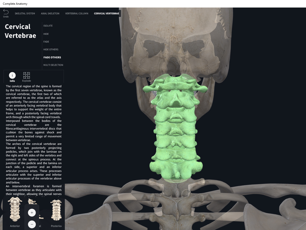
The vertebral column (spine or backbone) is a curved structure composed of bony vertebrae that are interconnected by a series of cartilaginous intervertebral discs. The vertebral column can be divided into the cervical, thoracic and lumbar vertebrae, the sacrum and coccyx. No two vertebrae are identical, however, each can fall under the category of typical or atypical vertebrae, depending on their structural composition.
What is the attachment point of the transverse ligament?
Located on the medial surface of each lateral mass is a tubercle known as the transverse ligament tubercle. As its name suggests, this tubercle provides the attachment point for the transverse ligament, which stabilizes the odontoid process of the axis (C2). The transverse ligament divides the vertebral canal (centre of ring) into an anterior and posterior compartment. The anterior third of the canal is occupied by the dens axis, while the posterior compartment is occupied by the spinal cord and its coverings.

Anatomy of The C1-C2 Vertebrae and Spinal Segment

Common Problems at C1-C2
- Problems at the C1-C2 vertebral levels may affect the vertebrae, the C2 spinal nerve, and/or the spinal cord. A few examples of problems at this cervical level include: 1. Arthritis. Arthritis in the C1-C2 joint is common in many of the systemic arthritic syndromes such as rheumatoid arthritis or other spondyloarthropathies (spinal arthritis syndromes). This condition is due to the high lev…
Common Symptoms and Signs Stemming from C1-C2
- Vertebral pain at C1-C2 can range anywhere from a dull ache to a sharp, burning pain in the neck. C1-C2 pain may either last for a short while or become chronic. If a C2 nerve root becomes inflamed or injured, additional symptoms may include: 1. Radiating pain up to the back and/or top of the head 2. Pain in the temple and/or behind the eyes and/or ears 3. Neck-tongue syndrome r…