Bone cells
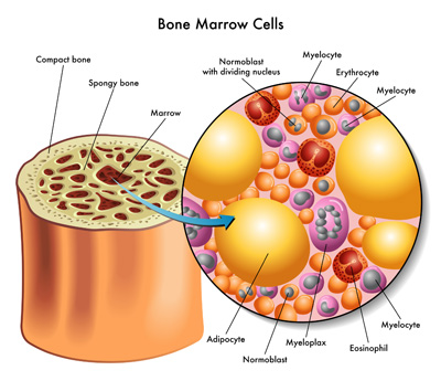
- Bone marrow. Bone marrow is found in almost all bones where cancellous bone is present. ...
- Extracellular matrix. Bones are essentially living cells embedded in a mineral-based organic matrix. ...
- Mechanical. Bones provide a frame to support the body. ...
- Synthesizing. Cancellous bone produces red blood cells, platelets, and white blood cells. ...
- Metabolic. ...
What is the main function of a bone cell?
What does bone do?
- Support. Bone provides a rigid framework as well as support for other parts of your body. ...
- Movement. Bones also play an important role in the movement of your body, transmitting the force of muscle contractions.
- Protection. Your bones also protect many of your internal organs. ...
- Blood cell generation and maintenance. ...
- Storage. ...
What does the body do with bone cells?
Three distinct types of bone cells are present in bone tissue, each with their own crucial function. Working together, osteoblasts, osteoclasts, and osteocytes are responsible for the proper development and maintenance of the skeleton, as well as regulating levels of minerals present in the bloodstream and throughout the body. Two related types of cells, lining cells and osteogenic cells, are derived from osteoblasts but have their own key functions for proper bone health.
What bone cells are responsible for breaking down bone tissue?
homeostatic maintenance of both density and blood concentration of calcium and phosphate ions. Two main types of cells are responsible for bone metabolism: osteoblasts (which secrete new bone), and osteoclasts (which break bone down).
What are some facts about bone cells?
Fun Facts About Bones
- The adult human body has 206 of them.
- There are 26 bones in the human foot.
- The human hand, including the wrist, contains 54 bones.
- The femur, or thighbone, is the longest and strongest bone of the human skeleton.
- The stapes, in the middle ear, is the smallest and lightest bone of the human skeleton.

What do bone cells produce?
Osteoblasts are cells that form bone tissue. Osteoblasts can synthesize and secrete bone matrix and participate in the mineralization of bone to regulate the balance of calcium and phosphate ions in developing bone. Osteoblasts are derived from osteoprogenitor cells.
Why is the bone cell important?
The principle function of osteoblasts is to synthesize the components that constitute the extracellular matrix of bone. These include structural macromolecules, such as type I collagen, which accounts for about 90% of the organic matrix, as well as numerous proteoglycans, non-collagenous and cell attachment proteins.
What is the main cells of the bone?
There are three types of cells that contribute to bone homeostasis. Osteoblasts are bone-forming cell, osteoclasts resorb or break down bone, and osteocytes are mature bone cells.
What is a bone cell definition?
Definition of bone cell 1 or bone corpuscle : any of the cells occupying the lacunae of bone : osteoblast. 2 : osteosclereid.
What do osteocytes do?
The potential functions of osteocytes include: to respond to mechanical strain and to send signals of bone formation or bone resorption to the bone surface, to modify their microenvironment, and to regulate both local and systemic mineral homeostasis.
How are bone cells adapted to their function?
The mechanisms for adaptation involve a multistep process called mechanotransduction, which is the ability of resident bone cells to perceive and translate mechanical energy into a cascade of structural and biochemical changes within the cells.
How do bones work?
Bones work with muscles and joints to hold our body together and support freedom of movement. This is called the musculoskeletal system. The skeleton supports and shapes the body and protects delicate internal organs such as the brain, heart and lungs. Bones contain most of our body's calcium supply.
How are bone cells specialized?
Bone Cells There are three types of specialized cells in human bones: osteoblasts, osteocytes, and osteoclasts. These cells are responsible for bone growth and mineral homeostasis. Osteoblasts make new bone cells and secrete collagen that mineralizes to become bone matrix.
What do osteoblasts do?
The primary role of osteoblasts is to lay down new bone during skeletal development and remodelling. Throughout this process osteoblasts directly interact with other cell types within bone, including osteocytes and haematopoietic stem cells.
What is another name for a bone cell?
a cell found in bone in any of its functional states; an osteoblast, osteoclast, or osteocyte.
How are bone cells specialized?
Bone Cells There are three types of specialized cells in human bones: osteoblasts, osteocytes, and osteoclasts. These cells are responsible for bone growth and mineral homeostasis. Osteoblasts make new bone cells and secrete collagen that mineralizes to become bone matrix.
What is the function of the osteoclasts?
Osteoclasts are the cells that degrade bone to initiate normal bone remodeling and mediate bone loss in pathologic conditions by increasing their resorptive activity. They are derived from precursors in the myeloid/monocyte lineage that circulate in the blood after their formation in the bone marrow.
How is it that bone cells and muscle cells differ in structure and function but come from the same original cell?
Answer and Explanation: Bone cells, muscle cells, and skin cells look different because (C) different genes are active in each kind of cell.
What is the function of osteoblasts?
The primary role of osteoblasts is to lay down new bone during skeletal development and remodelling. Throughout this process osteoblasts directly interact with other cell types within bone, including osteocytes and haematopoietic stem cells.
Bone Cells: Definitions & Functions
Bones have four kinds of cells. That may not seem like much, but they coordinate their activities to create a balanced system that modifies and maintains your entire skeleton. This process is similar to a construction company that redesigns homes.
Osteogenic Bone Cells
Let's begin by putting together the construction crews for your bones. Some members come from osteogenic cells, which are located in the surface lining of bones and in bone marrow. The term 'osteo' means bone and the suffix 'genic' refers to genesis, or the start of something new, such as these crews.
Osteoblast Bone Cells
Osteoblast cells are the creators of bone material. The suffix 'blast' is from the Greek word 'blastos,' which means to germinate or sprout. The function of osteoblasts is to make various proteins used in the matrix of bone.
Osteoclast Bone Cells
For bones to reshape themselves over time, their matrix must be broken down as well as built up. To accomplish this, osteoblasts team-up with osteoclasts, which break down bone material. The suffix 'clast' refers to destruction, so osteoclasts are the demolition experts.
What are the different types of bone cells?
These bone cells have distinct features, structure, and considered essential functions. These bone cells are Osteoclasts, Osteoblasts, and Osteocytes.
What is the most abundant bone cell?
History of Osteocytes. Osteocytes are the most abundant and long-lived bone cells with speculation of living for about 25 years. During the 1950s, Gordan and Ham extensively studied osteocytes. In prior days of osteocyte discovery, it was thought that osteocytes are dormant cells and do not perform any function.
What happens when osteoclasts are present in increased numbers than required?
On the other hand, when osteoclasts are present in increased numbers than required, the bone problem of Osteoporosis may develop.
How do osteoclasts affect bone structure?
Therefore, the number and amount of osteoclasts in the bone controls the bone structural integrity . Osteoclast resorbs bones by creating sealed compartments adjacent to the bone surface. Then, osteoclasts secrete acid phosphatases. These enzymes are acidic that functions to degrade the bone.
Why are osteocytes defined?
Initially, osteocytes were defined according to their morphology rather than their function. This was because their function remained unknown for decades. Later, it was recognized that they play many different yet important roles in bone development and maintenance.
What cells are osteoblasts derived from?
Osteoblasts are also derived from bone marrow precursor cells ( mesenchymal stem cells MSC). Osteogenic cells are undifferentiated and develop into Osteoblasts.
How many osteoclasts are there in the bone?
The occurrence of osteoclasts is quite scarce in the bony tissue. It is estimated that in an area of 1mm of the bony tissue, almost 2 to 3 osteoclasts are found. The structure of osteoclasts is related to their function.
How do cytokines affect bone?
Bone cells can be affected by inflammatory cytokines that alter their development and function under certain pathological conditions such as rheumatoid arthritis, a chronic inflammatory joint disease characterized by fibrosis and a destruction of cartilage and bone. Those cytokines can have opposite effects on osteoblasts and osteoclasts. For example, tumor necrosis factor-α (TNF-α) is known to inhibit osteoblast function and differentiation whereas interleukin-1 (IL-1), IL-6, TNF-α and others are all potent inducers of osteoclast differentiation and bone resorption. Thereby, one factor can stimulate bone resorption and inhibit bone formation at the same time. Similarly, estrogens can have an effect on those cytokines by decreasing there production and thus decreasing the number of osteoclasts in vivo and in vitro, mostly by decreasing the production of above mentioned factors (IL-1, IL-6 and TNF-α) (Charatcharoenwitthaya et al., 2007 ). At the same time, estrogens prevent osteoblast apoptosis and increase osteoblast lifespan ( Kousteni et al., 2001). In vitro labeling methods offer an easy and fast assessment of the effect of those cytokines on bone cells. For example, further insight into the effect of TNF-α on osteoblast differentiation has been given by using iTRAQ/MRM-LC-MS/MS, revealing 6 specific proteins (Periostin, Protein S100-A4, ATPase inhibitor, Cytochrome b5, SERCA3, and ELP2) that are affected by this cytokine (Xu et al., 2015). On the other hand, IL-4, a cytokine that inhibits the formation of osteoclasts by acting on their precursors, was shown to block signaling pathways, such as c-Jun N-terminal kinase (JNK), MAPK, inhibiting p38 and TNF-α signaling (Mohamed et al., 2005; Palmqvist et al., 2006; Bendixen et al., 2001; Freire et al., 2016; Abu-Amer, 2001 ).
How do bone cells communicate with each other?
Bone cells communicate with each other through gap junction channels formed by connexin proteins. Connexin43 (Cx43) is the most abundant connexin expressed in osteoblasts, osteoclasts and osteocytes. Connexins form hemichannels in unopposed cell members, allowing the communication of the cells with the extracellular environment. Connexins also function as scaffolding molecules through the interaction of C-terminus domain with intracellular signaling and structural proteins. Cx43 regulate bone cell differentiation and survival, and participate on the effect of mechanical, hormonal, and pharmacologic stimuli in these cells. Further, Cx43 is also involved in bone aging and primary and metastatic cancer in bone.
What cytokines inhibit osteoblasts?
Those cytokines can have opposite effects on osteoblasts and osteoclasts. For example, tumor necrosis factor-α (TNF-α) is known to inhibit osteoblast function and differentiation whereas interleukin-1 (IL-1), IL-6, TNF-α and others are all potent inducers of osteoclast differentiation and bone resorption.
How do bone cells maintain connections to the ECM?
Bone cells maintain connections to the ECM through tethering elements that also help transmit mechanical signals. Tethering elements provide linkage of ECM to osteocytes as outside-in connections that translate to the cell through changes in fluid shear stress and integrins (You et al., 2001, 2004; Thompson et al., 2011b). Within the pericellular space of the osteocyte canaliculi, electron microscopy images revealed that tethering elements are proteoglycan in nature. These elements are hypothesized to be related to perlecan, a protein present in the cortical bone. Mice deficient in perlecan have fewer tethering elements. Mathematical modeling of osteocyte canaliculi postulate increased integrins in the canaliculi through amplification related to mechanical force on the cell (Wang et al., 2008 ). The increase in the elements within the canaliculi and their location led to the classification of tethering elements as a mechanosensory proteins with the ability to spark and regulate downstream gene production ( McNamara et al., 2009; Litzenberger et al., 2010 ). Thus connection of proteins from the ECM to the cytoskeleton provides a necessary link for translation of biomechanical cues.
What are tethering elements?
Within the pericellular space of the osteocyte canaliculi, electron microscopy images revealed that tethering elements are proteoglycan in nature. These elements are hypothesized to be related to perlecan, a protein present in the cortical bone. Mice deficient in perlecan have fewer tethering elements.
What is CX43 involved in?
Further, Cx43 is also involved in bone aging and primary and metastatic cancer in bone. View chapter Purchase book. Read full chapter.
What is the inflammatory cytokine that affects bone cells?
Bone cells can be affected by inflammatory cytokines that alter their development and function under certain pathological conditions such as rheumatoid arthritis , a chronic inflammatory joint disease characterized by fibrosis and a destruction of cartilage and bone.
What is the only cartilage that remains in long bones?
After birth, the only cartilage that remains in long bones is the physis, also known as the epiphyseal plate or growth plate. This area will differentiate into bone from early childhood through puberty via endochondral osteogenesis. Five regions are recognized for the physis :
What is bone tissue?
Bone is a type of connective tissue that offers structural support for the body. It also serves as a calcium reservoir. As a type of connective tissue, bone has cells that are mesoderm derived. This lesson covers the development of bone during embryonic and post-embryonic development and describes the types of cells found in bone.
What are the cells that make up bone?
Osteoblasts. These cells originate from mesenchymal stem cells. They work to deposit collagen and minerals that will eventually form mature bone. When they’ve accomplished this, osteoblasts can become a cell on the bone surface, develop into an osteocyte, or die by a natural process called apoptosis.
What are the functions of bones?
In addition to providing a framework for your body, bones also serve many other important biological functions, such as protecting your internal organs from harm and storing essential nutrients. Read on to explore the diverse functions and types of bones.
What are the bones that connect muscle to bone called?
Sesamoids are bones that form within a tendon and bones surrounded by tendons, which connect muscle to bone. They help to protect the tendons from wear and tear and to relieve pressure when a joint is used. They give a mechanical advantage to the muscles and tendons in which they are located.
What are the most prevalent cells in bone tissue?
Osteocytes are trapped within the bone tissue and are the most prevalent cell type in mature bone tissue. They monitor things such as stress, bone mass, and nutrient content. They’re also important for signaling during bone remodeling, the process of bone resorption and generation of new bone tissue that can follow.
What are the bones that are long?
As their name implies, long bones are longer than they are wide. Some examples include: 1 femur (thigh bone) 2 humerus (upper arm bone) 3 bones of your fingers and toes
What is the human body made of?
In addition to that backbone, we also have an extensive skeletal system that’s made up of bones and cartilage as well as tendons and ligaments. In addition to providing a framework for your body, bones also serve many other important biological functions, ...
What is the outer shell of a bone?
Compact. The compact bone is the outer shell of the bone. It’s made up of many closely packed layers of bone tissue. Compact bone contains a central canal that runs the length of the bone, often called a Haversian canal. Haversian canals allow blood vessels and some nerves to reach into the bone.
What is the function of osteoclast?
This cell is associated with the process of growth and remodelling of bone. So its function is to resorb or destroy bone . Biology, Human, Bones, Bone Cells, Bone Cells in Human Body. Structure of Fungi (With Diagram) | Hindi | Microorganisms | ...
What is the function of the Golgi apparatus?
There is a well-marked Golgi apparatus. The cell secretes the bone matrix and thus calcification takes place. For its important function of calcification, bone-forming osteoblast possesses a rich concentration of enzyme, alkaline phosphatese in the cytoplasm.
What are the different types of bone cells?
There are three types of bone cells present in human body: 1. Osteoblast 2. Osteocyte 3. Osteoclast. Bone Cell # 1. Osteoblast: This is concerned with bone formation and is found in the growing surface where the bony matrix is deposited. This cell is strongly basophilic and cuboidal or pyramidal in shape and its nucleus is large ...
Which cell has affinity for basic dye?
The cytoplasm has got affinity towards basic dye due to presence of a large amount of rough-surfaced endoplasmic reticulum with its associated ribosomes. Bone Cell # 2. Osteocyte: It is the trapped or imprisoned osteoblast within the organic matrix. Osteoblast becomes trapped within its own secretory material-the matrix.
Where is the osteoclast found?
It is often found in a hollow pit, known as Howship’s lacuna, near the surface of bone.
What are the two groups of bone cells?
Bone cells fall broadly into two groups—those of mesenchymal lineage, and those of hematopoietic lineage. The bone forming cells—osteoblasts—are found as quiescent lining cells across all bone surfaces except where active remodeling is underway (Amizuka et al., 2014 ). Osteoblasts are incorporated into the bone matrix during the mineralization process and become entombed within the lacunar-canalicular network as osteocytes ( Prideaux et al., 2012 ). Osteoclasts, by contrast, are formed from mononuclear precursors during the process of removal of bone tissue ( Boyle et al., 2003 ). The coordinated action of replacing existing old bone tissue with new bone is known as remodeling. During this process, bone lining cells appear to lift and form a tent or canopy under which the remodeling events proceed ( Hauge et al., 2001 ). The factors controlling the site-specific removal of bone—the timing, the quantity, the speed of removal—are still not fully elucidated. It is clear however that the initiation of remodeling is likely to be an osteoblast/osteocyte activity ( Rumpler et al., 2012 ); that the removal of bone mineral and some bone matrix protein is largely accomplished by osteoclasts ( Sakamoto and Sakamoto, 1986 ), with the remainder of the matrix being removed by osteoblast lineage “reversal” cells ( Andersen et al., 2013) and then replacement of bone material by osteoid secreting and mineralising osteoblasts. The coordination of osteoclast and osteoblast activity may involve other hematopoietic lineage/immune cells ( Andersen et al., 2013 ).
How does PTH affect bone?
In bone, PTH affects several highly specialized cells (i.e., osteoblasts, osteocytes, and osteoclasts) that are critical in bone health and integrity and, as discussed later, are used in generating some of the homeostatic responses in mineral ion metabolism. Some of these effects reflect direct actions of PTH; others are indirect, mediated in an autocrine/paracrine manner through factors released by osteoblasts and osteocytes in response to PTH receptor activation that signal to other cells, osteoclasts, that lack these receptors (see later).
How does ECM affect bone cells?
Not only are bone cells in constant communication with each other, but the ECM can also influence the function of bone cells. Bone is a reservoir of factors ready to be released during resorption that can modify the bone coupling process or provide circulating growth factors. A number of transcription factors as described above have been identified that are specific for bone induction and development. Clearly, these growth factors and transcription factors are regulated by a number of circulating hormones such as parathyroid hormone, estrogen, and 1,25 (OH)2 D 3. As outlined in this chapter, another layer of complexity is added due to the fact that bone structure is also regulated by mechanical load or lack of mechanical loading. Understanding the normal physiology of bone and its diseases has led to the generation of therapeutics important in the prevention and treatment of bone disease and acceleration and initiation of bone repair. Studies are also underway to identify means to treat or reverse abnormal bone development based on studies of bone cell origin, function, and their regulation.
What are fetal bone cells used for?
Human fetal bone cells have been envisioned for use in bone tissue engineering and in the regeneration of adult skeletal tissue (Montjovent et al., 2004 ). To construct bioengineered bone structures, vascularized bone grafts theoretically have many advantages over non-vascularized free grafts. However, the availability of these grafts can be extremely limited. Kim and colleagues sought to determine whether new vascularized bone could be engineered by the transplantation of osteoblasts around existing vascular pedicles using biodegradable, synthetic polymer as a cell delivery vehicle ( Kim and Kim, 2005 ). They found that the degree of osteoid and bone formation progressed over time as blood vessels invaded the growing tissue. This tissue appeared to ultimately undergo morphogenesis to become organized trabeculated bone with a vascular pedicle. To date, there has been no description of human primary fetal bone cells successfully treated with differentiation factors. The characterization of these fetal bone cells is particularly important as the pattern of secreted proteins from osteoblasts has been shown to change as the bone tissue ages ( Montjovent et al., 2004 ). Regardless of the source of chondrocytes, all fetal cartilage constructs appear to resemble hyaline cartilage, both grossly and histologically, in vitro. Fuchs et al. showed that in vivo, engineered implants retained their hyaline characteristics for up to 10 weeks after implantation, but then remodeled into fibrocartilage by 12 weeks post-operatively ( Fuchs et al., 2003 ). Mononuclear inflammatory infiltrates surrounding residual polymer fibers were noted in all of the specimens but they were most prominently in the acellular controls. As a result, Fuchs et al. concluded that fetal bone tissue engineering may have some utility in the treatment of severe congenital chest wall defects at birth.
How do electromagnetic fields affect bone cells?
Electromagnetic fields regulate bone cell behavior in vivo and in vitro. For example, they alter cellular morphology, increase alkaline phosphatase activity, induce detachment and apoptosis, suppress and enhance osteoclastogenesis, induce proliferation, and regulate differentiation and mineralization depending on the parameters of stimulation. In terms of signaling, electromagnetic fields activate intracellular calcium, insulin-like growth factor (IGF), transforming growth factor β (TGF-β), PGE 2, and the RANKL-OPG system. The electrosensitivity of ensembles of bone cells linked by gap junctions is greater than uncoupled cells [71]. However, critical evaluation of both in vitro and in vivo experimental evidence has suggested that, although exogenous electromagnetic fields can stimulate bone cellular activity, the endogenous fields produced by habitual loading may not elicit a significant cellular effect. This has led many investigators to consider alternative loading-induced cell-level physical signals as mediators of bone mechanobiology.
Does T3 stimulate osteoblasts?
Studies on bone cell cultures have demonstrated that T3 stimulates osteoblasts by binding to nuclear receptors, and that activated osteoblasts stimulate osteoclast activity [476]. Through this mechanism, hyperthyroidism leads to increased bone resorption and bone mass loss [479,480]. Biochemical markers of bone metabolism reflect the increased bone turnover, with elevated indices of bone formation (serum BSAP, osteocalcin, propeptide of type I collagen) and resorption (collagen cross-links pyridinoline and deoxypyridinoline) [481]. In addition, in hyperthyroidism, the levels of TNF-alpha and IL-6 are elevated, further stimulating osteoclast activity [482].
What are the components of collagen?
Organic components, being mostly type 1 collagen. Inorganic components, including hydroxyapatite and other salts, such as calcium and phosphate. Collagen gives bone its tensile strength, namely the resistance to being pulled apart. Hydroxyapatite gives the bones compressive strength or resistance to being compressed.
How many types of bones are there in the human body?
There are five types of bones in the human body: Long bones: These are mostly compacted bone with little marrow and include most of the bones in the limbs. These bones tend to support weight and help movement. Short bones: Only a thin layer of compact bone, these include bones of the wrist and ankle.
What is the mineral that gives bones strength?
The mineral calcium phosphate hardens this framework, giving it strength. More than 99 percent of our body’s calcium is held in our bones and teeth. Bones have an internal structure similar to a honeycomb, which makes them rigid yet relatively light.
What is the largest bone in the human body?
The largest bone in the human body is the thighbone or femur, and the smallest is the stapes in the middle ear, which are just 3 millimeters (mm) long. Bones are mostly made of the protein collagen, which forms a soft framework. The mineral calcium phosphate hardens this framework, giving it strength.
What are the two types of bone?
Share on Pinterest. Bones are composed of two types of tissue: 1. Compact (cortical) bone: A hard outer layer that is dense, strong, and durable. It makes up around 80 percent of adult bone mass. 2. Cancellous (trabecular or spongy) bone: This consists of a network of trabeculae or rod-like structures.
Why are bones important?
Although they get less attention than other body parts, bones are more than just a protective scaffold on which the human body is built. Bones also maintain appropriate levels of many compounds and regulate hormonal pathways. Bones are the unsung heroes of anatomy.
What is the function of osteoclasts?
Osteoclasts: These are large cells with more than one nucleus. Their job is to break down bone. They release enzymes and acids to dissolve minerals in bone and digest them. This process is called resorption. Osteoclasts help remodel injured bones and create pathways for nerves and blood vessels to travel through.
Which type of cell breaks down bone material?
Osteoclast: cells in your body that break down bone material in order to reshape it. Osteocyte: a star shaped bone cell with long branching arms that connect it to its neighboring cells. Osteon: tube shaped structure in bones with an open space for blood vessels, veins, and nerves in the center.
What are the four main types of cells that make up bones?
Bones are made of four main kinds of cells: osteoclasts, osteoblasts, osteocytes, and lining cells. Notice that three of these cell type names start with 'osteo.'. This is the Greek word for bone. When you see 'osteo' as part of a word, it lets you know that the word has something to do with bones.
What is the name of the cell that breaks down bone?
Osteoclasts break down and reabsorb existing bone. The second part of the word, 'clast,' comes from the Greek word for 'break,' meaning these cells break down bone material. Osteoclasts are very big and often contain more than one nucleus, which happens when two or more cells get fused together.
What are lining cells?
Lining cells are very flat bone cells. These cover the outside surface of all bones and are also formed from osteoblasts that have finished creating bone material. These cells play an important role in controlling the movement of molecules in and out of the bone. Osteoclasts break down and reabsorb existing bone.
What is the process of making bone?
To make new bone, many osteoblasts come together in one spot then begin making a flexible material called osteoid. Minerals are then added to osteoid, making it strong and hard. When osteoblasts are finished making bone, they become either lining cells or osteocytes.
What is the inside of your bones called?
Bone Marrow. The inside of your bones are filled with a soft tissue called marrow . There are two types of bone marrow: red and yellow. Red bone marrow is where all new red blood cells, white blood cells, and platelets are made.
What are osteons made of?
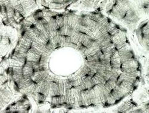
Looking at the osteons in bone (A) under a microscope reveals tube-like osteons (B) made up of osteocytes (C).
