A cervical spine X-ray is a safe and painless test that uses a small amount of radiation to take a picture of the bones in the back of the neck (cervical vertebrae). During the examination, an X-ray machine sends a beam of radiation through the neck, and an image is recorded on special film or a computer.
What is a normal spine X - ray?
Spine X-rays provide detailed images of the bones of the spine, and can be taken separately for the three main parts of the spine. Conditions that may show up in spine x-rays include fractures, tumors and arthritis. What is a spine X-ray? An X-ray is a test that uses radiation to produce images of the bones and organs of the body.
What is a normal cervical spine?
The neck is part of a long flexible column, known as the spinal column or backbone, which extends through most of the body. The cervical spine (neck region) consists of seven bones ( C1-C7 vertebrae ), which are separated from one another by intervertebral discs. These discs allow the spine to move freely and act as shock absorbers during activity.
What do the results of spine Xray mean?
The x-ray may also help in diagnosing the following:
- Fracture
- Arthritis
- Congenital abnormalities
- Spondylolisthesis (the slipping of the vertebra from the one that is below it)
- Scoliosis (abnormality in the spine curvature)
- Tumours
- Degeneration of the disks
What causes cloudy cervical spine X-ray?
What causes cloudy cervical spine x-ray?
- It may be due to improper exposure of radiation which leads to this x ray.
- It may be due to any inflammation of tissues including infection and abcess.
- Many tumours may cause cloudiness.
Can an X-ray show a tumor on the spine?
The soft tissues in the body (like blood, skin, fat, and muscle) allow most of the X-ray to pass through and appear dark gray on the film. A bone or a tumor, which is denser than soft tissue, allows few of the X-rays to pass through and appears white on the X-ray.
Can an X-ray show cervical spondylosis?
Neck X-ray. An X-ray can show changes in the spine, such as bone spurs, that indicate cervical spondylosis. Neck X-ray can also rule out rare and more serious causes for neck pain and stiffness, such as tumors, cancer, infections or fractures.
Can a neck X-ray show infection?
A neck X-ray can help detect a swollen epiglottis (epiglottitis), which is rare, or swelling in the tissues around the vocal cords (croup). It can also help doctors diagnose an infection in the area behind the throat (retropharyngeal abscess).
What are the warning signs of spondylosis?
Spondylosis SymptomsHeadaches.Loss of bladder control.Muscle spasms.Pain and soreness in the neck, shoulders, or lower back; pain may worsen with standing (if it originates in the lower back) or moving the head (if it originates in the neck)Stiffness.Tenderness.More items...•
What are the symptoms of neck spondylitis?
SymptomsPain in the neck that may travel to your arms or shoulders.Headaches.A grinding feeling when you move your neck.Weakness in your arms and legs.Numbness in your shoulders, arms, or hands.Stiffness in the neck.Trouble keeping your balance.Trouble controlling your bladder or bowels.
What are the symptoms of osteoarthritis in the neck?
Neck Osteoarthritis SymptomsNeck pain that worsens with activity performed when a person is upright.Neck pain that radiates to the arm or shoulder.Numbness, tingling, and weakness in the arms, hands, fingers, legs, or feet.Weakness in the legs, trouble walking, loss of balance.Loss of bladder or bowel control.More items...•
What is the difference between a neck X-ray and a cervical X-ray?
An X-ray is a form of radiation that passes through your body to expose a piece of film, forming an image of your body. A neck X-ray, also known as a cervical spine X-ray, is an X-ray image taken of your cervical vertebrae. This includes the seven bones of your neck that surround and protect the top section of your spinal cord.
How long does it take to get an X-ray of your neck?
The procedure is painless and generally takes 15 minutes or less. The technologist first has you lie flat on the X-ray table, and the X-ray machine then moves over your neck area. To keep the image from being blurry, you must stay very still and hold your breath for a few moments while the image is taken.
What is it called when your neck is dislocated?
If your neck is dislocated or fractured, your spinal cord may also be damaged. Neck injury caused by a sudden jerking of the head is commonly called whiplash. Your doctor may check the X-ray image for the following: chronic wear on the disks and joints of your neck, which is called cervical spondylosis.
What are the structures of the neck?
A neck X-ray also shows the nearby structures, including your: 1 vocal cords 2 tonsils 3 adenoids 4 trachea (windpipe) 5 epiglottis (the flap of tissue that covers your windpipe when you swallow)
How long does it take for a radiology technologist to develop X-rays?
The radiology technologist develops the X-rays and send them to your doctor within a few days.
Why do trachea appear white on X-rays?
trachea (windpipe) epiglottis (the flap of tissue that covers your windpipe when you swallow) Dense structures like bones appear white on X-rays because very little radiation can pass through them to expose the film on the other side. Soft tissues are less dense. That means more radiation can pass through them.
What is it called when you wear your neck?
chronic wear on the disks and joints of your neck, which is called cervical spondylosis. joints that are pushed out of their normal positions, which are called dislocations. abnormal growths on the bones, which are called bone spurs. spinal deformities.
What are X-rays of the spine, neck or back?
X-rays use invisible electromagnetic energy beams to make images of internal tissues, bones, and organs on film. Standard X-rays are performed for many reasons. These include diagnosing tumors or bone injuries.
What is the procedure to evaluate the spine?
X-rays of the spine may be performed to evaluate any area of the spine (cervical, thoracic, lumbar, sacral, or coccygeal). Other related procedures that may be used to diagnose spine, back, or neck problems include myelography (myelogram), computed tomography (CT scan), magnetic resonance imaging (MRI), or bone scans. Please see these procedures for additional information.
How many vertebrae are in the lumbar area?
The lumbar area consists of 5 vertebrae in the lower back. The sacrum has 5 small, fused vertebrae. The 4 coccygeal vertebrae fuse to form 1 bone, called the coccyx or tailbone. The spinal cord, a major part of the central nervous system, is located in the vertebral canal and reaches from the base of the skull to the upper part of the lower back.
What is the spinal cord?
The spinal cord is surrounded by the bones of the spine and a sac containing cerebrospinal fluid. The spinal cord carries sense and movement signals to and from the brain and controls many reflexes. ViewMedica 8. Start Picture-in-Picture. Get Embed Code.
How many vertebrae are in the spinal column?
The spinal column is made up of 33 vertebrae that are separated by spongy disks and classified into distinct areas:
What makes X-rays appear white?
The soft tissues in the body (like blood, skin, fat, and muscle) allow most of the X-ray to pass through and appear dark gray on the film. A bone or a tumor, which is denser than soft tissue, allows few of the X-rays to pass through and appears white on the X-ray. At a break in a bone, the X-ray beam passes through the broken area.
How are X-rays made?
X-rays are made by using external radiation to produce images of the body, its organs, and other internal structures for diagnostic purposes. X-rays pass through body tissues onto specially-treated plates (similar to camera film) and a "negative" type picture is made (the more solid a structure is, the whiter it appears on the film). ...
What is spine xray?
What is a spine X-ray? An X-ray is a test that uses radiation to produce images of the bones and organs of the body. Spine X-rays provide detailed images of the bones of the spine, and can be taken separately for the 3 main parts of the spine – cervical (neck), thoracic (mid back), and lumbar (lower back). During an X-ray, a focused beam of ...
What are the conditions that can be seen on a spine x-ray?
Conditions that may show up in spine x-rays include fractures, tumors and arthritis.
How do I prepare for a spine X-ray?
There is no special preparation for a spine X-ray. It is important to tell the technologist if you are or may be pregnant. X-rays generally are not used on pregnant women because of the possible risk of radiation exposure to the developing baby. Also, please notify the technologist if you have an insulin pump.
What will I feel during the test?
A spine X-ray is painless. You will not feel the radiation as it passes through your body. The X-ray room may be cool, because air conditioning is used to keep the equipment at a constant temperature.
How does an X-ray technologist take care of your body?
The technologist will position your body against the X-ray film in a way that produces the clearest image. Most X-rays of the upper or lower spine are taken while you are lying on the X-ray table, although sometimes they are taken while you are standing. The technologist may ask you to change positions or move your arms.
Why do you need a spine X-ray?
A spine X-ray may be ordered to evaluate a back or neck injury, or to help with the diagnosis and treatment of back or neck pain. Spine X-rays can help detect:
What to do when getting an X-ray of your neck?
You may be asked to open your mouth for an X-ray of your neck. This position moves your teeth out of the way and enables the technologist to get a clearer image of the upper bones of your neck. The technologist will ask you to be very still and hold your breath while the X-rays are passed through your body.
What is the standard view for cervical spine X-ray?
Typically there are three standard views provided when a cervical spine X-ray is performed, these include a lateral, antero-posterior (AP) and odontoid/open-mouth view. If you are struggling to see down to the level of C7/T1, a fourth “ swimmer’s view ” can be requested.
How to interpret cervical spine xray?
When interpreting a cervical spine X-ray, begin by confirming the patient’s details, reviewing the clinical history and ensuring the radiographs are adequate.
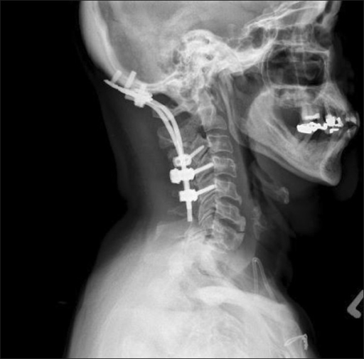
What is pre-vertebral soft tissue?
Pre-vertebral (i.e the area directly anterior to the vertebral bodies) soft tissue is best assessed using a lateral view. Soft tissue appears as a light grey opacity on cervical spine X-rays , located between the vertebral bodies and the darker-grey area that represents the trachea. Any widening of this space may represent a pre-vertebral haematoma and should significantly raise suspicion of a cervical fracture. It should be noted that this area naturally gets wider around the level of C4 so two different acceptable widths are used:
What is the color of the AP view of the C-spine X-ray?
Figure 3. AP view C-spine X-ray with red lines demonstrating alignment of lateral masses of C1 and C2, and purple lines demonstrating the alignment of the odontoid peg with the lateral masses of C1 1
Where is the spinolaminar line?
The spinolaminar line runs along the anterior edge of the spinous processes (at the junction of the spinous process and the laminae).
What does it mean when the lines intersect in the odontoid view?
The odontoid/open-mouth view has several intersecting lines which are sometimes referred to as a “meeting of corners ”. Irregularities in the areas where these lines intersect may indicate misalignment of the lateral masses of C1 and C2 (e.g. fracture, dislocation).
Where is the anterior longitudinal line?
Lateral view. The anterior longitudinal line runs along the anterior surface of the vertebral bodies. The posterior longitudinal line runs along the posterior surface of the vertebral bodies. The spinolaminar line runs along the anterior edge of the spinous processes (at the junction of the spinous process and the laminae). Figure 1. ...
What Happens in a CT Cervical Spine Scan?
Your CT cervical spine scan begins with screening questions. A radiology nurse or technician will ask you about any allergies you may have and will review your medical history. Next, they’ll ask you to remove all jewelry and piercings. You may need to wear a hospital gown.
What are the symptoms of cervical spine?
When you receive a doctor’s order for a medical test for your neck, you’ll see it written as a CT scan for the cervical spine. This is the top section of your spine that passes through your neck and ends at your brain. Issues with your cervical spine can cause: 1 Headaches 2 Dizziness 3 Pain in your neck, arms, shoulders, legs, and jaw 4 Numbness in your neck, hands, arms, and legs 5 Muscle spasms 6 Trouble with balance and walking 7 Loss of hand-eye coordination 8 Loss of bladder and bowel control
What is the top section of the neck that passes through the neck and ends at the brain?
When you receive a doctor’s order for a medical test for your neck, you’ll see it written as a CT scan for the cervical spine. This is the top section of your spine that passes through your neck and ends at your brain. Issues with your cervical spine can cause: Headaches. Dizziness.
How long does a CT scan take?
Once the scan begins, the machine sends out rotating X-ray beams to create the computerized images. You can expect the test to take about 10 to 15 minutes, but for some people, the CT scan may take up to 30 minutes. The radiology technician may ask you to hold your breath a few times to help avoid blurry images.
What is the best test for neck pain?
If you feel pain in your neck and shoulder, your doctor might want you to get a specific X-ray to see what’s causing the pain. This test is a computed tomography scan or what's more commonly called a CT scan.
What happens after a CT scan?
They feel that the benefit is far greater than the minor risks. After your CT scan, the imaging radiologist reviews the images and includes notes for your doctor.
What to ask a radiology nurse?
A radiology nurse or technician will ask you about any allergies you may have and will review your medical history. Next, they’ll ask you to remove all jewelry and piercings. You may need to wear a hospital gown. . If the doctor’s order requests contrast, you'll receive an injection at this point.
What is AP oblique cervical spine?
The AP oblique cervical spine projections are supplementary views to the standard AP , odontoid and lateral images in the cervical spine series and are always done bilaterally for comparison purposes. However, the PA oblique projection is preferred as it reduces radiation dose to the thyroid 1 compared to the AP oblique projection.
How to demonstrate intervertebral foramen of the C-spine open?
To demonstrate the intervertebral foramen of the c-spine open, it is necessary to achieve adequate rotation of the vertebral column, usually at 45°. If underrotated, the foramina will be narrowed and a sternoclavicular joint would be superimposed over the vertebral column 3.
What is the position of the patient's face in the image receptor?
the thorax and cervical spine is at 45° to the image receptor. the face is in a lateral position with the interpupillary line perpendicular to the image receptor.
What is the rotation angle of the C-spine?
To demonstrate the intervertebral foramen of the c-spine open, it is necessary to achieve adequate rotation of the vertebral column, usually at 45°.
Why should the patient's head be in a lateral position?
patient’s head should be in a lateral position to prevent mandibular superimposition over the vertebral bodies of the cervical spine. intervertebral foramina of the side positioned further from the image receptor should be demonstrated open.
Why do you need an x-ray for spinal fracture?
For this reason, x-rays are typically ordered when injury cause and symptom description suggest a person might be dealing with a spinal fracture or similar bone issue. This is most common when the cause of injury involves significant trauma to the area.
Why do you need an x-ray?
For this reason, x-rays are typically ordered when injury cause and symptom description suggest a person might be dealing with a spinal fracture or similar bone issue. This is most common when the cause of injury involves significant trauma to the area. For example, after a car accident, a heavy collision in sporting activities or following a fall, a spine specialist may order an x-ray to see if a fracture has occurred to any of the vertebrae in the spine. In most cases of trauma to the area, an x-ray can be a helpful diagnostic tool.

Purpose
Mechanism
- X-rays are made by using external radiation to produce images of the body, its organs, and other internal structures for diagnostic purposes. X-rays pass through body tissues onto specially-treated plates (similar to camera film) and a \"negative\" type picture is made (the more solid a structure is, the whiter it appears on the film). Instead of film, X-rays are now typically made by u…
Diagnosis
- X-rays of the spine may be performed to evaluate any area of the spine (cervical, thoracic, lumbar, sacral, or coccygeal). Other related procedures that may be used to diagnose spine, back, or neck problems include myelography (myelogram), computed tomography (CT scan), magnetic resonance imaging (MRI), or bone scans. Please see these procedures fo...
Structure
- The spinal column is made up of 33 vertebrae that are separated by spongy disks and classified into distinct areas:
Function
- The spinal cord, a major part of the central nervous system, is located in the vertebral canal and reaches from the base of the skull to the upper part of the lower back. The spinal cord is surrounded by the bones of the spine and a sac containing cerebrospinal fluid. The spinal cord carries sense and movement signals to and from the brain and controls many reflexes.
Treatment
- There may be other reasons for your health care provider to recommend an X-ray of the spine, neck, or back.