What causes mirror image artifact? These occur when an ultrasound beam is not reflected directly back to the transducer after hitting a reflective surface, but rather takes an indirect return journey. With mirror image artefact the interrogating ultrasound beam hits an angled, highly reflective surface, such a soft tissue / gas interface.
What causes a mirror image?
A mirror image is the result of light rays bounding off a reflective surface. Reflection and refraction are the two main aspects of geometric optics.
Is a mirror image real or not?
The mirror is the real you, but however, nobody can see their true self by looking into the mirror. Because the human eye cannot capture a face like a camera would, it scans through each part of your face and sends multiple packages of information about each section of your face to the brain.
How to create a mirror image effect?
- Import the footage you’d like to include in your project into the editor and place it on the timeline.
- Remove all the parts of the video clip you don’t want your mirror video to contain.
- Then click on the Visual Effects tab.
What makes a mirror reflect an image?
Some other contemporary artists use mirrors as the material of art :
- A Chinese magic mirror is an art in which the face of the bronze mirror projects the same image that was cast on its back. ...
- Specular holography uses a large number of curved mirrors embedded in a surface to produce three-dimensional imagery.
- Paintings on mirror surfaces (such as silkscreen printed glass mirrors)
What causes mirror artifact?
Mirror artifacts are produced by the reflection of ultrasound waves after they propagate through a structure and encounter a strong and smooth interface capable of acting as a mirror.
How do I stop mirror image artifact?
To avoid this artifact, change the position and angle of scanning to change the angel of insonation of the primary ultrasound beam.
What is mirror artifact in ultrasound?
Mirror image artefact is one of the beam path artefacts. These occur when an ultrasound beam is not reflected directly back to the transducer after hitting a reflective surface, but rather takes an indirect return journey.
Where is a mirror image artifact located?
Mirror image artifacts are commonly seen in sonographic imaging throughout the body. They can be seen on gray scale, color Doppler, power Doppler, and spectral Doppler.
How do you fix artifacts in ultrasound?
This type of artifact can often be corrected by gently pressing the probe against the rectal wall to force the gas away from the crystal face. If that does not work, the probe should be removed and the condom cover re-prepared. Copious gel should be applied to help minimize air between the covering and the probe.
When does mirror image artifact occur?
Mirror image artifacts occur when the transmitted pulse and returning echo reflect off of a highly reflective interface (an acoustic mirror) and change direction before returning to the transducer, thereby breaking this assumption.
How do I get rid of mirror image artifact in ultrasound?
To avoid this artifact, change the position and angle of scanning to change the angel of insonation of the primary ultrasound beam.
Why does mirror image happen in ultrasound?
Mirror image artifacts occur when the transmitted pulse and returning echo reflect off of a highly reflective interface (an acoustic mirror) and change direction before returning to the transducer, thereby breaking this assumption.
How do you get rid of refraction artifacts?
Refraction artifact should resolve if the transducer is moved such that the incident pulse is perpendicular to the interface.
Is ultrasound screen mirrored?
Most ultrasounds that you're going to use in a clinical setting will have a mirror-like effect. This happens when the left side of the body shows up on the left side of the screen. One common clinical ultrasound that uses straight shot imaging is transvaginal ultrasound.
What is an aliasing artifact?
Aliasing artifact, otherwise known as undersampling, in CT refers to an error in the accuracy proponent of analog to digital converter (ADC) during image digitization. Image digitization has three distinct steps: scanning, sampling, and quantization.
What is duty factor in ultrasound?
Duty Factor = Pulse Duration X Pulse Repetition Freq. Pulse Duration. Pulse Repetition Period. Spatial Pulse Length. distance in space traveled by ultrasound during one pulse.
What is mirror image artefact?
Mirror image artefact is one of the beam path artefacts. These occur when an ultrasound beam is not reflected directly back to the transducer after hitting a reflective surface, but rather takes an indirect return journey.
What happens when ultrasound pulses are caught between two surfaces?
Some of the pulse becomes caught between the two surfaces, bouncing forwards and backwards before returning in increments, between each reverberation, to the transducer. This reverberation causes a repetitive artefact on the ultrasound image.
What is mirror image artifact?
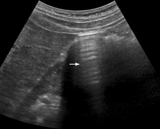
Mirror image artifacts occur when the ultrasound beam encounters a highly reflective nonperpendicular or curved boundary such as the diaphragm. The redirected beam encounters a specular reflector, producing a series of echoes that are reflected along the same path back to the transducer. As the field of view is scanned, the beam interacts with the same specular reflector to record its true position. Later echoes of the specular reflector from the redirected beam arrive and are mapped as distal objects on the opposite side of the strong reflector, appearing as a mirror image because of the double reflection. A common mirror image artifact occurs at the interface of the liver and the diaphragm in abdominal imaging. In one direction, the ultrasound beam correctly positions the echoes emanating from a lesion in the liver. As the ultrasound beam moves through the liver, echoes are strongly reflected from the curved diaphragm away from the main beam to interact with the lesion, generating echoes that travel back to the diaphragm and ultimately back to the transducer. The back-and-forth travel distance of these echoes creates artifactual anatomy that resembles a mirror image of the mass, placed beyond the diaphragm that would otherwise not have anatomy present in the image ( Fig. 2.7 ). Other strong reflectors that generate mirror image artifacts include the pericardium and bowel, which might be more difficult to detect due to the presence of other anatomic structures in the same area.
What causes refraction artifacts?
(A) Refraction artifacts arise from a change of direction of the ultrasound beam at a tissue boundary, with nonperpendicular incidence caused by a speed of sound difference and a change in wavelength.
What are ultrasound artifacts?
Ultrasound artifacts represent a false portrayal of image anatomy or image degradations related to false assumptions regarding the propagation and interaction of ultrasound with tissues, as well as malfunctioning or maladjusted equipment. Understanding how artifacts are generated and how they can be recognized is crucial, which places high demands on the knowledge of the sonographer and the interpreting physician. Most artifacts arise from violations of assumptions for creating the ultrasound image, including but not limited to (1) ultrasound travels at a constant speed in all tissues (1540 m/s); (2) ultrasound travels in a straight path; (3) reflections occur from the initial ultrasound beam with one interaction at a perpendicular incidence for each boundary; (4) attenuation of ultrasound echoes is uniform; (5) all of the energy emitted by the ultrasound transducer exists in the main beam; (6) the operator has transmit, receive gain, and other settings properly adjusted; and (7) all of the elements in the transducer array, as well as the remainder of the imaging system, are operating optimally.
Why are ultrasound artifacts important?
Fortunately, most ultrasound artifacts can be identified by the experienced sonographer because of obvious effects on the image or the transient nature of mismapped anatomy that appears and disappears during the scan. Some artifacts can be used to advantage as diagnostic aids in the characterization of tissue structures and composition.
How do reverberation artifacts occur?
Reverberation artifacts arise from multiple echoes generated between highly reflective and parallel structures that interact at a perpendicular angle to the ultrasound beam. These artifacts are often caused by reflections between a reflective interface and the transducer or between reflective interfaces, such as metallic objects (e.g., bullet fragments), calcified tissues, or air pocket/partial liquid areas of the anatomy, and are typically manifested as multiple equally spaced parallel lines at progressive depth with decreasing amplitude ( Fig. 2.5 ). Comet tail artifact is a form of reverberation between two closely spaced reflectors and appears as bright lines along the direction of ultrasound propagation. It is manifested as a tapering shape and decreasing width with depth of travel ( Fig. 2.6A ). Reverberation artifacts are useful in assessing the characteristic structures of tissues but can also hinder visualization of deeper anatomy. Effects of reverberation can be reduced by adjusting the transducer angle of incidence or by decreasing the distance between the reflective structure and the transducer. Harmonic imaging reduces these artifacts by receiving the first harmonic frequency and filtering out the fundamental frequency.
How does reverberation affect anatomy?
Effects of reverberation can be reduced by adjusting the transducer angle of incidence or by decreasing the distance between the reflective structure and the transducer.
What are artifacts used for?
Some artifacts can be used to advantage as diagnostic aids in the characterization of tissue structures and composition. The following descriptions represent some typical artifacts encountered in diagnostic ultrasound.
What causes a Gibbs artifact?
Artifacts are caused by a variety of factors that may be patient-related such as voluntary and physiologic motion, metallic implants or foreign bodies. Finite sampling, k-space encoding, and Fourier transformation may cause aliasing and Gibbs artifact.
What are MRI artifacts?
MRI artifacts. MRI artifacts are numerous and give an insight into the physics behind each sequence. Some artifacts affect the quality of the MRI exam while others do not affect the diagnostic quality but may be confused with pathology. When encountering an unfamiliar artifact, it is useful to systematically examine general features ...
What causes black boundary?
Characteristics of pulse sequences may cause black boundary, Moiré, and phase-encoding artifacts. Hardware issues may cause central point and RF overflow artifacts. Remember that artifacts are not all bad, and that occasionally they are intentionally exploited, e.g. susceptibility artifact.
