The mechanisms include:
- Degradation is when an enzyme changes the structure of the neurotransmitter so that it no longer fits the receptor.
- Diffusion is when the neurotransmitter begins to diffuse or drift away from the receptor.
- Reuptake is when the entire neurotransmitter molecule is taken back by the neuron’s axon, which initially releases neurotransmitters.
How are neurotransmitters released from the presynaptic terminal?
May 09, 2020 · What causes the release of neurotransmitter molecules? Storage of the neurotransmitter in storage granules or vesicles in the axon terminal. Calcium enters the axon terminal during an action potential, causing release of the neurotransmitter into the synaptic cleft.
How are neurotransmitters released from the vesicles?
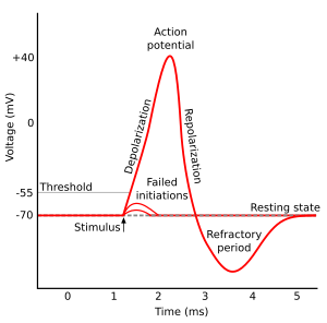
An increase in calcium causes the release of neurotransmitter molecules. When a neuron becomes depolarized and reaches threshold, it sends an... See full answer below.
What is the role of calcium in neurotransmitter release?
Jan 27, 2022 · A nerve impulse (at the end of the presynaptic axon) causes Ca2+ to rush inside the presynaptic axon, which causes the release of neurotransmitters in the synaptic cleft. … It is important for the neurotransmitter to be deactivated quickly in order to …
How are neurotransmitters released from the synaptotagmin complex?
The influx of calcium through the voltage-gated calcium channels initiates the exocytosis process that leads to neurotransmitter release. Calcium enters the cell and interacts with another vesicle-bound protein called synaptotagmin.

What stimulates the release of neurotransmitters?
0:102:002-Minute Neuroscience: Neurotransmitter Release - YouTubeYouTubeStart of suggested clipEnd of suggested clipCalcium seems to be involved with mobilizing vesicles to prepare them for neurotransmitter release.MoreCalcium seems to be involved with mobilizing vesicles to prepare them for neurotransmitter release. One way this occurs is through interaction between calcium.
What causes the release of the neurotransmitter quizlet?
A nerve impulse (at the end of the presynaptic axon) causes Ca2+ to rush inside the presynaptic axon, which causes the release of neurotransmitters in the synaptic cleft.
What causes the release of neurotransmitters and synaptic cleft?
Neurons talk to each other across synapses. When an action potential reaches the presynaptic terminal, it causes neurotransmitter to be released from the neuron into the synaptic cleft, a 20–40nm gap between the presynaptic axon terminal and the postsynaptic dendrite (often a spine).Nov 9, 2017
Which is the neurotransmitter that causes the release of calcium ions?
ACh is the neurotransmitter that binds at the neuromuscular junction (NMJ) to trigger depolarization, and an action potential travels along the sarcolemma to trigger calcium release from SR.
How does CD affect neurotransmission?
Cd may affect neurotransmission by different mechanisms. First of all it can suppress neurotransmitter release due to the blockade of CaV channels and, consequently, of Ca++ influx into synaptic terminals. This has first been well described for acetylcholine release at neuromuscular junction which was found to be inhibited by Cd in a Ca ++ dependent manner ( Cooper and Manalis, 1984; Toda, 1976 ). An additional mechanism of Cd interference with neurotransmission is the direct interaction of this ion with neurotransmitter receptors. This has been shown in the case of acetylcholine receptors which have been found to be activated by Cd in a subunit dependent manner ( Hsiao et al., 2001 ), of GABA-A receptors, which, on the contrary, are inhibited by Cd in a subunit dependent way ( Casagrande et al., 2003; Fisher and Macdonald, 1998 ). In addition, Cd blocks glycine ( Wang et al., 2006) and kainite ( Braitman and Coyle, 1987 ), metabotropic ( Vignes et al., 1996) and glutamate receptors.
Where are neurotransmitters stored?
In the axon terminals, neurotransmitters are stored in the synaptic vesicles. The mechanism of neurotransmission in the chemical synapses is illustrated in Fig. 12. The first step in neurotransmission begins with the arrival of the action potential to the axon terminal in the presynaptic neuron.
Why are snare proteins important?
Because SNARE interactions can be detected prior to neurotransmitter release, interactions between SNARE proteins are thought to represent the molecular underpinnings of vesicle priming. By extension, proteins that interact with the SNAREs are candidates for promoters and regulators of vesicle priming.
What are TRCs? What are their functions?
The TRCs are not neurons, even though they release neurotransmitters, because they have no dendritic of axonal processes. They make synapses onto dendritic processes of primary afferent sensory neurons whose cell bodies reside in three cranial nerve ganglia. The anterior two-thirds of the tongue and the palate are innervated by the facial nerve or cranial nerve VII. The cell bodies for the taste fibers are in the geniculate ganglia. The posterior third of the tongue is supplied by the glossopharyngeal nerve or cranial nerve IX. The cell bodies for this sensory nerve are located in the inferior glossopharyngeal ganglia. The vagus nerve, cranial nerve X, supplies the scattered taste receptors in the throat regions, including the glottis, epiglottis, and pharynx. The cell bodies of the vagus reside in the inferior vagal ganglia.
Which nerve innervates the palate?
The anterior two-thirds of the tongue and the palate are innervated by the facial nerve or cranial nerve VII. The cell bodies for the taste fibers are in the geniculate ganglia. The posterior third of the tongue is supplied by the glossopharyngeal nerve or cranial nerve IX.
Where are the sensory nerves located?
The cell bodies for this sensory nerve are located in the inferior glossopharyngeal ganglia. The vagus nerve, cranial nerve X, supplies the scattered taste receptors in the throat regions, including the glottis, epiglottis, and pharynx. The cell bodies of the vagus reside in the inferior vagal ganglia. Sensory fibers from all three of these cranial ...
What is the adrenal medulla?
The adrenal medulla is essentially a ganglion of the sympathetic nervous system that releases neurotransmitter into the blood instead of near local targets. Preganglionic sympathetic nervous fibers originating mainly in the thoracic spinal cord reach the adrenal medulla through the splanchnic nerves, and release acetylcholine onto chromaffin cells in the adrenal gland, causing release of epinephrine into the blood; the epinephrine is then transported to distant targets. Epinephrine is synthesized from tyrosine in the sequence tyrosine–dihydroxyphenylalanine–dopamine–norepinephrine–epinephrine. Circulating epinephrine and norepinephrine are degraded by catechol- O -methyl transferase (COMT) and monoamine oxidase (MAO). A variety of stimuli increase epinephrine secretion including hypoglycemia, hypovolemia, hypotension, fear and anxiety, pain, and trauma.