How fast do vesicles replenish?
How do synaptic vesicles release neurotransmitters?
What does the arrow on a frozen sample mean?
Where does synaptic fusion take place?
Where do vesicles fuse during asynchronous release?
Can multiple vesicles fuse?
How to capture exocytosis?
See 2 more
About this website

What causes synaptic vesicles to fuse?
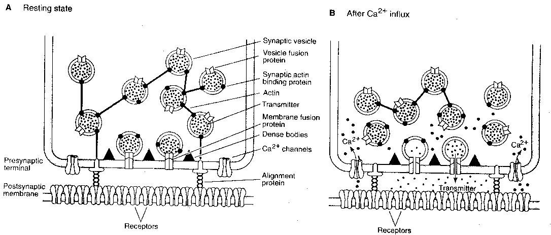
After an action potential, Ca2+ floods to the presynaptic membrane. Ca2+ binds to specific proteins in the cytoplasm, one of which is synaptotagmin, which in turn trigger the complete fusion of the synaptic vesicle with the cellular membrane.
How are synaptic vesicles activated?
The protein kinase then phosphoylates Synapsin, releasing the vesicle from its actin cage. The vesicle falls to the active zone at the presynaptic terminal. Once in the pre-synaptic terminal, calcium triggers a series of events that lead to neurotransmitter release.
Which ion causes synaptic vesicles to fuse with the membrane?
Calcium ionsCalcium ions entering the cell initiate a signaling cascade that causes small membrane-bound vesicles, called synaptic vesicles, containing neurotransmitter molecules to fuse with the presynaptic membrane.
What are active zones in synapses?
Active zones are the principal sites of synaptic vesicle fusion in synapses. The molecular components of the active zone are thought to serve a structural role by clustering synaptic vesicles around the active zone and increasing proximity between molecules on the synaptic vesicle membrane and the plasma membrane.
Which activity stimulates the movement of synaptic vesicles?
action potentialThe axon terminals contain vesicles filled with these neurotransmitters. When an impulse (action potential) arrives at the axon terminal, it stimulates the movement of the synaptic vesicles towards the membrane where they fuse with the plasma membrane and released their neurotransmitters in the synaptic cleft.
What triggers exocytosis of synaptic vesicles quizlet?
The influx of calcium ions into the " " triggers exocytosis of synaptic vesicles.
How do vesicles fuse with the membrane?
These two proteins may allow the vesicle and presynaptic membrane to recognize each other. Following docking, there is a second influx of calcium at the active zone, which causes the vesicle membrane to fuse to the presynaptic membrane, forming a temporary ion channel.
How do vesicles fuse with the cell membrane?
When the action potential reaches the nerve terminals, it causes an influx of Ca2+ through voltage-gated Ca2+ channels. The binding of Ca2+ ions to specific sensors then triggers the secretory vesicles (called synaptic vesicles) to fuse with the plasma membrane and release their contents to the extracellular space.
How do vesicles fuse with the presynaptic membrane?
The mechanism by which synaptic vesicles fuse with the presynaptic membrane is complex and relies on a group of proteins belonging to the SNARE (SNAP receptor) family. The vesicle and presynaptic membranes 'kiss' and the interaction between complimentary proteins creates a small fusion pore.
What is the function of the synaptic vesicles?
Abstract. Synaptic vesicles play the central role in synaptic transmission. They are regarded as key organelles involved in synaptic functions such as uptake, storage and stimulus-dependent release of neurotransmitter.
What proteins are involved in synaptic release?
There are five synaptic proteins that have been discovered, synapsin-Ia, synapsin-Ib, synapsin-IIa, synapsin-IIb, and synapsin-IIIa.
What happens at the presynaptic terminal?
In a presynaptic terminal, neurotransmitters are packaged into synaptic vesicles. When an action potential opens presynaptic voltage-gated Ca2+ channels, the neurotransmitters are released by Ca2+-triggered synaptic vesicle exocytosis into the synaptic cleft, where they activate postsynaptic receptors.
How do synaptic vesicles move?
After filling with transmitters, synaptic vesicles are moved to the active zone of the presynaptic plasma membrane by a translocation process that may be either diffusion-limited or dependent on molecular motors (step 2 in Fig.
How are synaptic vesicles filled with neurotransmitter?
Synaptic vesicles fill with neurotransmitter through a process driven by the vacuolar-type H+-ATPase.
How do vesicles move in neuron?
After budding from the Golgi, these vesicles use motor proteins called kinesins to travel down the axon along a cytoskeleton pathway made up of microtubules and actin, until they arrive at the nerve terminal (Pfeffer, 1999; Cai et al., 2007).
What is the function of synaptic vesicles?
Abstract. Synaptic vesicles play the central role in synaptic transmission. They are regarded as key organelles involved in synaptic functions such as uptake, storage and stimulus-dependent release of neurotransmitter.
What happens when an action potential reaches the presynaptic bouton?
When an action potential reaches the presynaptic bouton, the contents of the vesicles are released into the synaptic cleft and the released neurotransmitter travels across the cleft to the postsynaptic neuron (the lower structure in the picture) and activates the receptors on the postsynaptic membrane. The active zone is the region in the ...
How many clusters of synaptic vesicles are there in the synapse?
The synapse contains at least two clusters of synaptic vesicles, the readily releasable pool and the reserve pool. The readily releasable pool is located within the active zone and connected directly to the presynaptic membrane while the reserve pool is clustered by cytoskeletal and is not directly connected to the active zone.
How does the presynaptic bouton work?
The presynaptic bouton has an efficiently orchestrated process to fuse vesicles to the presynaptic membrane to release neurotransmitters and regenerate neurotransmitter vesicles. This process called the synaptic vesicle cycle maintains the number of vesicles in the presynaptic bouton and allows the synaptic terminal to be an autonomous unit. The cycle begins with (1) a region of the golgi apparatus is pinched off to form the synaptic vesicle and this vesicle is transported to the synaptic terminal. At the terminal (2) the vesicle is filled with neurotransmitter. (3) The vesicle is transported to the active zone and docked in close proximity to the plasma membrane. (4) During an action potential the vesicle is fused with the membrane, releases the neurotransmitter and allows the membrane proteins previously on the vesicle to diffuse to the periactive zone. (5) In the periactive zone the membrane proteins are sequestered and are endocytosed forming a clathrin coated vesicle. (6) The vesicle is then filled with neurotransmitter and is then transported back to the active zone.
What is the active zone of the presynaptic membrane?
The active zone is the region in the presynaptic bouton that mediates neurotransmitter release and is composed of the presynaptic membrane and a dense collection of proteins called the cytomatrix at the active zone (CAZ). The CAZ is seen under the electron microscope to be a dark (electron dense) area close to the membrane.
What are the active zones of a synapses?
The active zone is present in all chemical synapses examined so far and is present in all animal species. The active zones examined so far have at least two features in common, they all have protein dense material that project from the membrane and tethers synaptic vesicles close to the membrane and they have long filamentous projections originating at the membrane and terminating at vesicles slightly farther from the presynaptic membrane. The protein dense projections vary in size and shape depending on the type of synapse examined. One striking example of the dense projection is the ribbon synapse (see below) which contains a "ribbon" of protein dense material that is surrounded by a halo of synaptic vesicles and extends perpendicular to the presynaptic membrane and can be as long as 500 nm. The glutamate synapse contains smaller pyramid like structures that extend about 50 nm from the membrane. The neuromuscular synapse contains two rows of vesicles with a long proteinaceous band between them that is connected to regularly spaced horizontal ribs extending perpendicular to the band and parallel with the membrane. These ribs are then connected to the vesicles which are each positioned above a peg in the membrane (presumably a calcium channel). Previous research indicated that the active zone of glutamatergic neurons contained a highly regular array of pyramid shaped protein dense material and indicated that these pyramids were connected by filaments. This structure resembled a geometric lattice where vesicles were guided into holes of the lattice. This attractive model has come into question by recent experiments. Recent data shows that the glutamatergic active zone does contain the dense protein material projections but these projections were not in a regular array and contained long filaments projecting about 80 nm into the cytoplasm.
Which protein recruits in the periactive zone?
In the periactive zone, scaffolding proteins such as intersectin 1 recruit proteins that mediate endocytosis such as dynamin, clathrin and endophilin.
What is the active zone in microanatomy?
Anatomical terms of microanatomy. The active zone or synaptic active zone is a term first used by Couteaux and Pecot-Dechavassinein in 1970 to define the site of neurotransmitter release. Two neurons make near contact through structures called synapses allowing them to communicate with each other. As shown in the adjacent diagram, ...
What are the vesicles of the presynaptic neuron?
Inside the presynaptic neuron are synaptic vesicles, which fuse with the membrane in the active zone.
Which vesicles fuse with the membrane in the active zone?
b. Inside the presynaptic neuron are synaptic vesicles, which fuse with the membrane in the active zone.
What binds to receptors?
a. As neurotransmitter binds to its receptors
Where is the neurotransmitter released?
d. Neurotransmitter is released into the synaptic cleft.
What chapter is Neurotransmitters and Their Receptors?
Chapter 6: Neurotransmitters and Their Receptors A…
Where does a symlink act?
It acts on receptors in the postsynaptic membrane.
Does a synapse increase the magnitude of postsynaptic potential?
a. It would increase the magnitude of postsynaptic potential.
How fast do vesicles replenish?
Docking of vesicles to refill release sites must be rapid. A single action potential consumes some docked vesicles, bursts of action potentials would be expected to deplete all docked vesicles. Nevertheless, some central synapses can fire at a frequency of one kilohertz10. Studies using electrophysiology and electron microscopy indicate that recovery of the docked and readily-releasable vesicle pools is slow –about 3 seconds7,8,11. However, an emerging body of work suggests that vesicle replenishment constitutes several kinetically and molecularly distinct steps, some of which may occur on very fast timescales. In two notable recent examples, modeling based on physiological data predicted that vesicles reversibly transition from “replacement sites” to “docking sites” within milliseconds of an action potential12,13, and experiments with flash-and-freeze electron microscopy demonstrated that Synaptotagmin-1 mutants with docking defects can be reversed by binding calcium9. These fast vesicle docking events have been proposed to correspond to calcium-induced changes between loose and tight assembly of the SNARE complex, which may be both very fast and reversible14. However, there is currently no ultrastructural evidence for such fast and reversible docking steps at wild-type synapses.
How do synaptic vesicles release neurotransmitters?
Synaptic vesicles fuse with the plasma membrane to release neurotransmitter following an action potential, after which new vesicles must ‘dock’ to refill vacated release sites. To capture synaptic vesicle exocytosis at cultured mouse hippocampal synapses, we induced single action potentials by electrical field stimulation then subjected neurons to high-pressure freezing to examine their morphology by electron microscopy. During synchronous release, multiple vesicles can fuse at a single active zone. Fusions during synchronous release are distributed throughout the active zone, whereas fusions during asynchronous release are biased toward the center of the active zone. After stimulation, the total number of docked vesicles across all synapses decreases by ~40%. Within 14 ms, new vesicles are recruited and fully replenish the docked pool, but this docking is transient and they either undock or fuse within 100 ms. These results demonstrate that recruitment of synaptic vesicles to release sites is rapid and reversible.
What does the arrow on a frozen sample mean?
The arrow indicates a pit in the active zone, which is presumed to be a synaptic vesicle fusing with the plasma membrane.
Where does synaptic fusion take place?
Synaptic vesicle fusion takes place at a specialized membrane domain: the active zone 1. The active zone is organized into one or more release sites, individual units at which a single synaptic vesicle can fuse2. Ultrastructural studies demonstrate that some synaptic vesicles are in contact with the plasma membrane in the active zone and define the ‘docked’ pool3,4. Since both docking and physiological readiness require engaged SNARE proteins4–6, docked vesicles are thought to represent fusion-competent vesicles. In fact, previous studies demonstrate that docked vesicles are partially depleted following stimulation7–9. However, it is not clear how release sites are refilled by vesicles to sustain neuronal activity.
Where do vesicles fuse during asynchronous release?
Fusions during synchronous release occur throughout the active zone, but during asynchronous release are concentrated at the center of the active zone.
Can multiple vesicles fuse?
The presence of multiple vesicles docked at a synapse alone does not imply that multiple vesicles can fuse at an active zone. In fact, it was long thought that only one vesicle could fuse in response to an action potential21,33. These studies argued that responses at synapses are mostly, or even exclusively, uniquantal. For proponents of univesicular release, examples of recordings of multivesicular events were dismissed as being caused by multiple active zones impinging on the cell. Proponents of multiquantal release at single active zones argued that observations of uniquantal events were due to saturation of the postsynaptic receptor field, and multiquantal release could be observed under circumstances in which saturation could be avoided 34–36. By reconstructing synapses from serial sections immediately after a single action potential, we were able to capture multiple vesicles fusing in a single active zone. At 4 mM calcium, we observed up to 11 vesicles fusing in a single active zone. The probability of fusion at a release site appears to be low even in elevated calcium, but because active zones have ~10 docking sites, multiple vesicles can be consumed by a single action potential.
How to capture exocytosis?
1a). This device can be charged, then loaded into a high-pressure freezer and discharged with a flash of light to generate a 1 ms 10 V/cm stimulus before freezing at defined time points (see Methods).

Overview
Function
The function of the active zone is to ensure that neurotransmitters can be reliably released in a specific location of a neuron and only released when the neuron fires an action potential. As an action potential propagates down an axon it reaches the axon terminal called the presynaptic bouton. In the presynaptic bouton, the action potential activates calcium channels (VDCCs) that cause a local influx of calcium. The increase in calcium is detected by proteins in the active zon…
Structure
The active zone is present in all chemical synapses examined so far and is present in all animal species. The active zones examined so far have at least two features in common, they all have protein dense material that project from the membrane and tethers synaptic vesicles close to the membrane and they have long filamentous projections originating at the membrane and terminatin…
Neurotransmitter release mechanism
The release of neurotransmitter is accomplished by the fusion of neurotransmitter vesicles to the presynaptic membrane. Although the details of this mechanism are still being studied there is a consensus on some details of the process. Synaptic vesicle fusion with the presynaptic membrane is known to require a local increase of calcium from as few as a single, closely associated …
Synaptic vesicle cycle
The presynaptic bouton has an efficiently orchestrated process to fuse vesicles to the presynaptic membrane to release neurotransmitters and regenerate neurotransmitter vesicles. This process called the synaptic vesicle cycle maintains the number of vesicles in the presynaptic bouton and allows the synaptic terminal to be an autonomous unit. The cycle begins with (1) a regio…
Vesicle pools
The synapse contains at least two clusters of synaptic vesicles, the readily releasable pool and the reserve pool. The readily releasable pool is located within the active zone and connected directly to the presynaptic membrane while the reserve pool is clustered by cytoskeletal and is not directly connected to the active zone.
The releasable pool is located in the active zone and is bound directly to the presynaptic membr…
Periactive zone
The periactive zone surrounds the active zone and is the site of endocytosis of the presynaptic terminal. In the periactive zone, scaffolding proteins such as intersectin 1 recruit proteins that mediate endocytosis such as dynamin, clathrin and endophilin. In Drosophila the intersectin homolog, Dap160, is located in the periactive zone of the neuromuscular junction and mutant Dap160 deplete synaptic vesicles during high frequency stimulation.
Ribbon synapse active zone
The ribbon synapse is a special type of synapse found in sensory neurons such as photoreceptor cells, retinal bipolar cells, and hair cells. Ribbon synapses contain a dense protein structure that tethers an array of vesicles perpendicular to the presynaptic membrane. In an electron micrograph it appears as a ribbon like structure perpendicular to the membrane. Unlike the 'traditional' synapse, ribbon synapses can maintain a graded release of vesicles. In other words, the more d…