What color is a CSF leak?
The classic presentation of CSF leaks is the expression of clear, watery drainage from the nose. This occurs usually on one side; however if fluid drains into the back of the throat there may be a salty taste. Drainage also tends to increase when bending over or straining.
How do I know if my nasal drainage is CSF?
In either case, you'll have a runny nose (rhinorrhea) with thin, clear fluid. CSF coming out of your nose has two key differences from nasal mucus: If you wipe your nose with a handkerchief, drying nasal mucus will cause the cloth to stiffen, but CSF won't.
Does cerebrospinal fluid have color?
Color of the fluid—normal is clear and colorless. Changes in the color of the CSF are not diagnostic but may point to additional substances in the fluid. Yellow, orange, or pink CSF may indicate the breakdown of blood cells due to bleeding into the CSF or the presence of bilirubin.
Is CSF yellow?
Cerebrospinal Fluid The color of CSF is most usefully described as (1) pink or orange, (2) yellow, or (3) brown. These colors correspond to the major pigments derived from red cells: oxyhemoglobin, bilirubin, and methemoglobin. Oxyhemoglobin is red in color, but after dilution in the CSF it appears pink or orange.
Is CSF leak sticky?
Unlike mucus, which is thick and sticky, CSF is clear and watery.
How do I know if my brain is leaking fluid?
The most common symptom of a spinal CSF leak is a headache, while a cranial CSF leak causes symptoms such as clear fluid leaking from the nose or ear. Some CSF leaks may heal with conservative treatments such as bed rest. Many CSF leaks need a blood patch to cover the hole or surgery to repair the leak.
What does normal CSF look like?
Normal Results Appearance: clear, colorless. CSF total protein: 15 to 60 mg/100 mL. Gamma globulin: 3% to 12% of the total protein.
What is the color of CSF in meningitis?
Conditions associated with changes in the appearance of CSF include the following: Infectious meningitis – Turbid, milky, cloudy CSF samples. Hemorrhage or traumatic tap – Xanthochromic CSF samples with increased hemoglobin. Kernicterus - Xanthochromic CSF samples with increased bilirubin.
Is CSF leak an emergency?
CSF Leaks and Skull Base Defects This is considered a CSF leak. If a CSF leak is suspected, you should see a physician as soon as possible. If symptoms of meningitis (high fever, light sensitivity, neck stiffness) are suspected, you should go to the emergency room.
Can CSF be bright yellow?
A yellowish tinge to the CSF fluid is called xanthochromia. Xanthochromia is usually caused by red blood cell degeneration in the CSF as would be seen in subarachnoid hemorrhage (SAH).
What does Orange CSF mean?
If the CSF looks cloudy, it could mean there is an infection or a buildup of white blood cells or protein. If the CSF looks bloody or red, it may be a sign of bleeding or spinal cord obstruction. If it is brown, orange, or yellow, it may be a sign of increased CSF protein or previous bleeding (more than 3 days ago).
What is CSF fluid leak?
What is a CSF leak? A CSF leak is when the CSF escapes through a tear or hole in the dura, the outermost layer of the meninges, which surround the brain. The dura can be injured or punctured during a head injury or a surgical procedure involving the sinuses, brain or spine.
How can you tell the difference between mucus and cerebrospinal fluid?
How can you tell the difference between mucus and CSF? Mucus is sticky and viscous in nature, whereas CSF is non-sticky and a clear liquid.
Can post nasal drip be CSF leak?
SYMPTOMS OF A CEREBROSPINAL FLUID LEAK Watery postnasal drip that has a salty taste. Clear watery nasal drainage from only one side of the nose that is worse when getting up from a seated or lying down position.
How do you test for cerebrospinal fluid leak?
CT myelography. This test is considered the gold standard for diagnosing and locating CSF leaks. It uses a CT scan and a contrast dye to locate CSF leaks anywhere in the skull base. It provides the most precise location of a CSF leak and helps to determine the most appropriate treatment plan.
How do you differentiate CSF rhinorrhea and allergic rhinitis?
Diagnosis. While CSF rhinorrhea is a rare condition, if you have a chronic runny nose without other symptoms of nasal allergies (allergic rhinitis), such as sneezing, nasal congestion, and watery eyes, you may need to be evaluated for the condition.
Why is CSF yellow?
Because of the presence of albumin-bound bilirubin, elevated CSF protein levels can produce yellowish staining of the CSF; this generally needs a p...
What color is CSF fluid?
The fluid's color is typical, being clear and colorless. Color changes in the CSF are not diagnostic, but they may indicate the presence of other c...
What is abnormal CSF?
CSF typically contains a trace of protein and glucose, as well as a few white blood cells. Any disorder that interferes with the normal pressure or...
What does it mean when your blood is yellow?
Bilirubin, a byproduct of aged red blood cells, is responsible for the yellow hue. Jaundice can be a sign of a variety of health issues. If you hav...
What causes yellow blood?
Bilirubin, the yellow pigment responsible for jaundice, is a natural component of the pigment produced by the breakdown of "used" red blood cells....
What does "turn yellow" mean?
Jaundice is a disorder in which the skin, eye whites, and mucous membranes become yellow due to an excess of bilirubin, a yellow-orange bile pigmen...
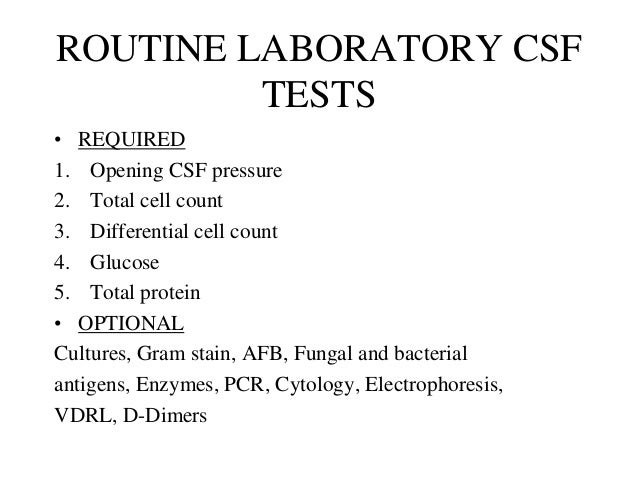
What is CSF used for?
How is it used? Cerebrospinal fluid (CSF) analysis may be used to help diagnose a wide variety of diseases and conditions affecting the brain and spinal cord (central nervous system).
What is CSF testing?
What is being tested? Cerebrospinal fluid (CSF) is a clear, watery liquid that flows around the brain and spinal cord, surrounding and protecting them. A CSF analysis is a group of tests that evaluate substances in CSF in order to diagnose conditions affecting the brain and spinal cord (central nervous system).
Why is CSF testing important?
Because CSF surrounds the brain and spinal cord, testing a sample of CSF can be very valuable in diagnosing a variety of conditions affecting the central nervous system.
Why is CSF protein small?
CSF protein – only a small amount is normally present in CSF because proteins are large molecules and do not cross the blood/brain barrier easily. Decreases in CSF protein are not generally considered significant. Increases in protein are most commonly seen with:
How many ounces of CSF are produced in a day?
About 17 ounces (500 mL) are produced each day. This rate of production means that all of the CSF is replaced every few hours. A protective blood-brain barrier separates the brain from the bloodstream and regulates the distribution of substances between the blood and the CSF.
How is CSF produced?
CSF is formed and secreted by the choroid plexus, a special tissue that has many blood vessels and that lines the small cavities or chambers (ventricles) in the brain. It is continually produced, circulated, and then absorbed into the blood. About 17 ounces (500 mL) are produced each day.
When is CSF analysis ordered?
CSF analysis may be ordered when a health practitioner suspects that a person has a condition or disease involving their central nervous system. A person's medical history may prompt the request for CSF analysis. It may be ordered when someone has suffered trauma to the brain or spinal cord, has been diagnosed with cancer that may have spread into the central nervous system, or has signs or symptoms suggestive of central nervous system involvement.
What is Xanthochromia in CSF?
Xanthochromia is the presence of bilirubin in the CSF.
What is bilirubin in CSF?
Bilirubin is the byproduct of the degradation of oxyhemoglobin by the enzyme oxygenase. There are many causes of xanthochromia such as acute intracerebral hemorrhage, brain tumors, infection, increased protein, and severe systemic jaundice. The finding of xanthochromia in the CSF is most commonly used in the diagnosis of subarachnoid hemorrhage (SAH) in the presence of a normal head computed tomography. [6]
What is the color of Xanthochromia?
Xanthochromia was originally a broad term used to describe the pigmentation of cerebrospinal fluid (CSF) as pink or yellow.[1] This color change is attributed to varying concentrations of pigmented compounds such as oxyhemoglobin, bilirubin, and methemoglobin that are typically the byproducts of red blood cell degradation.[2] The term now is more widely accepted to represent the yellow color created by the presence of bilirubin in the CSF. The presence of bilirubin resulting in yellow discoloration of the CSF is the contemporary definition of xanthochromia.[3][4][5]
What is the Creative Commons 4.0 license?
This book is distributed under the terms of the Creative Commons Attribution 4.0 International License (http://creativecommons.org/licenses/by/4.0/), which permits use, duplication, adaptation, distribution, and reproduction in any medium or format, as long as you give appropriate credit to the original author(s) and the source, a link is provided to the Creative Commons license, and any changes made are indicated.
Can a negative head CT show xanthochromia?
Standard practice in the diagnosis of subarachnoid hemorrhage with a negative head CT still includes the use of lumbar puncture to detect the presence of xanthochromia. This test is more cost effective and may reduce the need for CT angiography with contrast in many patients. [19],[21],[22] [Level IV]
How is CSF propelled?
CSF is propelled along the neuroaxis from the site of secretion to the site of absorption, mainly by the rhythmic systolic pulse wave within the choroidal arteries.
Where is CSF secreted?
CSF is secreted by the CPs located within the ventricles of the brain, with the two lateral ventricles being the primary producers. CSF flows throughout the ventricular system unidirectionally in a rostral to caudal manner.
What is CSF 2021?
Cerebrospinal fluid (CSF) is an ultrafiltrate of plasma contained within the ventricles of the brain and the subarachnoid spaces of the cranium and spine .[1] . It performs vital functions, including providing nourishment, waste removal, and protection to the brain.[2] .
What is the CSF turnover?
The reduction of CSF turnover may contribute to the accumulation of metabolites seen in aging and neurodegenerative diseases. The composition of CSF is strictly regulated, and any variation can be useful for diagnostic purposes.[1] Cerebrospinal fluid (CSF) is an ultrafiltrate of plasma contained within the ventricles of the brain and ...
What causes CSF to accumulate in the brain?
Hydrocephalus is a pathological condition in which CSF abnormally accumulates due to increased CSF production, blockage of flow, or decreased absorption. The ventricles distend to accommodate elevated CSF volumes, potentially causing damage to the brain by pressing its tissue against the boney skull. Hydrocephalus may be congenital or acquired. Blocked CSF flow throughout the ventricles is classified as non-communicating, or obstructive, hydrocephalus. The blockage is often a mass such as a tumor or an abscess located within a foramen. Because CSF secretion is constant, obstruction of flow will lead to CSF build up in front of the blockage. For example, stenosis of the cerebral aqueduct, one of the most common causes of obstructive hydrocephalus , leads to enlargement of both lateral ventricles as well as the third ventricle. If the flow of CSF becomes obstructed outside the ventricles, in either the subarachnoid space or site of absorption, it classifies as communicating, or non-obstructive, hydrocephalus.
How does CSF help the brain?
CSF assists the brain by providing protection, nourishment, and waste removal. CSF provides hydromechanical protection of the neuroaxis through two mechanisms. First, CSF acts as a shock absorber, cushioning the brain against the skull. Second, CSF allows the brain and spinal cord to become buoyant, reducing the effective weight of the brain from its normal 1,500 grams to a much lesser 50 grams. The reduction in weight lessens the force applied to the brain parenchyma and cerebral vessels during mechanical injury. Another function of CSF is to maintain homeostasis of the interstitial fluid of the brain. A stable environment for brain parenchyma is imperative for maintaining normal neuronal function.
What percent of CSF is produced by a network of modified ependymal cells?
The composition of CSF is strictly regulated, and any variation can be useful for diagnostic purposes. [1] Cellular. Seventy to eighty percent of CSF production is via a network of modified ependymal cells known as the choroid plexus (CP).[1] .
