What color is capsule after staining?
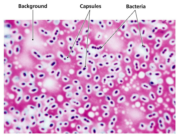
In a capsule stained microscope image, the bacterial cells will typically be stained purple, and the background of the slide should be darkly stained. Against this dark background, the capsules of the bacteria, if present, will appear as a clear halo around the cells.
What color is a positive capsule stain?
After the slide is air dried, it becomes possible to observe the stain that remained on the capsular layer. Here, one will see a dark violet color of the cell and a light violet color of the capsule.
What color is a capsule at the end of the capsule stain What color is the bacteria quizlet?
Selectively stains bacterial capsules. Capsule appears as a faint blue halo around a purple cell.
What stain is used to identify capsules?
THE CAPSULE STAIN - The Gin Stain This viscous surface layer is also known as the SLIME LAYER, the GLYCOCALYX or the EXTRACELLULAR POLYMERIC SUBSTANCE (EPS). Most bacterial capsules are composed of polysaccharide however some genera produce polypeptide capsules.
What is stained first in the capsule stain?
In this type of capsule staining procedure, the primary stain is crystal violet, and all parts of the cell take up the purple crystal violet stain. There is no mordant in the capsule staining procedure. A 20% copper sulfate solution serves a dual role as both the decolorizing agent and counterstain.
How do you identify a bacterial capsule?
Bacterial capsules are non-ionic, so neither acidic nor basic stains will adhere to their surfaces. Therefore, the best way to visualize them is to stain the background using an acidic stain and to stain the cell itself using a basic stain. We use India ink and Gram crystal violet.
What type of stain is a capsule stain quizlet?
Why is capsule stain considered differential stain? Because it's going to differentiate bacteria that has capsule and that dose not have.
What are the two things that are stained in a capsule stain quizlet?
Both Polypeptides and Polysaccharide.
What stain is added to the dried primary stain in a capsule stain quizlet?
A capsule stain was performed, using crystal violet as a primary stain, followed by a water rinse, and then nigrosin as a counterstain.
Why do capsules remain clear after staining?
A capsulated bacterium acquires a capsule around its body which appears as a clear halo. A Bacterial capsule is non-ionic in nature, so there is no or very minute possibility that the acidic or basic stains will adhere to their surfaces so it remains colorless.
Why is the capsule not stained in a capsule stain?
The capsule is a thick polysaccharide layer around the outside of the cell. It is nonionic,so the dyes that we commonly use will not bind to it. Two dyes, one acidic and one basic, are used to stain the background and the cell wall, respectively.
What do bacterial capsules look like under the microscope after staining What is stained and by which stain S Is this an example of positive or negative staining?
Usually in capsule staining, under the microscope, the bacterial cell wall appears violet in color and the capsule appears colorless against the dark background of the slide. The capsule staining uses a differential staining method (with acidic and basic dyes).
What does a positive capsule stain look like?
A positive capsule stain requires a mordant that precipitates the capsule. By counterstaining with dyes like crystal violet or methylene blue, bacterial cell wall takes up the dye. Capsules appear colorless with stained cells against dark background.
Why capsule staining is called negative staining?
We need to utilize negative staining techniques to view the outline of the capsule. Negative stains are repelled by the negatively charged bacterial cells preventing the cells from taking up the stain. Therefore, in negative staining the background (rather than the microorganism) is stained.
Which major type of stains do negative capsule stain belongs?
Capsule stain belongs to the category of differential stains, which is a staining technique used in various laboratories. The objective of such a staining process is to detect whether the capsule is present or not. As an essential differential stain, it involves two distinct stains such as acidic and basic dyes.
Which two stains are used in the capsule stain procedure?
Two methods are outlined here: Anthony's capsule stain and Maneval's capsule staining method. In Anthony's capsule stain, crystal violet is used as the primary stain, interacting with the protein material in the culture broth or added during the staining, and copper sulfate serves as the mordant.
Principle of Capsule Staining
Capsules stain very poorly with reagents used in simple staining and a capsule stain can be, depending on the method, a misnomer because the capsul...
Reagents Used For Capsule Staining
Crystal Violet (1%)Crystal Violet (85% dye content) = 1 gmDistilled Water = 100 mlNigrosinNigrosine, water soluble = 10 gmDistilled Water = 100 ml
Procedure of Capsule Staining
1. Place a small drop of a negative stain (India Ink, Congo Red, Nigrosin, or Eosin) on the slide. Congo Red is easier to see, but it does not work...
Examples of Capsule Positive and Negative
PositiveBacillus anthracis, Klebsiella pneumoniae, Streptococcus pneumonia Neisseria meningitidis Clostridium spp, Rhizaobium spp, etc.NegativeNeis...
Quality Control of Capsule Staining
Positive control: Klebsiella pneumoniae (ATCC e13883)Negative control: Alacilgenes denitrificans (ATCC 15173)
What is the purpose of capsule stain?
The main purpose of capsule stain is to distinguish capsular material from the bacterial cell. A capsule is a gelatinous outer layer secreted by bacterial cell and that surrounds and adheres to the cell wall. Most capsules are composed of polysaccharides, but some are composed of polypeptides. The capsule differs from the slime layer that most bacterial cells produce in that it is a thick, detectable, discrete layer outside the cell wall. The capsule stain employs an acidic stain and a basic stain to detect capsule production.
Why is a capsule stain misnomer?
Capsules stain very poorly with reagents used in simple staining and a capsule stain can be, depending on the method, a misnomer because the capsule may or may not be stained. Negative staining methods contrast a translucent, darker colored, background with stained cells but an unstained capsule. The background is formed with india ink ...
How does a capsule stain differ from a slime stain?
The capsule differs from the slime layer that most bacterial cells produce in that it is a thick, detectable, discrete layer outside the cell wall. The capsule stain employs an acidic stain and a basic stain to detect capsule production.
What is the best stain for a slide?
Place a small drop of a negative stain (India Ink, Congo Red, Nigrosin, or Eosin) on the slide.#N#Congo Red is easier to see, but it does not work well with some strains. India Ink generally works, but it has tiny particles that display Brownian motion that must be differentiated from your bacteria. Nigrosin may need to be kept very thin or diluted.
How long to dry crystal violet smear?
Allow to air dry (do not heat fix). Flood the smear with crystal violet stain (this will stain the cells but not the capsules) for about 1 minutes. Drain the crystal violet by tilting the slide at a 45 degree angle and let stain run off until it air dries .
Can a serum drop be used during smearing?
Capsules are fragile and can be diminished, desiccated, distorted, or destroyed by heating. A drop of serum can be used during smearing to enhance the size of the capsule and make it more easily observed with a typical compound light microscope.