One — the sixth cranial nerve — affects eye movement to the side, and the other — the seventh cranial nerve — affects facial movement. A stroke on one side of the pons will affect these nerves causing the eye and facial muscle weakness on the same side of the body as the injury.
What are the ten cranial nerves?
Cranial nerves; CN 0 – Terminal; CN I – Olfactory; CN II – Optic; CN III – Oculomotor; CN IV – ...
What cranial nerve is damaged?
The vestibulocochlear nerve, CN VIII, controls hearing and balance. Loss or a decrease of hearing may indicate nerve damage. Motor Function Nerves. Cranial nerves associated primarily with motor function are IV, VI, XI and XII. CN IV is the trochlear nerve, which controls inward and downward eye movement. Damage can cause double vision.
What is the Dirty mnemonic for cranial nerves?
- Scaphoid
- Lunate
- Triquetral
- Pisiform
- Trapezium
- Trapezoid
- Capitate
- Hamate
What are cranial nerve signs?
The symptoms of cranial nerve injuries or conditions vary depending on which nerve is affected. Contact your healthcare provider if you experience: Drooling with no known cause. Drooping in one side of your face. Facial pain or numbness. Muscle weakness or paralysis. Slurred speech. Tingling anywhere in your body. Vision loss.

What parts of the nervous system are affected by a stroke?
When a stroke occurs, nerve cells in the brain tissue become injured. As a result of this injury, nerve cells cannot communicate with other cells, and functions are impaired. If a stroke occurs on the right side of the brain, the left side of the body is affected, and vice versa.
Which cranial nerve is most commonly injured in trauma?
The most affected CN was the olfactory nerve (CN I), followed by the facial nerve (CN VII) and the oculomotor nerves (CNs III, IV, and VI). When more than 1 CN was involved, the most frequent association was between CNs VII and VIII.
Is cranial nerve palsy a stroke?
[4] Stroke and ocular motor CN palsy are both commonly associated with arteriosclerotic conditions such as diabetes mellitus, hyperlipidemia, and hypertension. [5] There have been two reports suggesting that ocular motor cranial nerve (CN) palsy may be an unrecognized risk factor of stroke.
What happens if cranial nerve 3 is damaged?
The oculomotor (third) cranial nerve plays an important role in the efferent visual system by controlling ipsilateral eye movements, pupil constriction, and upper eyelid elevation. Accordingly, damage to the third cranial nerve may cause diplopia, pupil mydriasis, and/or upper eyelid ptosis.
What happens if cranial nerve 8 is damaged?
CN VIII pathology can result from direct trauma, congenital malformations, tumor formation, infection, and vascular injury. Presenting symptoms include vertigo, nystagmus, tinnitus, and sensorineural hearing loss.
Can MRI show cranial nerve damage?
An MRI may be able help identify structural lesions that may be pressing against the nerve so the problem can be corrected before permanent nerve damage occurs. Nerve damage can usually be diagnosed based on a neurological examination and can be correlated by MRI scan findings.
How long does it take for cranial nerves to heal?
If your nerve is bruised or traumatized but is not cut, it should recover over 6-12 weeks. A nerve that is cut will grow at 1mm per day, after about a 4 week period of 'rest' following your injury. Some people notice continued improvement over many months.
Can damaged cranial nerves heal?
Treatment. If a cranial nerve is completely cut in two, it cannot be repaired. However, if it is stretched or bruised but the nerve remains intact, it can recover. This takes time and can cause a variety of unpleasant symptoms including tingling and pain.
What are the symptoms of cranial nerve damage?
Individuals with a cranial nerve disorder may suffer from symptoms that include intense pain, vertigo, hearing loss, weakness or paralysis. These disorders can also affect smell, taste, facial expression, speech, swallowing, and muscles of the neck.
What does the 4th cranial nerve control?
The fourth cranial nerve controls the actions of one of the external eye muscles, the superior oblique muscle. This muscle runs from the back of the eye socket to the top of the eye. It passes through a loop of tissue near the nose known as the trochlea. It turns the eye inward and downward.
What does the 3rd cranial nerve control?
The oculomotor nerve (the third cranial nerve; CN III) has three main motor functions: Innervation to the pupil and lens (autonomic, parasympathetic) Innervation to the upper eyelid (somatic) Innervation of the eye muscles that allow for visual tracking and gaze fixation (somatic)
How do you check cranial nerve 3?
Inability to follow and object in direction of CN III (the quickest test is to observe upward gaze which is all CN III; the eye on the affected side does not look upward) Inability to open the eyelid. CN III dysfunction causes the eyelid on the affected side to become "droopy".
What happens if cranial nerve 9 is damaged?
Glossopharyngeal nerve lesions produce difficulty swallowing; impairment of taste over the posterior one-third of the tongue and palate; impaired sensation over the posterior one-third of the tongue, palate, and pharynx; an absent gag reflex; and dysfunction of the parotid gland.
What would be the effect of damage to cranial nerve VII?
Activation of CN VII causes the entire left side of the face to move. Bell's Palsy, or a true left CN VII paralysis causes paralysis of the entire left side of the face. This includes loss of the ability to wrinkle the left forehead on the affected side.
What is cranial nerve injury?
Cranial neuropathies are caused by damage to one or more cranial nerves. These are nerves that arise directly from the brain. They affect movement and feeling in the eyes and face.
What causes damage to the vestibulocochlear nerve?
Researchers think the most likely cause is a viral infection of the inner ear, swelling around the vestibulocochlear nerve (caused by a virus), or a viral infection that has occurred somewhere else in the body.
What are the cranial nerves?
Overview of the Cranial Nerves. Twelve pairs of nerves—the cranial nerves—lead directly from the brain to various parts of the head, neck, and trunk. Some of the cranial nerves are involved in the special senses (such as seeing, hearing, and taste), and others control muscles in the face or regulate glands.
Where do cranial nerves pass?
Twelve pairs of cranial nerves emerge from the underside of the brain, pass through openings in the skull, and lead to parts of the head, neck, and trunk. A cranial nerve disorder may result when the following are damaged or malfunction: Areas of the brain that control cranial nerves (called centers, or nuclei), ...
How do you know if you have cranial nerve disorders?
Symptoms. Symptoms of cranial nerve disorders depend on which nerves are damaged and how they were damaged. Cranial nerve disorders can affect smell, taste, vision, sensation in the face, facial expression, hearing, balance, speech, swallowing, and muscles of the neck. For example, vision may be affected in various ways:
What nerve causes facial pain?
If the 8th cranial nerve (auditory or vestibulocochlear nerve) is damaged or malfunctions, people may have problems hearing and/or have vertigo —a feeling that they, their environment, or both are spinning. Cranial nerve disorders can also cause various kinds of facial or head pain.
How to test cranial nerve function?
When doctors suspect a cranial nerve disorder, they ask the person detailed questions about the symptoms. They also test the function of the cranial nerves by asking the person to do simple tasks, such as to follow a moving target with the eyes.
What nerves control the movement of the eye?
The muscles are controlled by the following cranial nerves: 3rd cranial nerve. 4th cranial nerve. 6th cranial nerve.
What nerves are damaged in the eye?
If any of the three cranial nerves that control eye movement (3rd, 4th, or 6th cranial nerve) is damaged, people cannot move their eyes normally. Symptoms include double vision when looking in certain directions.
What is a stroke in the brain?
Tables (2) Videos (1) A stroke occurs when an artery to the brain becomes blocked or ruptures, resulting in death of an area of brain tissue due to loss of its blood supply (cerebral infarction) and symptoms that occur suddenly. Most strokes are ischemic (usually due to blockage of an artery), but some are hemorrhagic (due to rupture of an artery).
Why do people have no symptoms of stroke?
When the large arteries that supply the brain are blocked, some people have no symptoms or have only a small stroke. But others with the same sort of blockage have a massive ischemic stroke. Why? Part of the explanation is collateral arteries. Collateral arteries run between other arteries, providing extra connections. These arteries include the circle of Willis and connections between the arteries that branch off from the circle. Some people are born with large collateral arteries, which can protect them from strokes. Then when one artery is blocked, blood flow continues through a collateral artery, sometimes preventing a stroke. Other people are born with small collateral arteries. Small collateral arteries may be unable to pass enough blood to the affected area, so a stroke results.
What is the term for the death of an area of brain tissue?
Ischemic Stroke An ischemic stroke is death of an area of brain tissue (cerebral infarction) resulting from an inadequate supply of blood and oxygen to the brain due to blockage of an artery. Ischemic stroke... read more
How long does it take for a stroke to resolve?
Transient ischemic attacks resemble ischemic strokes except that no permanent brain damage occurs and the symptoms typically resolve within 1 hour.
What arteries protect you from stroke?
These arteries include the circle of Willis and connections between the arteries that branch off from the circle. Some people are born with large collateral arteries, which can protect them from strokes. Then when one artery is blocked, blood flow continues through a collateral artery, sometimes preventing a stroke.
What are the risk factors for stroke?
Risk factors for stroke 1 High blood pressure 2 High cholesterol levels 3 Diabetes 4 Insulin resistance (an inadequate response to insulin), which occurs in type 2 diabetes 5 Cigarette smoking 6 Obesity , particularly if the excess weight is around the abdomen 7 Consumption of too much alcohol 8 Lack of physical activity 9 An unhealthy diet (such as one that is high in saturated fats , trans fats, and calories) 10 Depression or other mental stresses 11 Heart disorders that increase the risk of blood clots forming in the heart, breaking off, and traveling through the blood vessels as emboli (such as a heart attack or an abnormal heart rhythm called atrial fibrillation ) 12 Infective endocarditis (infection of the heart's lining and usually of the heart valves) 13 Use of cocaine or amphetamines 14 Inflammation of blood vessels ( vasculitis )
How does the body protect itself from stroke?
The body can also protect itself against strokes by growing new arteries. When blockages develop slowly and gradually (as occurs in atherosclerosis), new arteries may grow in time to keep the affected area of the brain supplied with blood and thus prevent a stroke.
How many areas of the brain are affected by stroke?
There are 9 major areas of the brain that can be affected by stroke. Each area of the brain controls different functions, and everyone’s brain is wired a bit differently.
What happens to the brain after a stroke?
During a stroke, the affected areas of the brain do not receive enough oxygen-rich blood. As a result, brain tissue begins to die. Depending on the area of the brain affected by stroke, this damage will cause changes in certain sensory, motor, or cognitive functions.
What Is a Stroke?
A stroke occurs when the supply of blood in the brain becomes compromised. This can happen by either a blood clot obstructing an artery and stopping blood flow to an area of the brain (called an ischemic stroke) or an artery in the brain bursting and leading to bleeding inside the brain (called a hemorrhagic stroke ).
What is the difference between a subcortical stroke and a cortical stroke?
For example, cortical strokes often impact higher level functioning; and it’s uncommon for subcortical strokes to result in language difficulties.
What is a stroke in the brain called?
Strokes in these regions are known as a cortical strokes. Aside from the cerebrum, there are subcortical structures that lie deep within the brain. Strokes in these areas of the brain are also known as subcortical strokes. The arteries that supply the subcortical areas of the brain are smaller and more delicate.
What are the effects of a frontal lobe stroke?
Effects of a frontal lobe stroke (a type of cortical stroke) include motor impairments, problem solving and judgement issues, behavioral changes, and difficulty with speech ( aphasia, dysarthria, or apraxia of speech), among others. Learn more about frontal lobe stroke ». 2. Parietal Lobe Stroke.
Why is stroke recovery unique?
The stroke recovery process is unique to each individual because every stroke is different. The most important thing to do is never give up hope.
What are the functions of the cranial nerves?
Their functions are usually categorized as being either sensory or motor. Sensory nerves are involved with your senses, such as smell, hearing, and touch. Motor nerves control the movement and function of muscles or glands. Keep reading to learn more about each of the 12 cranial nerves and how they function.
How many cranial nerves are there?
What are cranial nerves? Your cranial nerves are pairs of nerves that connect your brain to different parts of your head, neck, and trunk. There are 12 of them, each named for their function or structure. Each nerve also has a corresponding Roman numeral between I and XII.
What is the function of the oculomotor nerve?
The oculomotor nerve has two different motor functions: muscle function and pupil response. Muscle function. Your oculomotor nerve provides motor function to four of the six muscles around your eyes. These muscles help your eyes move and focus on objects.
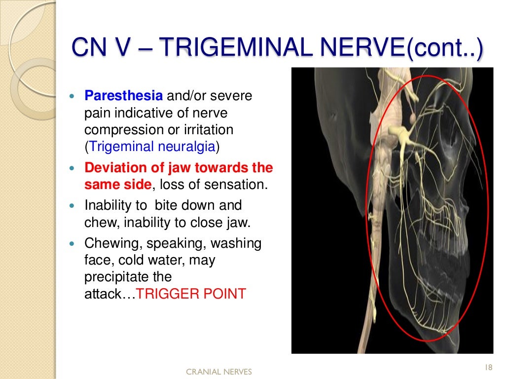
How many divisions does the trigeminal nerve have?
The trigeminal nerve has three divisions, which are:
Which nerve is located in the ophthalmic, maxillary, and mandibular divisions?
The sensory root of your trigeminal nerve branches into the ophthalmic, maxillary, and mandibular divisions. The motor root of your trigeminal nerve passes below the sensory root and is only distributed into the mandibular division. VI. Abducens nerve.
Which nerve transmits sensory information to your brain regarding smells that you encounter?
The olfactory nerve transmits sensory information to your brain regarding smells that you encounter.
Where does the trigeminal nerve originate?
It also controls the movement of muscles within your jaw and ear. The trigeminal nerve originates from a group of nuclei — which is a collection of nerve cells — in the midbrain and medulla regions of your brainstem.
How does a stroke affect the brain?
The effects of a stroke depend primarily on the location of the obstruction and the extent of brain tissue affected . The effects of a stroke depend on several factors, including the location of the obstruction and how much brain tissue is affected.
Which side of the brain is affected by a stroke?
If the stroke occurs in the right side of the brain, the left side of the body will be affected, producing some or all of the following:
What happens if you have a stroke?
Effects of Stroke. The brain is an extremely complex organ that controls various body functions. If a stroke occurs and blood flow can't reach the region that controls a particular body function, that part of the body won't work as it should. If the stroke occurs toward the back of the brain, for instance, it's likely that some disability involving ...
Why does one side of the brain affect the other side of the body?
However, because one side of the brain controls the opposite side of the body, a stroke affecting one side will result in neurological complications on the side of the body it affects. Every stroke is unique, but strokes tend to affect people in common ways.
Can a brain stem stroke leave you in a locked state?
When stroke occurs in the brain stem, depending on the severity of the injury, it can affect both sides of the body and may leave someone in a ‘locked-in ’ state. When a locked-in state occurs, the patient is generally unable to speak or achieve any movement below the neck. How a Brain Stem Stroke Impacts Recovery.
Is every stroke unique?
Every stroke is unique, but strokes tend to affect people in common ways.
Overview
A number of cranial nerves send electrical signals between your brain and different parts of your neck, head and torso. These signals help you smell, taste, hear and move your facial muscles.
Function
Your cranial nerves play a role in controlling your sensations and motor skills.
Anatomy
Two of your cranial nerve pairs originate in your cerebrum. The cerebrum is the largest portion of your brain that sits above your brainstem. These two pairs of cranial nerves include:
Conditions and Disorders
Some conditions or injuries can damage parts of the brain where cranial nerves are located. In some cases, a condition may damage only one cranial nerve. Trauma or surgery could injure or sever a nerve.
Care
You can keep your brain, cranial nerves and entire nervous system healthier with a few lifestyle changes. You can:
Why do people have no symptoms of stroke?
When the large arteries that supply the brain are blocked, some people have no symptoms or have only a small stroke. But others with the same sort of blockage have a massive ischemic stroke. Why? Part of the explanation is collateral arteries. Collateral arteries run between other arteries, providing extra connections. These arteries include the circle of Willis and connections between the arteries that branch off from the circle. Some people are born with large collateral arteries, which can protect them from strokes. Then when one artery is blocked, blood flow continues through a collateral artery, sometimes preventing a stroke. Other people are born with small collateral arteries. Small collateral arteries may be unable to pass enough blood to the affected area, so a stroke results.
How does the body protect itself from stroke?
The body can also protect itself against strokes by growing new arteries. When blockages develop slowly and gradually (as occurs in atherosclerosis), new arteries may grow in time to keep the affected area of the brain supplied with blood and thus prevent a stroke. If a stroke has already occurred, growing new arteries can help prevent a second stroke (but cannot reverse damage that has been done).
What arteries connect to the cerebral arteries?
In the skull, the vertebral arteries unite to form the basilar artery (at the back of the head). The internal carotid arteries and the basilar artery divide into several branches, including the cerebral arteries. Some branches join to form a circle of arteries (circle of Willis) that connect the vertebral and internal carotid arteries.
What causes an ischemia in the brain?
Ischemic stroke usually results when an artery to the brain is blocked, often by a blood clot and/or a fatty deposit due to atherosclerosis.
How long does it take for a brain to die from an ischemic stroke?
As a result, brain cells are deprived of blood. Most brain cells die if they are deprived of blood for 4.5 hours.
Which arteries carry blood to the brain?
Supplying the Brain With Blood. Blood is supplied to the brain through two pairs of large arteries: Internal carotid arteries , which carry blood from the heart along the front of the neck. Vertebral arteries, which carry blood from the heart along the back of the neck.
What tests are done to determine the cause of a stroke?
Other imaging tests (computed tomography and magnetic resonance imaging) and blood tests are done to identify the cause of the stroke. Treatment may include drugs to break up blood clots or to make blood less likely to clot and procedures to physically remove blood clots, followed by rehabilitation.
