Heart sounds represent reverberations of the heart chambers and exiting major arteries caused by turbulence or altered blood flow patterns within the chambers. The common causes are the reversal of flow direction and subsequent rapid closing of the cardiac valves or rapid entry of blood into the ventricles.
What are the four heart sounds?
The phonocardiogram signal (PCG) is the signal generated after conversion of the sound noises coming from the heart into an electrical signal, it groups together a set of four cardiac noises (S1, S2, S3, S4) which are in direct correlation with cardiac activity.
What sound does a normal heart make?
The healthy, normal rhythm of the heart makes a lub dub sound. The lub is the first part of the heartbeat, also known as S 1. The dub is the second half of the heartbeat, also known as S 2. The S 1 heart sound occurs when the bicuspid/mitral and tricuspid valves contract and close in order to keep the blood flowing in one direction.
What are the 4 stages of the cardiac cycle?
What are the 4 stages of the cardiac cycle?
- Atrial Systole.
- Early Ventricular Systole.
- Ventricular Systole.
- Early Ventricular Diastole.
- Late Ventricular Diastole.
What is S1 and S2 heart sounds?
The two major sounds of the normal heart sound like "lub dub". The "lub" is the first heart sound, commonly termed S1, and is caused by turbulence caused by the closure of mitral and tricuspid valves at the start of systole. The second heart sound, "dub" or S2, is caused by the closure of the aortic and pulmonic valves, marking the end of systole.

What does S1 S2 S3 and S4 heart sounds represent?
These two phases constitute the heartbeat. In a healthy adult, the heart makes two sounds, commonly described as 'lub' and 'dub. ' The third and fourth sounds may be heard in some healthy people, but can indicate impairment of the heart function. S1 and S2 are high-pitched and S3 and S4 are low-pitched sounds.
What sound is heart during each cardiac cycle?
The two distinct sounds are heard, a low, slightly prolonged “lub” (first sound) occurring at the beginning of ventricular contraction or systole and a sharper, higher-pitched “dup” (second sound), caused by the closure of aortic and pulmonary valves at the end of systole.
What does the R S1 interval represent in the cardiac cycle?
Heart Sounds The first heart sound (S1) represents closure of the atrioventricular (mitral and tricuspid) valves as the ventricular pressures exceed atrial pressures at the beginning of systole (point a).
What does the lub and dub sounds of your heartbeat represent?
When the valves between the upper chambers (atria) and lower chambers (ventricles) close, a "lub" sound is heard. When the valves in the pulmonary and aortic arteries leaving the heart close, a "dub" sound is heard followed by a longer pauseLub-DubLub-Dub.
What do S1 and S2 sounds represent?
S1 is normally a single sound because mitral and tricuspid valve closure occurs almost simultaneously. Clinically, S1 corresponds to the pulse. The second heart sound (S2) represents closure of the semilunar (aortic and pulmonary) valves (point d).
What are the significance of four heart sounds?
Types Of Heart SoundsAttributesFirst soundFourth soundSite of auscultationAudible by over the Mitral and Tricuspid sites–SignificanceIndicates ventricular systole Depicts condition of the myocardium Shows compliance of atrioventricular valvesDepicts end of ventricular filling occurring just before first heart sound4 more rows
What does T wave represent?
The T wave on the ECG (T-ECG) represents repolarization of the ventricular myocardium. Its morphology and duration are commonly used to diagnose pathology and assess risk of life-threatening ventricular arrhythmias.
Why does the second heart sound occur after the T wave?
Why does the second heart sound occur after the T wave? The T wave represents ventricular repolarization. This is followed by ventricular relaxation, during which the pressure in the ventricles falls. When the pressure in the left ventricle falls below the pressure in the aorta, the aortic valve closes.
What are the 4 phases of cardiac cycle?
(a) Left ventricular pressure–volume (P-V) loop, the segments of which correspond to events of the cardiac cycle: diastolic ventricular filling along the passive P-V curve (phase I), isovolumetric contraction (phase II), ventricular ejection (phase III), and isovolumetric relaxation (phase IV).
Is diastole a dub or lub?
The first sound LUB is produced when the atrioventricular valves i.e. tricuspid and bicuspid valves close at the start of ventricular systole. The second sound DUP is produced at the beginning of ventricular diastole when the pulmonary and aortic semilunar valves close.
Is the lub sound systolic or diastolic?
The 1st heart sound, S1 (lub), marks the beginning of systole (end of systole). Related to the closure of the mitral and tricuspid valves.
Why is the first heart sound louder than the second?
The intensity of the first sound is primarily related to the position of the AV valves at the onset of ventricular systole. The first sound is usually louder in subjects with a short PQ interval than in those with a long PQ interval.
During which phase of the cardiac cycle is the second heart sound heard?
The vibrations of the second heart sound occur at the end of ventricular contraction and identify the onset of ventricular diastole and the end of mechanical systole.
What are S3 and S4 heart sounds?
The third and fourth heart sound (S3 and S4) are two abnormal heart sound components which are proved to be indicators of heart failure during diastolic period.
What is S2 and S3 heart sound?
S3 is a low-pitched sound; this is helpful in distinguishing a S3 from a split S2, which is high pitched. A S3 heart sound should disappear when the diaphragm of the stethoscope is used and should be present while using the bell; the opposite is true for a split S2.
Which heart sound is longer lub or dub?
Solution : The first heart sound (lub) is caused by sudden closure of bicuspid and tricuspid valves and is longer than the second heart sound (dup) which is caused by closure of semilunar valves.
Q.1. What does the cardiac cycle explain?
Ans: The cardiac cycle describes the human heart's activity from the start of one heartbeat to the start of the next. It is divided into two parts:...
Q.2. What are the phases of the cardiac cycle?
Ans: The cardiac cycle is split into 7 phases, including atrial contraction, isovolumetric contraction, rapid ejection, reduced ejection, isovolume...
Q.3. What is the significance of the cardiac cycle?
Ans: The heart's primary function is to circulate blood throughout the body in a cycle known as the cardiac cycle. The cardiac cycle is the electri...
Q.4. What is the systole and diastole of the heart?
Ans: The cardiac cycle is divided into two phases: diastole and systole. They happen as the heart beats, moving blood through a system of blood ves...
Q.5. What is the difference between the information contained in a phonocardiogram and an electrocar...
Ans: ECG is produced by electrical activities of the heart, and PCG is produced by mechanical activities of the heart; the two have different crite...
How many heart sounds are there in a cardiac cycle?
Heart Sounds. Heart sounds are caused primarily from the turbulence in the blood flow created by the closure of the valves. While there are 4 heart sounds per cardiac cycle, only the 1st and 2nd heart sounds are loud enough to be auscultated.
Why does the heart make a 3rd sound?
The 3rd heart sound happens due to the blood turbulence during rapid ventricular filling, while the 4th heart sound happens due to blood turbulence during the atrial systole, both of which are not long enough to be auscultated.
How long does atrial diastole last?
Atrial systole starts after the P wave of the ECG and lasts 0.1 seconds, which is then followed by atrial diastole that lasts 0.7 seconds.
What is the S1 heart sound?
S1 HEART SOUND is a long booming sound caused by the closure of the atrioventricular valves soon after ventricular sistole begins.
What are the two major events of the cardiac cycle?
The cardiac cycle can be divided into 2 major events: systole and diastole, both of which sub-divide into smaller phases. Systole refers to the contraction of the heart muscle, whilst diastole refers to the relaxation of the heart muscle. Both are equally important for the normal functioning of the heart, as diastole allows filling ...
What increases the pressure within the chamber while relaxation lowers the pressure?
contraction increases the pressure within the chamber while relaxation lowers the pressure
Why is diastole important?
It is important to note that: blood flows from higher to lower pressure.
What is the heart sound represented by?
The heart sounds are represented by the phonocardiogram during the cardiac cycle.
What is the heart cycle?
Cardiac Cycle: The heart’s primary function is to circulate blood throughout the body in a cycle. Every day, the human heart beats around 100,000 times. The cardiovascular system involves systemic and pulmonary circulation and is responsible for the transport of various substances in human beings, and is composed of the heart, arteries, veins, and blood capillaries. The heart’s valves control blood flow, resulting in structured blood propulsion to the next chamber. The cardiac cycle is a series of heart contractions that pressurise distinct chambers of the heart, forcing blood to flow in one direction. Read on more about the cardiac cycle, meaning, duration, and phases for better understanding.
What is the process of the heart contracting?
The heart contracts (systole) during each cardiac cycle, pushing blood out and pumping it around the body; this is followed by a relaxation phase (diastole), during which the heart fills with blood—the atria contract at the same moment, pumping blood into the ventricles through the atrioventricular valves. A monosyllabic “lub” sound is produced as the atrioventricular valves close. The ventricles contract at the same moment after a slight delay, pumping blood through the semilunar valves into the aorta and the artery carrying blood to the lungs (via the pulmonary artery). The semilunar valves close, producing a monosyllabic “dup” sound.
What is the aortic pressure graph?
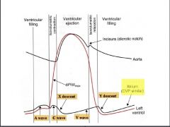
The aortic pressure graph depicts the variation in aortic pressure throughout the cardiac cycle. The graph begins with a moderate inclination, then a notch, and finally a smaller incline. Before starting afresh, the graph concludes with a slow fall.
How many waves are there in the ventricular pressure curve?
They are, nevertheless, best interpreted in combination. The ventricular pressure curve has two waves: a small wave at the beginning, followed by a return to baseline pressure, and then a much greater wave.
What is the cardiac cycle?
The cardiac cycle events are the sequence of events that occur during a heartbeat. The (Sinoatrial) SA node possesses the properties of automaticity and rhythmicity. As a result, it causes action potentials that spread throughout the atrial and ventricular muscle fibres. As a result, depolarization and repolarization occur. Following that, the heart undergoes several changes, which are repeated from beat to beat.
What is another important aspect of cardiac muscles?
Another significant aspect of cardiac muscles is their ability to contract.
How are heart sounds created?
Heart sounds are created from blood flowing through the heart chambers as the cardiac valves open and close during the cardiac cycle. Vibrations of these structures from the blood flow create audible sounds — the more turbulent the blood flow, the more vibrations that get created. The same variables determine the turbulence of blood flow as all fluids. These are fluid viscosity, density, velocity, and the diameter of the column through which the fluid is traveling. Auscultation of the heart sounds with a stethoscope is a cornerstone of physical medical exams and a valuable first-line tool to evaluate a patient. Some sounds are very characteristic of significant pathological lesions that have major pathophysiological consequences, and these first present on auscultation. These type of lesions can be heard in systole, diastole, or continuously through the cardiac cycle.
How to evaluate heart sounds?
The classic tool for evaluating heart sounds is the stethoscope . The stethoscope has been around for decades with many changes in design, but the function has always remained the same-to amplify the noise created by the heart and blood for better evaluation. The basic components are a headset with earpieces connected to a chest piece via tubing. The chest piece can act as a bell for low-frequency sounds and a diaphragm for high-frequency sounds. Most chest pieces incorporate both the bell and diaphragm, usually through a two-sided model, or a one-sided model where changing the amount of pressure applied to the chest piece allows for switching between each. The headpiece and earpieces are designed to optimize hearing by creating a seal around the ear canal to decrease ambient noise. The external ear canal travels in an anterior angle towards the tympanic membrane. The angle of the headset facilitates alignment with the external ear canal anatomy to create a complete seal. The correct size of the earpieces is also important for creating a proper seal. [11]
What are some examples of diastolic murmurs?
Examples of diastolic murmurs are aortic and pulmonic valve regurgitation (AR & PR), tricuspid and mitral valve stenosis (TS & MS), S3 sounds, and S4 sounds. Diastolic heart sounds are more clinically significant because all diastolic murmurs are pathologic, except for some S3.[18] The mechanism for sound creation is the same in AR, PR, MS, and TS as their systole counterparts. Turbulent flow from the stenosis is due to a pressure gradient the obstruction creates. The sound created in regurgitation murmurs is from regurgitant flow through the incompetent valve. The AR and PR sounds have a blowing character that occurs in early diastole and decreases in intensity as the phase progresses, resulting in a decrescendo configuration. AR has a high pitch while PR has a low to medium pitch.[8] MS occurs in mid to late diastole and begins with a loud opening snap followed by a rumble. TS has a similar sound, but it is softer and best heard in the tricuspid area. The S3 heart sound correlates to conditions of increased left atrial volume and/or increased ventricular filling pressure. The exact mechanism for the creation of the S3 has been more controversial than most of the other heart sounds. [19][20][21]It is classically taught this sound is created from blood filling a volume-overloaded ventricle., like during an acute heart failure exacerbation. Recent research suggests that mitral valve annulus diameter is one of the more important factors in creating the sound.[21] The sound can be physiologic in some children and athletes. It is a low frequency early diastolic sound best heard at the cardiac apex in the left lateral decubitus position. The S4 sound is created when someone has a less compliant ventricle. As the atria contracts in late diastole against a stiffened ventricle, it must increase its force-production, which creates turbulent blood flow. It is the hallmark of diseases that decrease ventricular compliance, like left ventricular hypertrophy. [8]
What is the S2 heart sound?
The S2 heart sound is produced with the closing of the aortic and pulmonic valves in diastole. [8][10]The aortic valve closes sooner than the pulmonic valve, and it is the louder component of S2; this occurs because the pressures in the aorta are higher than the pulmonary artery. Unlike the S1, under normal conditions, the closure sound of the aortic and pulmonic valves can be discernable, which occurs during inspiration due to the increase in venous return. The increase in volume means the right ventricle will take longer to pump out blood, which slightly delays the pressure increase in the pulmonic artery that leads to the pulmonic valve closure. So, the later sound in a physiologic split S2 is the closure of the pulmonic valve.[8] S2 can provide a lot of useful clinical information. Some have referred to it as the “auscultatory anchor point” pointing to its use as a reliably discernable sound that orients the listener to the other sounds. [11]
What is the role of the heart valves in preventing backward regurgitation?
The heart valves permit forward flow of blood while preventing backward regurgitant flow.[4] During systole, the tension provided by the chordae tendineae keep the atrioventricular valve leaflets together. The rise in pressure pushes the aortic and pulmonic valves open, allowing blood to flow forward. As the ventricle stops contracting and pressures fall in diastole, elastic recoil of the great arteries will cause blood to fall back toward the heart. The sinus-like leaflets will begin to fill with blood, which will distend the valve cusp toward one another for closure. Tension on the chordae tendineae also decreases. The atria fill with blood then contract, causing the atrioventricular valves to open so the ventricles can fill with blood. [5]
Why does S1 sound louder?
[8][9]This structural and hemodynamic change creates vibrations that are audible at the chest wall. The mitral valve closing is the louder component of S1. It also occurs sooner because of the left ventricle contracts earlier in systole. Thus, changes in the intensity of S1 are more attributable to forces acting on the mitral valve. Such causes include a change in left ventricular contractility, mitral structure, or the PR interval. However, under normal resting conditions, the mitral and tricuspid sounds occur close enough together not to be discernable. The most common reasons for a split S1 are things that delay right ventricular contraction, like a right bundle branch block. [8]
What is auscultation of heart sounds?
Auscultation of heart sounds is a foundational component in clinical physical examination. An abundant amount of on-going research has been produced on the proper technique and interpretation of heart auscultation. Heart sounds and murmurs have been described in terms of their timing in the cardiac cycle, intensity, how intensity changes during the cardiac cycle, sound wave shape, pitch, location where the sound is audible, radiation, rhythm, and response to physical exam maneuvers. These different characteristics are utilized to differentiate between physiologic and pathologic sounds.
What is the cardiac cycle?
The cardiac cycle, otherwise known as a “complete heartbeat”, involves the rhythmic muscular contractions of the heart through systole and diastole of both the atria and ventricles. Each phase of the cycle can be represented by waves on an electrocardiogram and consist of seven distinct phases: 1.
Why is it important to understand the cardiac cycle?
In order to assess the various stages of the cardiac cycle it is important to understand the process of the cardiac cycle and how to enhance the noticeable sounds of the heart that indicate a proper functioning cardiovascular system.
What is the difference between systole and diastole?
Systole consists of isovolumetric contraction and ventricular ejection (phases 2-4). Diastole consists of isovolumetric relaxation and ventricular filling (phases 5-1).
What is the P wave of an AP?
Represented by the P-wave of the electrocardiogram, it is characterized by electrical depolarization of the atria. An AP fires at the SA node leading to atrial depolarization, which initiates contraction of the muscle of the atria and pressure within the atrial chambers consequently increases. This pressure increase then causes the atrioventricular valves (mitral and tricuspid valves) to open, forcing blood into the ventricles. Approximately 70 percent of blood goes into the ventricle and the remaining 30 percent is squeezed out through atrial contraction (P-wave).
What percentage of blood goes into the ventricle?
Approximately 70 percent of blood goes into the ventricle and the remaining 30 percent is squeezed out through atrial contraction (P-wave). Following atrial contraction, the pressure of the atrial chamber begins to decline creating a pressure gradient forcing the atrioventricular valves to close.
Where is the S2 sound?
S2 – The second heart sound (dub). This is best heard at the base of the heart at the end of ventricular systole.
Is ventricular volume constant?
As there is no net movement of volume in or out of the ventricles, ventricular volume remains constant and contraction is said to be “isovolumetric”.”. This indicates individual cardiac muscle fibres are lengthening and shortening through excitation-contraction coupling.
What creates the heart sounds?
Blood flow creates vibrations in the heart chambers and valves which produce audible sounds that can be heard through a stethoscope. Smooth, low-resistance blood flow is called a laminar flow. When the flow is rough with high resistance it is known as a turbulent flow.
What is the first sound of the heart called?
The first sound S1 is generated by vibrations created by the closing of these two valves. Normally the mitral valve closes just before the tricuspid valve, and when the two different sounds are detectable, it is called a “split S1.”. A split S1 may be indicative of certain conditions affecting the heart.
How does the heart function?
The heart is a muscular organ and has four chambers that receive and pump blood :
How does a stethoscope help the heart?
Using a stethoscope to assess different sounds the heart makes is an important diagnostic tool. Heart sounds are generated by blood flowing in and out of the heart’s chambers through the valves as they open and close. Listening to the heart sounds through a stethoscope (auscultation) is one of the first steps a physician takes in evaluating ...
What is the purpose of the heart valve?
Heart valves ensure that the flow of the blood is in only one direction, by opening and closing as the heart pumps blood. The four heart valves are. Tricuspid valve separating right atrium and right ventricle.
When the aortic valve closes just before the pulmonic valve, it may generate a split?
When the aortic valve closes just before the pulmonic valve, it may generate a split S2. This may indicate impairment in the heart function.
Why does my heart beat irregularly?
AFib symptoms like heart racing, fluttering, and irregular heart beat may be caused by heart disease, obesity, alcohol use, thyroid disease, and other conditions. AFib medications may include blood thinners, drugs to control heart rate or convert the heart to a normal rhythm. AFib surgery is also a treatment possibility.
What are the two phases of the heart?
All these events are “organized” in two phases: diastole (when the heart fills with blood) and systole (when the heart pumps the blood) During these two phases, many different events are observed and we will describe them in the following paragraphs.
What are the two phases of the cardiac cycle?
This stimulus causes a series of events in the atria and the ventricles. All these events are “organized” in two phases: diastole (when the heart fills with blood)
What is the atrial pressure wave?
The atrial pressure wave shows the change in the atrial pressure during systole and diastole. There are three significant pressure changes represented by the letters a, v, and c. The pressure change generated as the atria fill with blood is represented by the ‘v’ wave towards the end of the atrial pressure wave. There is a slight decline in the atrial pressure that corresponds with the opening of the atrioventricular valve. This is followed by the ‘a’ wave which represents the contraction of the atria. The ‘a’ wave is followed by a downward slope as the atrioventricular valves close. This is followed by another increase labeled as the ‘c’ wave. This represents bulging of the atrioventricular valves into the atria during ventricular contraction.
What causes the aortic valve to open?
The increase in ventricular pressure during systole causes the aortic valve to open. The pressure generated in the ventricle is then transmitted to the aorta. The walls of the aorta are able to dilate due to their high elasticity in order to accommodate the sudden, dramatic increase in pressure. These pressure changes are represented by the first and largest wave on the aortic pressure graph .
What is the period of contraction of the ventricles?
Ventricular systole refers to the period of contraction of the ventricles. The electrical impulse arrives at the atrioventricular node (AV node) shortly after the atria are depolarized. There is a small delay at the AV node, which allows the atria to complete contracting before the ventricles are depolarized. The action potential passes to the AV node, down the bundle of His, and subsequently to the left and right bundle branches (conductive fibers that travel through the interventricular septum and branches to supply the ventricles). These fibers carry the electrical impulses through their respective ventricular territories, leading to ventricular contraction.
What is the alternating state of the atria and ventricles?
Both the atria and the ventricles undergo alternating states of systole and diastole. In other words, when the atria are in diastole, the ventricles are in systole and vice versa. This article will discuss the phases of the cardiac cycle and the underlying physiological principles that govern the process.
How does ventricular pressure change?
The first increase in the ventricular pressure occurs as the atria contract to pump residual blood into the ventricle. This increase doesn’t last for a long time and the ventricular pressure soon returns to baseline. At this time more blood is being pumped into the ventricle, bringing it to its end-diastolic or preload volume. At the beginning of systole, the atrioventricular valves are closed and the ventricle is in isovolumetric contraction. So there is a sharp increase in pressure but the volume remains the same. Once the ventricular pressure overcomes the aortic pressure, the aortic valves open and there is a sudden fall in ventricular volume. As the volume decreases, the ventricular pressure begins to fall as well. Eventually, the ventricle stops contracting, re-enters the diastolic phase, and begins isovolumetric relaxation.