
What does the accessory nerve innervate?
The accessory nerve (Fig. 1), named after Thomas Willis (Willis, 1965) cranial nerve XI, innervates the sternocleidomastoid (SCM) and trapezius muscles.
What happens when accessory nerve is damaged?
The spinal accessory nerve originates in the brain and enables motion in the trapezius and sternomastoid muscles in the neck. A spinal accessory nerve injury can be caused by trauma or damage during surgery, resulting in shoulder pain, "winging" of the shoulder blades and weakness of the trapezius muscle.
What muscles are controlled by the accessory nerve?
The spinal component of the accessory nerve provides motor control of the sternocleidomastoid and trapezius muscles. The trapezius muscle controls the action of shrugging the shoulders, and the sternocleidomastoid the action of turning the head.
What causes damage to spinal accessory nerve?
The spinal accessory nerve may get damaged due to neck trauma, wrenching injury to arm or neck, or even after surgical procedures such as lymph node biopsy, parotid surgery, carotid surgery and jugular vein cannulation.
Where is the spinal accessory nerve particularly at risk of damage?
Cranial nerve XI, the spinal accessory nerve (SAN), is vulnerable to injury, owing to its long and superficial course in the posterior cervical neck. An important landmark in the neck, the SAN is considered to contribute most motor innervation to the trapezius muscle.
How do you test the accessory nerve?
0:395:09Accessory nerve examination | Eleventh cranial nerve - YouTubeYouTubeStart of suggested clipEnd of suggested clipYou apply resistance with the palm of your hand observe the contraction of the contralateralMoreYou apply resistance with the palm of your hand observe the contraction of the contralateral sternocleidomastoid.
Where is the accessory nerve?
the spinal cordAfter the rootlets fuse, the spinal accessory nerve travels cranially behind the dentate ligaments of the spinal cord. They travel parallel to the spinal cord and enter the cranial vault via the foramen magnum.
What would happen if there were nerve damage to the sternocleidomastoid?
Interruption of the nerve supply to the sternocleidomastoid muscle results in an asymmetric neckline, while weakness of the trapezius muscle can produce a drooping shoulder, winged scapula, and a weakness of forward elevation of the shoulder.
What happens when the axillary nerve is damaged?
Axillary nerve dysfunction is nerve damage that can lead to a loss of movement or sensation in the shoulder. Conditions associated with axillary nerve dysfunction include fracture of the humerus (upper arm bone), pressure from casts or splints, and improper use of crutches.
What happens if cranial nerve XI is damaged?
Supranuclear lesions of the eleventh nerve cause moderate, often transient, impairment of function of the sternocleidomastoid and trapezius muscles, due to the bilateral innervation. In the spinal cord the nuclei can be involved in amyotrophic lateral sclerosis, syringomyelia, polio, and intraspinal tumors.
What nerve causes shoulder drop?
Axillary nerve dysfunction (AND) is a condition marked by a loss of movement or sensation in the shoulder area. It's also known as neuropathy of the axillary nerve. Excessive stress or damage to the axillary nerve, which serves the deltoid muscles and skin of the shoulder, causes AND.
What is the function of accessory nerve?
Function. Associated Conditions. Rehabilitation. The accessory nerve provides motor function (movement) to two muscles essential to neck and shoulder movement, the sternocleidomastoid (SCM) and the trapezius, as well as to the larynx (voice box) and other structures in the throat.
Where is the accessory nerve located?
The spinal component of the accessory nerve is made up of roots from the sixth and seventh cervical vertebrae, which are in your neck. Once the nerve is formed, it runs up to enter the cranial cavity through an opening called the foramen magnum, which is a large opening near the back of the skull.
What nerve is involved in innervation of the SCM?
A possible relationship inside the skull with the facial nerve (CN VII), causing the involvement of CN VII in the innervation of the SCM
What are the symptoms of accessory nerve damage?
Diseases that impair nerve function in general. Lesions left behind from surgery in the region. Symptoms of damage to the accessory nerve include: Weakness, wasting, and loss of function in the muscles it innervates.
How do nerve roots connect to the body?
Fibers from multiple nerve roots can combine to form a single nerve. From their roots, they travel outward to the structures they supply nerve function to, which is called innervation. Most nerves send out multiple branches along the way, which innervate muscles, skin, and other tissues throughout the body.
Which part of the accessory nerve is the cranial component?
The cranial component then joins the vagus nerve and follows it along its course in the throat. It’s often referred to as the internal branch of the accessory nerve. When it sends out branches, it does so via the vagus nerve, so it’s considered a part of that nerve, as well.
What nerves are replaced in quadriplegia?
Replace the phrenic nerve in people with quadriplegia (paralysis of all four limbs).
Where is accessory nerve palsy located?
Accessory nerve palsy is one complication that most often occurs after surgery has been performed on the neck’s posterior triangle, a triangle-shaped area that lies between the sternocleidomastoid and trapezius on each side of the neck . Additionally, there are three types of accessory nerve schwannoma tumors that occur in some people: intracisternal, spinal canal, and intrajugular; they can be removed with surgery from beneath the base of the skull. Schwannoma tumors are tumors that grow in the tissue that covers nerves, the nerve sheath.
Which part of the spinal accessory nerve is coiled?
It is coiled in appearance. It is divided into spinal and cranial divisions, but its cranial part is often disregarded. The spinal accessory nerve provides motor function to the sternocleidomastoid muscle, which extends the neck and the trapezius, as well as the upper back and shoulder.
What nerve controls the movement of certain neck muscles?
Accessory nerve. The accessory nerve is a cranial nerve that controls the movement of certain neck muscles. It is coiled in appearance. It is divided into spinal and cranial divisions, but its cranial part is often disregarded.
What is accessory nerve?
t. e. The accessory nerve is a cranial nerve that supplies the sternocleidomastoid and trapezius muscles. It is considered as the eleventh of twelve pairs of cranial nerves, or simply crani al nerve XI, as part of it was formerly believed to originate in the brain. The sternocleidomastoid muscle tilts and rotates the head, ...
What type of information does the spinal accessory nerve carry?
As the trapezius and sternocleidomastoid muscles are derived from the pharyngeal arches, some investigators believe the spinal accessory nerve that innervates them must carry specific special visceral efferent (SVE) information . This is in line with the observation that the spinal accessory nucleus appears to be continuous with the nucleus ambiguus of the medulla. Others consider the spinal accessory nerve to carry general somatic efferent (GSE) information . Still others believe it is reasonable to conclude that the spinal accessory nerve contains both SVE and GSE components.
What nerves are left behind after leaving the skull?
After leaving the skull, the cranial component detaches from the spinal component. The spinal accessory nerve continues alone and heads backwards and downwards. In the neck, the accessory nerve crosses the internal jugular vein around the level of the posterior belly of digastric muscle.
What nerve is responsible for winging the scapula?
Injury can cause wasting of the shoulder muscles, winging of the scapula, and weakness of shoulder abduction and external rotation. The accessory nerve is derived from the basal plate of the embryonic spinal segments C1–C6.
How is accessory nerve tested?
The accessory nerve is tested by evaluating the function of the trapezius and sternocleidomastoid muscles. The trapezius muscle is tested by asking the patient to shrug their shoulders with and without resistance. The sternocleidomastoid muscle is tested by asking the patient to turn their head to the left or right against resistance.
Where does the accessory nerve go in the neck?
In the neck, the accessory nerve crosses the internal jugular vein around the level of the posterior belly of digastric muscle , in front of the vein in about 80% of people, and behind it in about 20%, and in one reported case, piercing the vein.
Which nerve connects to the vagus nerve?
Traditionally, the accessory nerve is described as having a small cranial component that descends from the medulla and briefly connects with the spinal accessory component before branching off of the nerve to join the vagus nerve.
What are the parts of the accessory nerve?
Traditionally, the accessory nerve is divided into spinal and cranial parts.
How to examine accessory nerve?
The accessory nerve is examined by asking the patient to rotate their head and shrug their shoulders, both normally and against resistance. Simply observing the patient may also reveal signs of muscle wasting in the sternocleidomastoid and trapezius in cases of long-standing nerve damage.
What nerve is the cranial part of?
Immediately after leaving the skull, cranial part combines with the vagus nerve (CN X) at the inferior ganglion of vagus nerve (a ganglion is a collection of nerve cell bodies). The fibres from the cranial part are then distributed through the vagus nerve. For this reason, the cranial part of the accessory nerve is considered as part of the vagus nerve.
What causes accessory nerve damage?
The most common cause of accessory nerve damage is iatrogenic. Procedures such as cervical lymph node excision biopsy or central line insertion can cause trauma to the nerve.
Which muscle innervates the spinal cord?
Outside the cranium, the spinal part descends along the internal carotid artery to reach the sternocleidomastoid muscle, which it innervates. It then moves across the posterior triangle of the neck to supply motor fibres to the trapezius.
Which part of the spinal cord innervates the sternocleidomastoid and trapezius?
The fibres from the cranial part are then distributed through the vagus nerve. For this reason, the cranial part of the accessory nerve is considered as part of the vagus nerve. The spinal accessory nerve innervates two muscles - the sternocleidomastoid and trapezius.
Where does the accessory nerve exit?
The nerve traverses the posterior cranial fossa to reach the jugular foramen. It briefly meets the cranial portion of the accessory nerve, before exiting the skull (along with the glossopharyngeal and vagus nerves).
Spinal Accessory Nerve
A nerve is a bundle of fibers that transmits signals to the brain, spinal cord, muscles, and organs. There are 12 pairs of cranial nerves, or nerves that emerge from the brain and extend into the body. The nerves are paired because they serve the right and left sides of the body.
Accessory Nerve Anatomy
There are two types of nerves. Sensory nerves provide sensation, and motor nerves provide movement. The spinal accessory nerve is considered a motor nerve. The accessory nerve is unique because it has two compartments that originate in different parts of the body. One portion originates in the brain, and the other portion starts in the spine.
Accessory Nerve Innervation
Innervate means to supply an organ or body part with nerves. With the accessory nerve innervation, the spinal portion of the accessory nerve innervates two important muscles: the sternocleidomastoid muscle and the trapezius muscle.
Spinal Accessory Nerve Test
A spinal accessory nerve test can be performed to assess damage to the nerve. Because the spinal accessory nerve innervates the sternocleidomastoid and trapezius muscles, the doctor will examine both muscles.
What nerve crosses the posterior triangle of the neck?
Isolated lesions of the spinal accessory nerve are rare. Surgical injury is one cause. The eleventh nerve crosses the posterior triangle of the neck lying on the levator scapulae, and it is quite vulnerable to surgical procedures in that area, such as biopsy or exploration.
What are the parts of the eleventh nerve?
The eleventh nerve has two parts. The smaller cranial part arises from cells in the nucleus ambiguus and ultimately is distributed with the vagus nerve. This portion innervates the pharyngeal muscles. The main part, the spinal portion, arises from a long column of nuclei situated in the ventral part of the medulla and extending to the fifth cervical segment or lower. Supranuclear innervation is not well known. It has been characterized by authors as being ipsilateral, contralateral, or bilateral. It begins in the precentral gyri and descends in the corticobulbar tract. As the fibers leave the cord they join together and ascend through the foramen magnum, then leave through the jugular foramen with the vagus nerve. The nerve descends in the neck near the jugular vein and supplies the sternocleidomastoid and trapezius muscles, joined by motor or sensory contributions from the upper cervical nerves. Some recent insights into the supranuclear contributions are discussed in the Clinical Significance section below.
What is the clinical significance of a supranuclear lesions of the eleventh nerve?
Supranuclear lesions of the eleventh nerve cause moderate, often transient, impairment of function of the sternocleidomastoid and trapezius muscles, due to the bilateral innervation.
Which neurons innervate the trapezius?
Motor neurons at C1–2 innervate the sternocleidomastoid, while neurons at C3–4 innervate the trapezius. This innervational pattern can account for isolated weakness of one muscle or the other. The supranuclear innervation to motor neurons for each of the muscles appears to take different courses.
Where do the XI and XI nerves travel?
Nerves IX, X, and XI travel together in the jugular foramen. They may be compressed by tumors and aneurysms (Vernet's syndrome). The XII nerve may also be involved in more extensive lesions occurring in the posterior later-ocondylar space (syndrome of Collet-Sicard); causes include parotid tumors, carotid body tumors, adenopathy of whatever cause, and tuberculosis involving the lymph nodes. Sarcoidosis is another cause. A similar set of etiologies can damage the same four nerves (IX, X, XI, XII) in the posterior retroparotid space (Villaret's syndrome).
Where is the spinal innervation located?
The main part, the spinal portion, arises from a long column of nuclei situated in the ventral part of the medulla and extending to the fifth cervical segment or lower. Supranuclear innervation is not well known. It has been characterized by authors as being ipsilateral, contralateral, or bilateral.
What are the functions of the trapezius?
This nerve supplies the sternocleidomastoid and trapezius muscles,#N#which have the following functions: 1 Rotation#N#of head away from the side of the contracting sternocleidomastoid muscle. 2 Tilting of the head toward the contracting sternocleidomastoid#N#muscle. 3 Flexion of the neck by both sternocleidomastoid#N#muscles. 4 Elevation of the shoulder by the#N#trapezius. 5 Drawing the head back so the face is upward by#N#the trapezius muscles.
Which nerve is responsible for vision?
The optic nerve is the sensory nerve that involves vision.
What are the functions of the cranial nerves?
Their functions are usually categorized as being either sensory or motor. Sensory nerves are involved with your senses, such as smell, hearing, and touch. Motor nerves control the movement and function of muscles or glands. Keep reading to learn more about each of the 12 cranial nerves and how they function.
What is the function of the oculomotor nerve?
The oculomotor nerve has two different motor functions: muscle function and pupil response. Muscle function. Your oculomotor nerve provides motor function to four of the six muscles around your eyes. These muscles help your eyes move and focus on objects.
How many cranial nerves are there?
What are cranial nerves? Your cranial nerves are pairs of nerves that connect your brain to different parts of your head, neck, and trunk. There are 12 of them, each named for their function or structure. Each nerve also has a corresponding Roman numeral between I and XII.
Which nerve is located in the ophthalmic, maxillary, and mandibular divisions?
The sensory root of your trigeminal nerve branches into the ophthalmic, maxillary, and mandibular divisions. The motor root of your trigeminal nerve passes below the sensory root and is only distributed into the mandibular division. VI. Abducens nerve.
Which nerve transmits sensory information to your brain regarding smells that you encounter?
The olfactory nerve transmits sensory information to your brain regarding smells that you encounter.
What nerves are involved in the sense of taste?
Facial nerve. The facial nerve provides both sensory and motor functions, including: moving muscles used for facial expressions as well as some muscles in your jaw. providing a sense of taste for most of your tongue. supplying glands in your head or neck area, such as salivary glands and tear-producing glands.
Overview
Structure
Function
- Treatment and management of problems with the accessory nerve are based on what’s causing the dysfunction. For direct damage to the nerve itself, treatment may involve physical therapy (electrostimulation and strength exercises), an osteopathic approach to improve the movement of tissues impaired due to scarring, or nerve transfer.6
Clinical significance

History
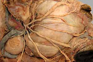
Additional images
External links
The accessory nerve, also known as the eleventh cranial nerve, cranial nerve XI, or simply CN XI, is a cranial nerve that supplies the sternocleidomastoid and trapezius muscles. It is classified as the eleventh of twelve pairs of cranial nerves because part of it was formerly believed to originate in the brain. The sternocleidomastoid muscle tilts and rotates the head, whereas the trapezius muscle, connecting to the scapula, acts to shrug the shoulder.