What is a full mouth X-ray?
Full mouth x-rays provide a series of pictures of the upper and lower jaws in a single x-ray. This type of x-ray is used to detect oral health issues that require treatment that your dentist cannot see with their eyes. Your dentist can see the position of fully emerged teeth and teeth just below the surface.
What can show up on a dental Xray?
Bones and teeth Fractures and infections. In most cases, fractures and infections in bones and teeth show up clearly on X-rays. Arthritis. X-rays of your joints can reveal signs of arthritis. Dental decay. Dentists use X-rays to take pictures of the teeth and jaw and check for cavities. Osteoporosis.
What is the best way to see the inside of mouth?
Panoramic x-rays are the best way to see the entire inside of the mouth. Full mouth x-rays provide a series of pictures of the upper and lower jaws in a single x-ray. This type of x-ray is used to detect oral health issues that require treatment that your dentist cannot see with their eyes.
What does an X-ray show you?
Overview. An X-ray is a quick, painless test that produces images of the structures inside your body — particularly your bones. X-ray beams pass through your body, and they are absorbed in different amounts depending on the density of the material they pass through. Dense materials, such as bone and metal, show up as white on X-rays.
What is an open mouth xray?
The open mouth odontoid radiograph (x-ray) is used to assess for the presence of an upper cervical spine injury. Common injuries to the upper cervical spine include: Dens Fracture (i.e., C2 Odontoid Fracture) Jefferson's Fracture (i.e., C1 Burst Fracture)
What can an xray of your throat show?
The x-ray is used to evaluate neck injuries and numbness, pain, or weakness that does not go away. A neck x-ray can also be used to help see if air passages are blocked by swelling in the neck or something stuck in the airway. Other tests, such as MRI, may be used to look for disk or nerve problems.
What is the central ray for AP open mouth projection?
Open Mouth - Cervical X-ray X-ray examination of cervical spine projecting C1 and C2 with the patient in open mouth.
How do you get a good Odontoid X-ray?
0:362:42Odontoid Positioning tutorial - YouTubeYouTubeStart of suggested clipEnd of suggested clipThe bottom of the top teeth. Front teeth with the mastoid tips. If you can do that draw that lineMoreThe bottom of the top teeth. Front teeth with the mastoid tips. If you can do that draw that line straight through perpendicular to the IR.
Why is it called a hangman's fracture?
Schneider introduced the term "hangman's fracture" in 1965, and reported on the biomechanics and other similarities of the cervical fractures seen following judicial hangings and those caused by motor vehicle accidents.
What is a C1 fracture?
A Jefferson fracture is another name for a bone fracture of the front and back arches of the C1 vertebra. The C1 vertebra is the top one, closest to your skull. C1 fractures represent about 2 percent of all vertebral fractures, according to a 2013 review.
What is the Odontoid view?
The odontoid or 'peg' projection, also known as the open mouth AP projection (or radiograph), is an AP projection of C1 (atlas) and C2 (axis) with the patient's mouth open.
What is Uncovertebral arthropathy?
Uncovertebral arthrosis is thought to be the result of dehydration/reduction of the intervertebral disc, leading to an increased load between the cervical vertebrae and hence the uncovertebral joints. It typically is seen in the lower cervical vertebrae due to the increased load at these levels.
How do you take waters view?
It is commonly used to get a better view of the maxillary sinuses. An x-ray beam is angled at 45° to the orbitomeatal line. The rays pass from behind the head and are perpendicular to the radiographic plate....Waters' viewMethod of obtaining Waters' viewSpecialtyRadiology1 more row
Is the Odontoid C1?
The odontoid process is the pivot for the C1 vertebra, which carries the cranium (head).
What is a CT scan of the throat?
A neck CT scan uses a special X-ray machine to make images of the soft tissues and organs of the neck, including the muscles, throat, tonsils, adenoids, airways, thyroid, and other glands. The blood vessels and upper spinal cord are also seen. A person getting a CT scan lies on a table.
What is soft tissue in throat?
The soft palate, also known as the velum, is the soft tissue at the back of the throat that closes the nasal passages during swallowing.
What is dental X-ray?
You might be familiar with dental X-rays wherein the dentist places a piece of plastic inside your mouth for you to bit down on and takes multiples images that show one or a couple of your teeth. This is just one type of X-ray. There are many others, one of them being a panoramic dental X-ray. This type of X-ray captures your entire mouth in ...
How does a dental X-ray work?
Instead of relying on film placed inside your mouth, the X-ray technician will direct the X-ray machine to project a beam through your mouth onto a film that’s rotating opposite the X-ray tube.
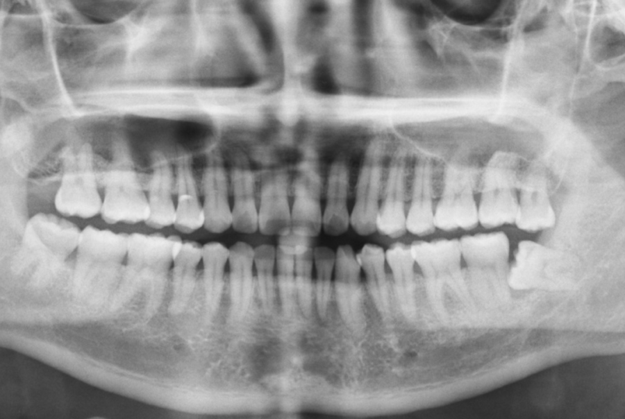
Why Use a Panoramic X-Ray?
A panoramic dental X-ray creates an image of your entire mouth, including the upper and lower jaws, all the teeth, temporomandibular (TMJ) joints, and even your nasal area and sinuses. This makes it possible for your dentist to notice any of the following problems:
Why do dentists take panoramic X-rays?
A dental professional may also ask you to take a panoramic dental X-ray to plan treatments like braces, implants, or dentures. Another advantage of this type of X-ray is that it creates a flat representation of your jaw’s curved structure, making it easy for your dentist to analyze every part.
How long does an X-ray last?
The X-ray lasts between 12-20 seconds, so it’s quite easy to perform. In preparation for the procedure, the dental technician will ask you ...
Can you have a panoramic X-ray if you have braces?
If you or your child need braces, or if you have an impacted tooth, a panoramic X-ray can be invaluable. Now that you’re armed with all that you need to know about this type of X-ray, you’re well-equipped to face any dental problem you might have and go back to smiling again!
Is it safe to take an intraoral X-ray?
While a panoramic dental X-ray is still perfectly safe, don’t hesitate to talk to your dentist if you have any concerns about taking one.
Why do dentists take x-rays in Dayton?
Taking x-rays allows your dentist in Dayton to see any issues that may not yet have signs or symptoms, making them easier to treat before they lead to pain or more complicated treatments.
What is a bitewing x-ray?
Bitewing x-rays are the ones that can show cavities or areas of decay that may not yet be visible, or are lurking in hard-to-see areas such as in between teeth or fillings. These x-rays may also show bone density health and any deterioration caused by gum disease.
What is the best way to see your wisdom teeth?
Panoramic X-rays. Panoramic x-rays are used for the most complete view of your entire mouth. The images don’t only show all your teeth, but also your sinuses, jaw joints, and jaw bones. These photos can help determine if wisdom teeth are impacted and can even help diagnose a tumor.
What is periapical x-ray?
You probably have had them taken and may not have even known it. These x-rays show images of the entire tooth, including the roots. The images allow your dentist to look at each tooth individually to make sure each one is structurally sound and bone levels are healthy and strong. Periapical x-rays can also help catch cysts and abscesses.
Why do dentists use X-rays?
These X-rays are used with low levels of radiation to capture images of the interior of your teeth and gums. This can help your dentist to identify problems, like cavities, tooth decay, and impacted teeth. Dental X-rays may seem complex, but they’re actually very common tools that are just as important as your teeth cleanings.
What happens after dental xrays?
After dental X-rays. When the images are ready — instantly in the case of digital X-rays — your dentist will review them and check for abnormalities. If a dental hygienist is cleaning your teeth, the dentist may go over the results of the X-rays with you after your cleaning is done. The exception is if the hygienist discovers any significant ...
What is the most common type of dental X-ray?
There are several types of dental X-rays, which record slightly different views of your mouth. The most common are intraoral X-rays, such as: Bitewing. This technique involves biting down on a special piece of paper so that your dentist can see how well the crowns of your teeth match up.
Why do you need X-rays for dental?
If you’re a new patient, you’ll probably undergo dental X-rays so that your new dentist can get a clear picture of your dental health. This is especially important if you don’t have any X-rays from your previous dentist.
When to use extraoral x-rays?
Extraoral X-rays may be used when your dentist suspects there might be problems in areas outside of the gums and teeth, such as the jaw. A dental hygienist will guide you through each step of the X-ray process. They might step outside of the room briefly while the images are being taken.
Why do children need X-rays?
Children may need to have dental X-rays more often than adults because their dentists might need to monitor the growth of their adult teeth. This is important because it can help the dentist determine if baby teeth need to be pulled to prevent complications, such as adult teeth growing in behind baby teeth.
What is the technique that captures all of your teeth in one shot?
Occlusal. This technique captures all of your teeth in one shot.
What is the odontoid view?
Cervical spine (odontoid view) The odontoid or 'peg' projection, also known as the open mouth AP projection (or radiograph ), is an AP projection of C1 (atlas) and C2 (axis) with the patient's mouth open.
Which ray should be angled cephalic?
If teeth are superimposed over the upper aspect of the dens, the head needs to be hyperextended or in the case of trauma, the central ray should be angled cephalic.
Why should the patient's shoulders be at equal distances from the image receptor?
patient’s shoulders should be at equal distances from the image receptor to avoid rotation, the head facing straight forward. at the last instant, the patient is instructed to open their mouth as wide as possible.
Who interprets X-ray results?
A radiologist typically views and interprets the results and sends a report to your doctor, who then explains the results to you. In an emergency, your X-ray results can be made available to your doctor in minutes. By Mayo Clinic Staff.
What is the purpose of X-rays?
An X-ray is a quick, painless test that produces images of the structures inside your body — particularly your bones . X-ray beams pass through your body, and they are absorbed in different amounts depending on the density of the material they pass through. Dense materials, such as bone and metal, show up as white on X-rays.
What can be measured with X-rays?
Osteoporosis. Special types of X-ray tests can measure your bone density.
How long does it take to get an X-ray?
An X-ray procedure may take just a few minutes for a simple X-ray or longer for more-involved procedures, such as those using a contrast medium.
What is the contrast medium on X-rays?
The air in your lungs shows up as black. Fat and muscle appear as shades of gray. For some types of X-ray tests, a contrast medium — such as iodine or barium — is introduced into your body to provide greater detail on the images.
How do X-ray techs work?
A technologist positions your body to obtain the necessary views. He or she may use pillows or sandbags to help you hold the position. During the X-ray exposure, you remain still and sometimes hold your breath to avoid moving so that the image doesn't blur.
Why are X-rays not safe?
Some people worry that X-rays aren't safe because radiation exposure can cause cell mutations that may lead to cancer. The amount of radiation you're exposed to during an X-ray depends on the tissue or organ being examined. Sensitivity to the radiation depends on your age, with children being more sensitive than adults.
What does an X-ray show?
If someone has lung cancer, the X-ray will show a visible mass or nodule that will appear as a white spot on the lungs. If an X-ray detects a tumor, the image will then be closely studied to reveal whether it’s malignant or benign.
What Can You Tell from an X-ray?
From fractures and infection to breast cancer, X-ray technology can bring light to many different health issues affecting various parts of the body.
How Do You Prepare for an X-ray?
Before your X-ray, it is a good idea to ask your doctor or nurse if there are any specific instructions. This depends on the type of X-ray you need.
How Long Does It Take to Get Results from an X-ray?
Most X-rays are saved in a digital form so they can be viewed on-screen within minutes. The radiologist will take a look at the results and interpret what’s going on before sending the report to your doctor.
What does it mean when a material is white on an X-ray?
If a material is dense, like bone or metal, it shows up as white on an X-ray. The air inside your lungs appears black on an X-ray, while fat and muscle show up as shades of gray. In certain situations, when greater detail of the image is needed, a contrast medium like iodine or barium is used. We’re sharing everything you need to know in case you ...
How long does a bone X-ray last?
The procedure could last anywhere from a few minutes for a bone X-ray to more than an hour if your procedure is more involved. If your child is getting an X-ray, you may be able to stay with him or her during the procedure as long as you agree to wear a lead apron to protect you from any unnecessary radiation.
What is the name of the test that shows the presence of cancer in the breast?
Breast cancer: X-rays of the breast, called mammograms, examine breast tissue to reveal any signs of cancer that may exist.
Why is it important to read cervical radiographs?
A more systematic approach to reading cervical radiographs can significantly reduce the chances of missing an important injury.
How to visualize facet joints?
A – Alignment and adequacy: First, visualize the spine from the base of the skull to the C7-Th1 junction. Next, check if the x-ray is a real lateral view, or if it is slightly rotated. Facet joints are best visualized when we have a proper lateral projection. (see Figure 3).
What is C Spine X-ray interpretation?
Although current guidelines lead us to use CT scan for a suspected c-spine injury, c-spine x-rays are still valuable in some low resource settings and patient groups who are susceptible to radiation. Therefore, this chapter will summarize the basics of c-spine x-ray interpretation.
Can a C2 x-ray be done with the mouth open?
The main goal is to picture the odontoid process of the C2 and the C1. It can be done with the mouth either open or closed. Two things are assessed when inspecting the odontoid x-ray: the distance between the odontoid process and the lateral masses of the C1 should be equal.