How to interpret corneal topography?
May 18, 2021 · A topography scan measures the distortion and its effect on vision. Growths. The size of pterygia or other growths can be monitored with topography. Astigmatism and keratoconus. Topography can help find astigmatism and early cases of keratoconus and track their progression. Contact lens fitting. Topography scans help find what type of contact lens …
What type of topography should Carlos choose?
Nov 15, 2021 · Corneal topography is a computer assisted diagnostic tool that creates a three-dimensional map of the surface curvature of the cornea. These details are used to diagnose, monitor, and treat various eye conditions. They are also used in fitting contact lenses and for planning surgery, including laser vision correction.
How to perfom corneal topography?
Apr 27, 2020 · What does corneal topography measure? Corneal topography is a procedure used to monitor and measure changes that may occur to the shape and integrity of the cornea of your eye. A corneal topographer projects a series of illuminated rings, referred to as a Placido disc, onto the surface of the cornea, which are reflected back into the instrument.
What are the differences between corneal opacity and cataract?
Jan 21, 2021 · Topography measures the curvature of the surface of the eye and creates a colored “map” of the cornea. Can keratoconus be treated? Mild to moderate keratoconus can be treated with eyeglasses or contact lenses. This will likely be a long-term treatment, especially if your cornea becomes stable with time or from cross-linking.
How do you interpret corneal topography results?
Warmer colors represent steeper corneal curvature while cooler colors represent flatter areas. For the elevation maps (anterior and posterior float), warmer colors denote where the cornea is elevated above the best fit sphere and cooler colors denote where the cornea is depressed below the best fit sphere.Oct 19, 2016
What is normal corneal topography?
Central Keratometry (K Central). This is the average value of corneal power for the rings with diameters of 2, 3 and 4 mm. Values below 47.2 D are considered normal, while values between 47.2 and 48.7 D are considered probable keratoconus. Values above 48.7 D are clinical keratoconus [14, 74].Feb 22, 2016
Is corneal topography necessary?
Computer-assisted corneal topography is considered not medically necessary to detect or monitor disea ses of the cornea. Computerized Corneal Topography is considered not medically necessary if performed pre- or post-operatively in relation to a non-covered procedure (i.e., refractive surgery).
How accurate is corneal topography?
Results: All of the topography systems performed reasonably well in measuring the aspheric surface, with root mean square elevation error ranging from 1.2 to 14.3 microm.
How do you read a corneal tomography?
7:319:54Quick guide to reading corneal tomography: Part 6 - YouTubeYouTubeStart of suggested clipEnd of suggested clipBut what are the progression criteria that we should depend on in order to say that the case isMoreBut what are the progression criteria that we should depend on in order to say that the case is progressive or not actually there is a lot of criteria. You can find them in researchers.
What is considered irregular astigmatism?
What is irregular astigmatism? Irregular astigmatism is similar to regular astigmatism in that the curvature of the eye's surface is not perfectly round, but where it differs is that instead of the curvature being evenly shaped (mostly in one direction), it is uneven, or curved in multiple directions.
What is mapping of the eye?
Corneal topography is a non-invasive imaging method that maps the surface of your cornea. This three-dimensional map traces the unique curvatures of your eye to determine your vision quality, diagnose certain eye diseases, and better-fit contact lenses to your eye.
Can your cornea grow back?
The cornea can recover from minor injuries on its own. If it is scratched, healthy cells slide over quickly and patch the injury before it causes infection or affects vision. But if a scratch causes a deep injury to the cornea, it will take longer to heal.
What is the difference between tomography and topography?
Results: Topography is the study of the shape of the corneal surface, while tomography allows a three-dimensional section of the cornea to be presented.
What is the cause keratoconus?
The definitive cause of keratoconus is unknown, though it is believed that the predisposition to develop the disease is present at birth. A common finding in keratoconus is the loss of collagen in the cornea.
What is the corneal topography?
Corneal topography, also known as corneal mapping, is a diagnostic tool that provides 3-D images of the cornea. The cornea is the outer layer of the eye, responsible for about 70 percent of the eye’s focusing power.
Why is the corneal map important?
This map is used to determine the true shape of the cornea and is crucial for selecting the best contact lens design for an irregular cornea. This map display is especially important when deciding between a scleral gas permeable (GP) lens or a corneal contact lens.
Is topography more accurate than other maps?
However, since it collects the averages of the data to produce a smooth map, it is considered less accurate than the other maps.
Why do we do corneal topography?
A corneal topography scan provides insight into any curvature or surface abnormalities that may be present. Such abnormalities can indicate diseases, scarring and other conditions that may affect the health of your cornea. Corneal topography is used for several reasons, including the following: Diagnosing, monitoring and treating eye conditions ...
What is the name of the tool that creates a color-coded, three-dimensional map of the cornea
Corneal topography (also known as computerized corneal mapping or computer-assisted videokeratoscopy) is a diagnostic tool that creates a color-coded, three-dimensional map of the surface of the cornea — the eye’s outermost layer.
What is corneal crosslinking?
Corneal crosslinking is a procedure performed to strengthen the corneal tissue of people who have keratoconus. In these cases, corneal topography scans are also used postoperatively to monitor the eye. In refractive surgeries such as LASIK, the cornea is reshaped to correct refractive errors such as myopia (nearsightedness).
What is astigmatism after corneal transplant?
Astigmatism, especially after corneal transplant surgery (keratoplasty). Keratoconus (detecting early signs of the disease and monitoring progression). If you have an eye disease that requires steady monitoring, your eye doctor will let you know how frequently you’ll need a corneal topography exam.
What is the best way to fit contact lenses?
Contact lens fitting via corneal topography is recommended for people who need contact lenses after a refractive surgery, as well as those who have astigmatism or keratoconus — a progressive eye disease that causes the cornea to thin and bulge into a cone-like shape.
What are the conditions that ophthalmologists monitor?
Ophthalmologists may recommend an exam to detect and/or monitor the following: Corneal growths such as pterygium (surfer’s eye). Corneal abrasions and ulcers. Corneal trauma, or scarring near the cornea that can result in a change in shape. Astigmatism, especially after corneal transplant surgery (keratoplasty).
Do you have to remove contacts before a corneal topography?
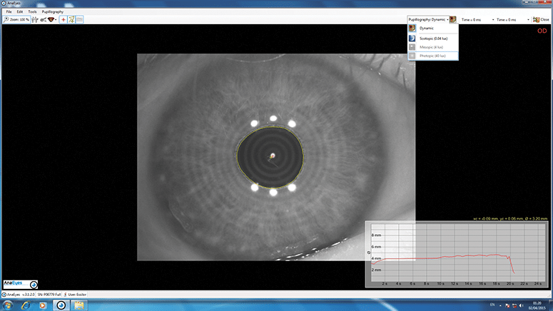
Contact lenses should be removed before a scan. During a corneal topography scan, you will be seated in front of a large, bowl-like instrument (called a corneal topographer) with lighted circles inside. This structure has a forehead and chin rest, which are used to keep your head and eyes aligned during the scan.
Which error is associated with steeper central corneal curvature?
4) Refractive error. Myopia is associated with steeper central corneal curvature. Hyperopia is associated with flatter central corneal curvature. However, these are not hard and fast rules, as axial length plays a big role in the overall myopia/hyperopia of the eye.
What are the colors of topographic maps?
Colored Maps: You will see a rainbow of colors on every topographic map. These range from warm colors ( red, orange, yellow ), to neutrals ( green) to cool colors ( blue, purple ). On our representative Pentacam images below, you will see four different types of maps.
Can refractive surgery cause corneal ectasia?
Refractive surgery itself can induce corneal ectasia. In preoperative planning, percent tissue altered (PTA) is used to estimate the risk of inducing a cornea ectasia. Generally, a PTA < 40% is accepted as a lower risk in a normal eye. 5.
What is corneal topography used for?
Corneal topography is most commonly used for the following purposes. Refractive surgery: To screen candidates for normal corneal shape, patterns and ruling out suspicious or keratoconic patterns .
What is the history of corneal topography?
History of Corneal topography. In early 17th century, Schiener used reflection of marbles from the cornea as perhaps the earliest corneal topography. Placido’s disc was a major advancement in the late 19th century. Placido disc has stood the test of time and the current placido based topographers work on the same principle ...
What is the non-planar shape of cornea?
The non planar shape of cornea can potentially lead to spurious results and therefore the use of schiempflug principle in corneal imaging is a welcome new change. Theodre Scheimpflug , an Austrian army man worked extensively on a method for correcting arial skew distortion in perspective photographs. Even though the technique was described before him, his development of the principle let his name to be associated with the principle. In an ideal scenario , the lens plane and the image plane are parallel. Therefore a linear object will form a plane of focus parallel to the lens plane and thus can be focused totally on the image plane (Figure 3a). Consider a situation , when the object is not parallel to the prospective image plane .It will not be possible to focus all the image on a plane parallel to image plane (Figure 3b). Thus this may lead to image distortion. However, according to the Scheimpflug principle, when a planar subject is not parallel to the image plane , an oblique tangent can be drawn from the image, object and lens planes, and the point of intersection is called schiempflug intersection (Figure 3c) .Using this orientation, A careful manipulation of the image plane and the lens plane can however lead to a focused and sharp image on the non parallel object.
What is the primary objective of the cornea?
The primary optical aim of cornea is refraction and focusing of the light rays as it acts as a covering lens overall. However , all non-ideal refracting surfaces reflect some light off them. This is the principle used for Purkinje imaging as well in the Placido discs.
Why is topography important for keratoconus?
In cases with established keratoconus, the role of topography is paramount for monitoring progression and doing a timely collagen cross linking , and in contact lens fitting.
Why is topography printout so daunting?
A modern topography printout can be quite daunting for the beginner because of the volumes of data it contains . This is a major shift from the rather simplistic videokeratographs and keratometers. A stepwise approach is very handy in interpretation. Step 1.
Can corneal topography be affected by corneal artifacts?
It should always be kept in mind that corneal topography can be effected by corneal artifacts and therefore interpretative value is decreased in cases such as nebulomacular corneal opacities, dry eye , corneal neovascularisations and corneal scars.
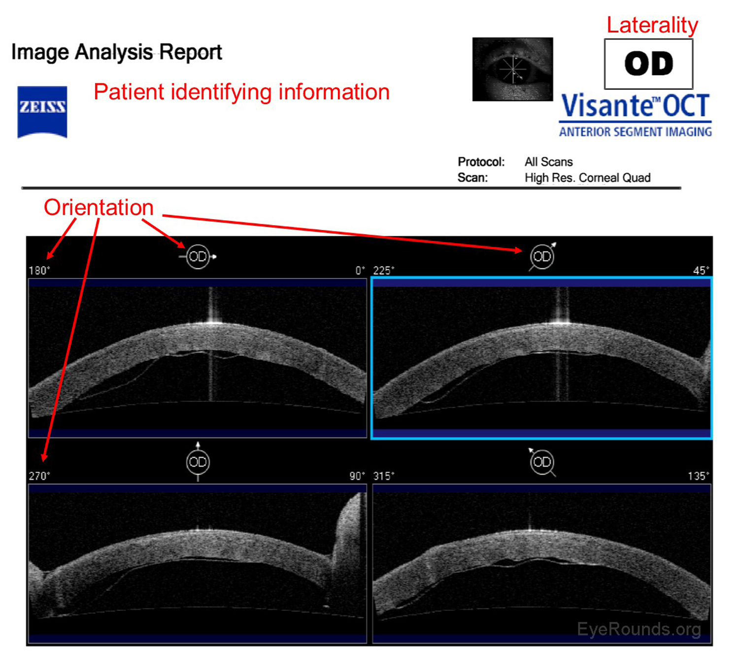
What is corneal topography?
Basic Principles. Corneal topography is used to characterize the shape of the cornea, similar to how one would characterize a mountain using a topographic map. Originally, corneal topography was only used to describe the anterior surface of the cornea.
What is the purpose of imaging the cornea?
Imaging techniques for assessing the structure and function of the cornea and anterior segment are crucial for diagnosing and treating a wide variety of ocular diseases. There is a huge variety of diagnostic testing available to ophthalmologists, and learning how to interpret these tests can seem daunting. For those beginning training in ophthalmology, the utilization of common diagnostic tests provides quicker and more accurate diagnosis and management of corneal diseases. The goal of this tutorial is to explain the basics of the most commonly used corneal imaging techniques at the University of Iowa, including an overview of how they work and how each modality is used in clinical practice.
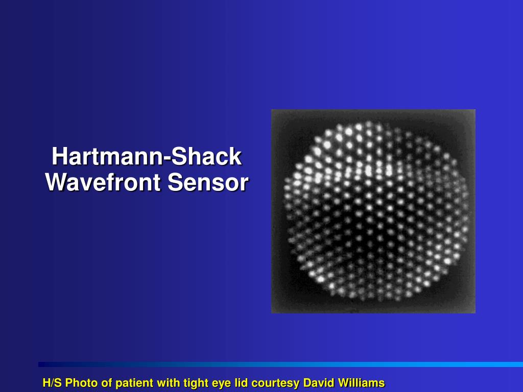
What is confocal microscopy?
Confocal microscopy is an imaging technique that allows in vivo examination of corneal structures at high magnification and resolution. Building off of imaging principles developed for neuronal imaging, confocal microscopy was first used to study the cornea in the 1990s (9-10). The device ( e.g., NIDEK Confoscan, Heidelberg HRTII) allows characterization of each of the five corneal layers by simultaneously illuminating and imaging a single point of tissue (Figure 14) (11). The point light source and the camera are in the same plane, hence the name "confocal." Modern confocal microscopes scan small regions of tissue, illuminating and imaging thousands of points of tissue to create the final confocal image (10). By scanning different thickness levels of certain tissues in the anterior segment, significant information about structure and function at the cellular level can be gained.
What is the most common corneal ectasia?
Keratoconus, the most common corneal ectasia, is a progressive corneal condition characterized by central thinning and steepening of the cornea. Early keratoconus often looks normal on slit lamp examination, and manual keratometry, which assesses the central 3 mm, may give an insufficient assessment.
What is a Zeiss Atlas?
The Zeiss Atlas and NIDEK OPD-Scan are Placido disc-based topographers. As shown in Figure 3, the Zeiss Atlas report includes a Placido disc image and several maps that provide information regarding tangential curvature, axial curvature, and elevation. A tangential, or instantaneous, map is very similar to an axial map. It is a slightly more accurate way of characterizing the corneal curvature but appears more "noisy" and irregular. Axial maps are less sensitive at measuring the corneal curvature and, thus, are used mainly for screening purposes (4-5).
Why is corneal topography important?
Corneal topography is an established and important technology for measuring the shape and power of the cornea. It allows practitioners not only to fit contact lenses to match the power and shape of the cornea, but it also helps them to evaluate intricacies of the contact lens relationship with the ocular surface.
What is the part of the ocular surface that is actually reflecting light?
These systems are highly dependent on the tear film, which is the part of the ocular surface that is actually reflecting light. This allows for a noninvasive measure of tear film quality, but it can also hinder accuracy when measuring corneal power and shape.
What happens when you wear ortho K lenses?
When ortho-k lenses for myopia are worn, tissue is moved from the central cornea to the periphery. As a result, the central cornea will become thinner and the peripheral cornea thicker. The sensitive global pachymetry measurements can show if there is asymmetry in the movement of tissue centrally to peripherally.
Why use axial maps?
Axial maps are ideal for base curve selection of a corneal or soft contact lens because the average of the central curvature is portrayed. For specific information about the corneal shape and power, other displays will be more helpful. Tangential Display Map.
Is corneal power map accurate?
One positive of this map is that it is the best way to get a quick overview of the corneal power. It can be also misleading, however, because it averages the data to create a “smooth” map, making it less accurate than other power maps (i.e., tangential).
Is tangential map useful for corneal topography?
Some indications of corne al topography will benefit from use of the tangential map display more than others. For example, tangential maps may be beneficial in orthokeratology (ortho-k), especially when evaluating the shape of the peripheral cornea, as this display provides the most accurate peripheral data. 3.
Types of Corneal Topography
- There are three different types of technologies used for corneal topography: Placido disc topography Placido disc reflection systems measure the curvature, irregularities, tear film quality, foreign bodies, and other parts of theanterior cornea. The reflection is highly dependent on the tear film which reflects the light, and can be either small-co...
Types of Topographic Maps
- Axial display map This is the most traditional way of viewing a topography image, as it is known for its overview of the corneal power. However, since it collects the averages of the data to produce a smooth map, it is considered less accurate than the other maps. Axial maps are a helpful tool for selecting the base curve of a soft contact lens because the average of the centra…
What Should I Expect During A Corneal Topography Test?
- A corneal topography test is quick and painless. During the test, you will sit in front of a lighted bowl that contains a pattern of rings, and rest your head against a bar. A series of data points will be collected, and a color coded image of your corneal shape will be generated on a computer screen. The images will contain different colors to differentiate elevations— similar to a topogra…
When Is Corneal Topography used?
- Corneal topography can be used for a variety of reasons: 1. Keratoconus 2. Planning refractive surgery 3. Monitoring ocular health post refractive surgery 4. Determining appropriate intraocular lens for cataract surgery 5. Evaluating and treating astigmatismpost-keratoplasty 6. Detecting corneal conditions such as pterygia, corneal scars, and Salzmann nodules 7. Monitoring ocular …