
See additional information. Cranial nerve IV: The fourth cranial nerve, the trochlear nerve, is the nerve supply to the superior oblique muscle of the eye, one of the muscles that moves the eye. Paralysis of the trochlear nerve results in rotation of the eyeball upward and outward (and, therefore, double vision ).
What is the purpose of cranial nerves?
What is the function of the cranial nerves?
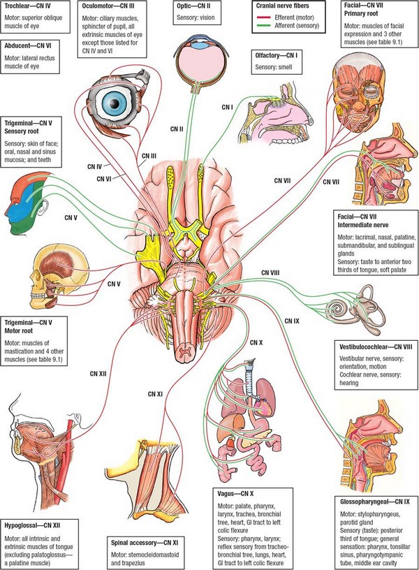
- I Olfactory (Smell)
- II Optic (Sight)
- III Oculomotor (Moves eyelid and eyeball and adjusts the pupil and lens of the eye)
- IV Trochlear (Moves eyeballs)
- V Trigeminal (Facial muscles incl.
- VI Abducens (Moves eyeballs)
- VII Facial (Taste, tears, saliva, facial expressions)
- VIII Vestibulocochlear (Auditory)
What cranial nerve is chiefly responsible for eye movements?
The internal anatomy of the brain has which structure White matter surrounded by gray matter The right and left hemisphere of the cerebrum are connected by the Corpus callosum Which structure in the diencephalon relays information to the cerebrum Thalamus What cranial nerve is chiefly responsible for eye movements Oculomotor
Which cranial nerves are pure motor in function?
- The olfactory nerve (CN I) contains special sensory neurons concerned with smell.
- The optic nerve (CN II) contains sensory neurons dedicated to vision.
- The oculomotor nerve (CN III) provides motor function for all eye muscles except those supplied by cranial nerves IV and VI.
Which cranial nerve is responsible for equilibrium?
vestibulocochlear nerve: Also known as the auditory vestibular nerve, this is the eighth of twelve cranial nerves, and it is responsible for transmitting sound and equilibrium (balance) information from the inner ear to the brain.
What happens if cranial nerve IV is damaged?
Diseases or injuries to the fourth cranial nerve can cause the superior oblique muscle to be paralyzed. The name for this condition is fourth nerve palsy. Other names for it are superior oblique palsy and trochlear nerve palsy. You may have fourth nerve palsy from birth, or you may develop it later.
What is cranial IV?
Cranial nerve 4, also called the trochlear nerve, controls the movement of the superior oblique muscle. This muscle moves the eye down and rotates the top of the toward the nose. It also helps pull the eye outward when the eye is looking downward.
What muscles does cranial IV innervate?
Synonyms: Cranial nerve IV, CN IV , show more... The trochlear nerve is a purely motor nerve, responsible for providing general somatic efferent (GSE)/motor innervation to just one muscle, the superior oblique muscle of the eye, on the contralateral side of its associated nucleus.
What does cranial nerve 4 assess?
3rd, 4th, and 6th Cranial nerves Extraocular movements controlled by these nerves are tested by asking the patient to follow a moving target (eg, examiner's finger, penlight) to all 4 quadrants (including across the midline) and toward the tip of the nose; this test can detect nystagmus and palsies of ocular muscles.
What is the main function of CN VI?
Cranial nerve six (CN VI), also known as the abducens nerve, is one of the nerves responsible for the extraocular motor functions of the eye, along with the oculomotor nerve (CN III) and the trochlear nerve (CN IV).
How do you test the function of the trochlear nerve?
This muscle depresses and abducts the eyeball when working independently. The extraocular muscles, on the other hand, synergistically move the eye. As a result, the trochlear nerve is tested by having the patient look 'down and in,' as the superior oblique contributes the most to this motion.
What does cranial nerve 4 palsy look like?
Fourth cranial nerve palsy may affect one or both eyes. Because the superior oblique muscle is paretic, the eyes do not adduct normally. Patients see double images, one above and slightly to the side of the other; thus, going down stairs, which requires looking down and inward, is difficult.
What is CN IV palsy?
Patients with acquired CN IV palsy typically present with acute onset, binocular vertical or oblique diplopia that may have a torsional component. The diplopia is typically worse in down gaze (consequently, they will often complain of difficulty reading) and lateral gaze toward the contralateral side.
How do you test for trochlear nerve damage?
To detect excyclotropia one must ask the patient whether there is a tilted double image in down gaze. Bilateral trochlear nerve palsy causes a change of vertical deviation between right and left gaze and between head-tilt to the right and to the left shoulder.
How should nurse assess cranial nerves III IV and VI?
Cranial Nerve III, IV, and VI – Oculomotor, Trochlear, AbducensTest eye movement by using a penlight. Stand 1 foot in front of the patient and ask them to follow the direction of the penlight with only their eyes. ... Test bilateral pupils to ensure they are equally round and reactive to light and accommodation .
How is the trochlear nerve cranial nerve IV classified according to function?
Trochlear nerve (CN IV) Cranial nerve 4 is a general somatic motor nerve. The trochlear nerve originates from the midbrain and enters the orbit through the superior orbital fissure, supplying one extraocular muscle thus playing a role in eye movement.
Does 4th nerve palsy cause headaches?
This could be a symptom of a fourth nerve palsy or another serious condition. Having double vision and a sudden severe headache could be a symptom of a stroke. Call 911 right away if you think you may be having a stroke. It could be from a blood clot (ischemic stroke) or a ruptured blood vessel (hemorrhagic stroke).
What are the 12 cranial nerve and their function?
Table: Overview of the 12 cranial nerves (CNs)NerveCNTypeOlfactoryISensory Nervous System: HistologyOpticIISensory Nervous System: HistologyOculomotorIIIMotor Nervous System: HistologyTrochlearIVMotor Nervous System: Histology8 more rows•Sep 5, 2022
Which nerve Innervates the sternocleidomastoid muscle?
the XI nerveThe muscles innervated directly by the XI nerve are the trapezius and the sternocleidomastoid, in addition to the laryngeal musculature (in collaboration with the vagus nerve), such as the palatal, pharyngeal, laryngeal muscles.
Are any skeletal muscles innervated by cranial nerves?
Skeletal muscles that arise from the branchial arches are innervated by fibres of cranial nerves V, VII, IX, and X; these are classified as special visceral efferent fibres.
Which one of these muscles is innervated by CN V?
Trigeminal nerve (CN V)TypeMixed (motor and sensory)Field of innervationMotor: Muscles of mastication, mylohyoid, anterior belly of digastric, tensor tympani muscles Sensory: Scalp, face, orbit, paranasal sinuses, anterior two-thirds of the tongue2 more rows
What are the functions of the cranial nerves?
Their functions are usually categorized as being either sensory or motor. Sensory nerves are involved with your senses, such as smell, hearing, and touch. Motor nerves control the movement and function of muscles or glands. Keep reading to learn more about each of the 12 cranial nerves and how they function.
How many cranial nerves are there?
What are cranial nerves? Your cranial nerves are pairs of nerves that connect your brain to different parts of your head, neck, and trunk. There are 12 of them, each named for their function or structure. Each nerve also has a corresponding Roman numeral between I and XII.
What is the function of the oculomotor nerve?
The oculomotor nerve has two different motor functions: muscle function and pupil response. Muscle function. Your oculomotor nerve provides motor function to four of the six muscles around your eyes. These muscles help your eyes move and focus on objects.
How many divisions does the trigeminal nerve have?
The trigeminal nerve has three divisions, which are:
Which nerve is located in the ophthalmic, maxillary, and mandibular divisions?
The sensory root of your trigeminal nerve branches into the ophthalmic, maxillary, and mandibular divisions. The motor root of your trigeminal nerve passes below the sensory root and is only distributed into the mandibular division. VI. Abducens nerve.
Which nerve transmits sensory information to your brain regarding smells that you encounter?
The olfactory nerve transmits sensory information to your brain regarding smells that you encounter.
Where does the trigeminal nerve originate?
It also controls the movement of muscles within your jaw and ear. The trigeminal nerve originates from a group of nuclei — which is a collection of nerve cells — in the midbrain and medulla regions of your brainstem.
Which nerve controls the facial muscles?
The trochlear nerve controls an extraocular muscle. The trigeminal nerve is responsible for sensory enervation of the face and motor enervation to muscles of mastication (chewing). The abducent nerve enervates a muscle, which moves the eyeball. The facial nerve enervates the muscles of the face (facial expression).
Which nerve enervates the muscles of the face?
The abducent nerve enervates a muscle, which moves the eyeball. The facial nerve enervates the muscles of the face (facial expression). The vestibulocochlear nerve is responsible for the sense of hearing and balance (body position sense). The glossopharyngeal nerve enervates muscles involved in swallowing and taste.
Which nerve enervates the sternocleidomastoid muscles and the trapezius muscles?
The vagus nerve enervates the gut ( gastrointestinal tract ), heart and larynx. The accessory nerve enervates the sternocleidomastoid muscles and the trapezius muscles. The hypoglossal nerve enervates the muscles of the tongue.
Which nerve controls the sense of smell?
The olfactory nerve carries impulses for the sense of smell. The optic nerve carries impulses for the sense of sight. The occulomotor nerve is responsible for motor enervation of upper eyelid muscle, extraocular muscle and pupillary muscle. The trochlear nerve controls an extraocular muscle.
What is the name of the nerve that carries impulses for the sense of smell?
The following are the list of cranial nerves, their functions, and tumor examples: The olfactory nerve carries impulses for the sense of smell.
Why is cranial nerve anatomy important?
Cranial nerves anatomy is essential for almost any medical specialty since they control so many body functions, such as rolling your eyes when you’re annoyed by something. So let’s break the stigma of them being hard to understand, and learn this important neuroanatomy topic once and for all.
What nerve controls the head?
The vagus nerve controls a large number of functions, including gland secretion, peristalsis, phonation, taste, visceral and general sensation of the head, thorax and abdomen. This cranial nerve is frequently tested in anatomy exams. Use our content to swot up on the vagus nerve and ace your cranial nerve exams!
How many cranial nerves are there?
Test your knowledge about the cranial nerves by taking this quiz which is specially designed to cover the most important anatomy facts about the 12 cranial nerves!
What is the function of a nerve?
The function of a nerve is to carry sensory and/or motor information between the body and the brain. If the information goes from the brain to the periphery, then it is an efferent (motor) nerve. If it travels from the periphery to the brain, then it is an afferent (sensory) nerve. Nerves that do both are mixed nerves.
What is the CN IV?
Trochlear nerve (CN IV) Cranial nerve 4 is a general somatic motor nerve. The trochlear nerve originates from the midbrain and enters the orbit through the superior orbital fissure, supplying one extraocular muscle thus playing a role in eye movement. Key facts about the trochlear nerve (CN IV) Type. GSE.
Which nerve innervates the olfactory mucosa?
Cranial nerve 1 is a special somatic afferent nerve which innervates the olfactory mucosa within the nasal cavity. It carries information about smell to the brain.
Which nerves carry information about movement and position?
Information of movement and position (proprioception) from somatic structures like muscles, tendons, and joints is carried by general somatic afferent nerves. Lastly, be aware that there is no special somatic efferent classification.
Which nerve controls the position of the eyeballs?
Cranial nerv es III (CNIII) (oculomotor), IV (trochlear), and VI (abducens) control the position of the eyeballs; CNIII influences the position of the eyelids and the size of the pupils. In addition to their value in localizing lesions, these three oculomotor nerves (sensory function is limited to proprioception) can reveal subtle changes in general ...
What nerves are involved in oculomotor activity?
Motor activity affecting the direction of gaze, the position of the eyelids, and the size of the pupils are served by cranial nerves III, IV, and VI. Unusual oculomotor activity is often encountered in psychiatric patients and can be quite informative. Evaluation techniques include casual observation and simple tests that require no equipment in addition to the sophisticated methods used in specialty clinics and research labs. This article reviews pupil size, extraocular movements, nystagmus, lid retraction, lid lag, and ptosis. Beyond screening for diseases and localizing lesions, these tests yield useful information about the individual’s higher cortical function, extrapyramidal motor functioning, and toxic/pharmacologic state.
Which part of the optic tract is responsible for sending efferent projections through the ciliary ganglion?
The optic tract synapses in the pretectal nucleus, which projects equally to the Edinger-Westphal nucleus (part of CNIII) on both sides. The Edinger-Westphal nucleus sends efferent projections through CNIII to the ciliary ganglion, then to the pupil.
Where is the nucleus of CNIII?
The nucleus is in the midbrain.
Which cranial nerve exits dorsally?
There are several clinically significant features of the trochlear nerve anatomy. It is the thinnest, and longest cranial nerve. Additionally, the fourth cranial nerve exits dorsally, crosses the midline, and innervates the contralateral SOM. The trochlear nucleus is in the midbrain, dorsal to the medial longitudinal fasciculus at the level ...
Which nerve has the longest intracranial course?
The trochlear nerve has the longest intracranial course of all of the cranial nerves. There are four anatomic regions which can be responsible for non-isolated CN IV palsies :
What is the name of the eye that rotates outward?
Thus, a trochlear nerve palsy causes an ipsilateral higher eye (i.e., hypertropia) and excyclotorsion (the affected eye deviates upward and rotates outward). Patients may report vertical and/or torsional diplopia that is usually worse on downgaze and gaze away from the affected side.
What is a CN IV palsy?
Congenital Trochlear nerve palsy is a common cause of congenital cranial nerve (CN) palsy. Patients with congenital CN IV palsies may compensate for diplopia with variable head positioning; chin-down head posture is seen in bilateral CN IV palsy and contralateral head tilt is typically seen in unilateral CN IV palsy. Later in life, these patients may experience decompensation of their previously well controlled CN IV palsy from the gradual loss of fusional amplitudes that occurs with aging or after illness or other stress event. Congenital CN IV palsies can have very large hypertropias in the primary position (greater than 10 prism diopters) despite the lack of diplopia or only intermittent diplopia symptoms. These large vertical fusional ranges characteristic of congenital cases.
What causes trochlear nerve palsy?
Trochlear nerve palsy can also occur as part of a broader syndrome related to causes like trauma, neoplasm, infection, and inflammation. These etiologies are further categorized based on the anatomic location of involvement (midbrain, subarachnoid space, cavernous sinus, orbit).
How long does it take for 4th nerve palsy to resolve?
Fourth nerve palsy secondary to microvascular disease will frequently resolve within 4-6 months spontaneously. Idiopathic In a small subset of patients with acquired trochlear palsy, no etiologic cause can be established even after extensive testing.
Which nerve passes adjacent to the trigeminal nerve?
The trochlear nerve passes adjacent to the ophthalmic division of the trigeminal nerve and the two share a connective tissue sheath. The trochlear nerve gains entry to the orbit via the superior orbital fissure, passes outside the tendinous ring of Zinn and innervates the SOM.
Which nerve is responsible for the movement of the eye?
It is the smallest nerve to service the eye. CN-IV passes through the superior orbital fissure, and it provides motor function, or movement. It serves the superior oblique eye muscle and connects to the annular tendon. As a result, it processes brain signals to move the eyes up and down, and also outwards.
What is the trochlear nerve?
The trochlear nerve is also known as cranial nerve IV (CN-IV). It is the only cranial nerve that emerges dorsally from the brain (near the back ), giving it the longest pathway. It is the smallest nerve to service the eye.
How to check cranial nerves?
Your healthcare provider can check your cranial nerves by doing a medical exam. They will look at the position of your eyes at rest and then have you follow an object with your eyes. Your healthcare provider may also check how your pupils react to light, measure the pressure in your eye, and look at the backs of your eyes. They may ask to look at old photos of you to try to find out when the problem started.
Which cranial nerve controls the actions of one of the external eye muscles?
The fourth cranial nerve controls the actions of one of the external eye muscles, the superior oblique muscle. This muscle runs from the back of the eye socket to the top of the eye. It passes through a loop of tissue near the nose known as the trochlea. It turns the eye inward and downward.
What is fourth nerve palsy?
Three nerves control how your eyes move, where your eyelids are, and how large your pupils are. These 3 nerves are:
How is fourth nerve palsy treated?
Treatment of fourth nerve palsy depends on its cause. Idiopathic fourth nerve palsies tend to go away on their own. Palsies caused by injury can also get better with time. If something is pressing on the fourth cranial nerve, you may need surgery to ease the pressure.
What are possible complications of fourth nerve palsy?
Fourth nerve palsies that don’t go away on their own cause a change in how your eyes work with one another (comitant). This means that the other eye moves with the affected eye. That way, the image separation you see stays the same no matter which way you are looking.
What nerves are involved in paralysis?
It enters the eye socket through an opening at the back and then travels to the superior oblique muscle. Diseases or injuries to the fourth cranial nerve can cause the superior oblique muscle to be paralyzed. The name for this condition is fourth nerve palsy.
How does a child with 4th nerve palsy look?
Children with fourth nerve palsy may develop a change in how their face looks (asymmetry), especially if they tend to keep their head tilted. The muscles on one side of the face do not develop the same as those on the other side, so the two sides start to appear different over time.
What is the 4th cranial nerve?
This nerve is the fourth set of cranial nerves (CN IV or cranial nerve 4). It is a motor nerve that sends signals from the brain to the muscles. CN IV works with the oculomotor nerve and other eye muscles to control eye movement.
Which cranial nerve enables you to look down?
The trochlear nerve is one of 12 sets of cranial nerves. It enables movement in the eye’s superior oblique muscle. This makes it possible to look down. The nerve also enables you to move your eyes toward your nose or away from it.
Why does the trochlear nerve not function?
When the trochlear nerve doesn’t function as it should, it’s often due to fourth nerve palsy. The condition is called trochlear nerve palsy. A palsy occurs when illness or injury paralyzes nerves that control muscle movement.
How did the trochlear nerve get its name?
The trochlear nerve gets its name from the Latin word pulley, “trochleae.” A pulley is a device that lifts an object.
What is the term for an abnormal growth of the trochlear nerve?
Cavernous sinus syndrome, when an abnormal growth affects the trochlear nerve.
What happens if you look down on a 4th nerve palsy?
Contact your healthcare provider if you experience symptoms of fourth nerve palsy. They often include vision changes that worsen when you look down. These include blurry or double vision.
What is the name of the condition where one eye turns inward?
Esotropia, a form of strabismus in which one or both eyes turn inward (crossed eyes).
