Is Cresyl violet a Nissl stain?
Cresyl violet: a red fluorescent Nissl stain Cresyl violet is widely used by neurobiologists to visualize Nissl substance in bright-field microscopy. Here we describe a method for using this dye as a red fluorescent Nissl stain.
How do you use Cresyl violet to stain paraffin?
Cresyl Violet Staining for paraffin embedded sections. Cresyl Violet Acetate solution is used to stain Nissl substance in the cytoplasm of neurons in paraformaldehyde or formalin-fixed tissue. The neuropil will be stained a granular purple-blue.
What stain is used to stain Nissl substance?
Cresyl Violet Stain Solution (0.1%) ab246816 is designed for staining Nissl substance in neurons on formalin fixed, paraffin-embedded tissue. Nissil granules are purple, nuclei of neuroglia and endothelial cells are slightly bluer than Nissl granules. Nissil Substance: Violet. Neuroglia Nuclei: Violet to Dark Blue. 1.
What is the Cresyl violet method?
The Cresyl Violet method uses basic aniline dye to stain RNA blue, and is used to highlight important structural features of neurons.

What does cresyl violet bind to?
These dyes bind avidly to RNA and DNA and thus highlight the heterochromatin of the nucleus and the rough endoplasmic reticulum of the cytoplasm (Fig. 48-5A).
Does cresyl violet stain nucleus?
Nuclei and nissl bodies (rough endoplasmic reticulum) are stained blue purple with cresyl violet.
What type of stain is a cresyl violet?
standard histological stainCresyl violet is a standard histological stain for neurons. Sections are first mounted onto gelatin-coated slides and dried overnight. Neonatal brains do not need to be delipidized, and after a rinse in H2O slides are immersed in stain for 3–5 min.
Does cresyl violet stain RNA?
Cresyl violet or NissI staining uses a basic aniline dye that stains RNA, and thus the endoplasmic reticulum and ribosomes blue [2].
Which structure does the cresyl violet stains most intensely?
Nissl stains use a variety of dyes (e.g. thionin, cresyl violet, fluorescent compounds) to show charged structures (Nissl bodies) in the soma of neurons and glia. The Nissl stain is most intense in nucleoli and in the rough endoplasmic reticulum of neurons.
Is cresyl violet A fluorescence?
Cresyl Violet is a fluorescent compound with an excitation peak at 598 nm and an emission peak at 621 nm. It can be excited using a 633 nm laser paired with a 660/10 nm bandpass filter, a configuration that can be found, for example, in the BD FACSVerse™.
What is crystal violet stain used for?
Crystal violet or gentian violet, also known as methyl violet 10B or hexamethyl pararosaniline chloride, is a triarylmethane dye used as a histological stain and in Gram's method of classifying bacteria.
How do you make cresyl violet stain?
* Cresyl violet stain: add 1.25 g cresyl violet acetate and 0.75 mL glacial acetic acid to 250 mL of warm dH2O, then cool and filter. + 70% acid ethanol: 2 mL glacial acetic acid in 200 mL 70% ethanol.
Does cresyl violet stain glia?
Alternatively, when nerve tissue is stained using the cresyl fast violet technique, the Nissl bodies, nuclear membranes and nucleoli stain intensely blue or violet, while the cytoplasm, ganglia cells, and glia stain weakly blue.
What stain is used for RNA?
Molecular Probes® fluorescent nucleic acid gel stains—SYBR® Gold, SYBR® Green I, SYBR® Green II, and SYBR® Safe dyes—are highly sensitive reagents for staining RNA in electrophoresis gels.
What structures does crystal violet stain?
The gram stain utilizes crystal violet as the primary stain. This basic dye is positively charged and, therefore, adheres to the cell membranes of both gram negative and positive cells.
What stain is used for nuclear DNA?
DAPIA simple-to-use fluorescent stain, 4',6-diamidino-2-phenylindole (DAPI), visualizes nuclear DNA in both living and fixed cells. DAPI staining was used to determine the number of nuclei and to assess gross cell morphology.
Which stain is used for nucleus staining?
Haematoxylin stains basophilic substances. Hence, hematoxylin is commonly used to stain the nucleus.
What stain stains the nucleus?
Staining the nucleus. The bulk of the content inside the nucleus is nucleic acid, so nucleic acid stains are the obvious choice for nuclear staining.
Which dye can stain nucleus?
PureBlu™ Nuclear Staining Dyes are designed to specifically stain the nuclei of cells in fixed and unfixed samples for fluorescence microscopy and cell imaging applications. Based on the well-recognized DAPI and Hoechst 33342 chemistries, PureBlu Dyes are offered in a ready-to-reconstitute, high-purity powder format.
Does crystal violet stain nucleus?
In biomedical research, crystal violet can be used to stain the nuclei of adherent cells. In this application, crystal violet works as a intercalating dye and allows the quantification of DNA which is proportional to the number of cells. In forensics, crystal violet was used to develop fingerprints.
What are the three types of interneurons in the striatum?
The striatum additionally contains three types of relative small- to medium-sized (perikaryal diameter 10–25 μm) GABAergic interneurons: (1) parvalbumin (PV)-positive interneurons (fast-spiking), (2) nitric oxide synthase (NOS)/somatostatin-expressing interneurons, and (3) calretinin-expressing interneurons ( Bernácer, Prensa, & Giménez-Amaya, 2012 ). The 2–5 profusely branched primary dendrites receive major inputs from the cortex, the specific and unspecific thalamic nuclei, and dopaminergic input from the SNc (in the rat also from GPe neurons); they give inhibitory input to the MSNs (for review, see Holt et al., 1997 ).
How many striatal neurons are there in the human putamen?
More specifically, in the human putamen, 80 million neurons per hemisphere were estimated stereologically ( Schmitt, Eggers, & Haug, 1995 ).
How many interneurons are there in the human body?
At least four populations of interneurons exist, with three of them being GABAergic interneurons and one being cholinergic. The large cholinergic interneurons (aspiny type II neurons, diameter 20–35 μm, choline acetyltransferase (ChAT)-immunoreactive) only account for about 1% of all striatal neurons.
What is cresyl violet?
Cresyl Violet. Cresyl violet is a standard histological stain for neurons. Sections are first mounted onto gelatin-coated slides and dried overnight. Neonatal brains do not need to be delipidized, and after a rinse in H2 O slides are immersed in stain for 3–5 min. Three components are made and then mixed: 0.3 g of cresyl echt violet (Roboz) ...
How to screen brain malformations?
The adult brains were embedded into Tissue-Tek OCT® (Optimal Cutting Temperature) compound, snap -frozen in 2-methylbutane/dry ice bath, and cryosectioned into 10 μm sections with a cryostat. The sections were mounted onto FisherBrand plus slides and fixed in 4% paraformaldehyde for 15 min. After rinsing with water, the slides were stained with 1% hematoxylin for 2 min and rinsed with running water. The slides were then stained with 2% eosin for 1 min, rinsed with water, dehydrated with an ascending ethanol series, and cover slipped with a xylene-based mounting medium.
What is the phasically active firing rate of MSNs?
In monkeys, MSNs are phasically active neurons (PANs). Phasically active neurons have a very low spontaneous discharge rate (0.5–1 spike s − 1) but a relatively high phasic firing rate associated with behavioral tasks, such as movements, preparation for movements, and the performance of learned tasks.
What do the arrows on the STN mean?
The arrows indicate the direction of the projection and the principal transmitters used by the respective projection neurons: green, glutamatergic; red, GABAergic; yellow, dopaminergic.
What is cresyl violet staining?
Cresyl Violet Staining for paraffin embedded sections. Cresyl Violet Acetate solution is used to stain Nissl substance in the cytoplasm of neurons in paraformaldehyde or formalin-fixed tissue.
What color is the neuropil?
The neuropil will be stained a granular purple-blue. This stain is commonly used to identify the neuronal structure in brain and spinal cord tissue. The Cresyl Violet method uses basic aniline dye to stain RNA blue, and is used to highlight important structural features of neurons.
Why is the cytoplasm blue?
The Nissl substance (rough endoplasmic reticulum) appears dark blue due to the staining of ribosomal RNA, giving the cytoplasm a mottled appearance. Individual granules of extra-nuclear RNA are named Nissl granules (ribosomes). DNA present in the nucleus stains a similar color.
Images
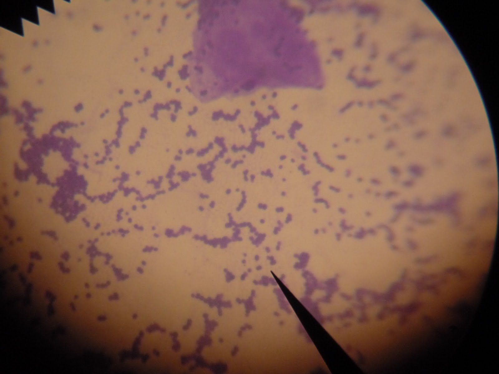
ab246816 Cresyl Violet Stain Solution (0.1%) staining formalin-fixed-paraffin embedded human brain.
Protocols
To our knowledge, customised protocols are not required for this product. Please try the standard protocols listed below and let us know how you get on.
References (1)
Publishing research using ab246816? Please let us know so that we can cite the reference in this datasheet.
What is Cresyl violet?
Cresyl violet: a red fluorescent Nissl stain. Cresyl violet is widely used by neurobiologists to visualize Nissl substance in bright-field microscopy. Here we describe a method for using this dye as a red fluorescent Nissl stain.
Is cresyl violet fluorescent?
Unlike the bright-field staining technique, fluorescent cres yl is compatible with other fluorescent dyes and tracers …. Cresyl violet is widely used by neurobiologists to visualize Nissl substance in bright-field microscopy. Here we describe a method for using this dye as a red fluorescent Nissl stain.
What color are nissl granules?
Nissl granules are purple, nuclei of neuroglia and endothelial cells are slightly bluer than Nissl granules. Nissl Substance: Violet. Neuroglia Nuclei: Violet to Dark Blue. Working Solutions: If preferred, reagent may be diluted up to 1:10 with Deionized water just before use. 1.
How to remove a stain from a tissue?
1. Deparaffinize sections and hydrate to distilled water. 2. Apply Cresyl Violet Acetate solution (or working solution) to tissue for 3-5 minutes. 3. Rinse quickly in 1 change of distilled water. 4. Dehydrate quickly in absolute alcohol. (Alcohol may remove stain from tissue over time).
What is a LFB kit?
Newcomer Supply Luxol Fast Blue (LFB) – Cresyl Violet Stain Kit, with included microwave modification, is for the demonstration of myelin and Nissl substance in central nervous system and peripheral nerve tissues.
How to make Cresyl Violet stain?
Combine, mix well and filter. Directly before use, heat filtered solution to 57°C in microwave; hold in oven. Stain in heated Cresyl Violet Working Solution; 6 minutes in oven. Rin se in distilled water. Dehydrate quickly to maintain Cresyl Violet Stain in two changes each of 95% and 100% ethyl alcohol.
What is a complimentary control slide?
COMPLIMENTARY POSITIVE CONTROL SLIDES: Enclosed are two complimentary unstained positive control slides for initial verification of staining techniques and reagents. Verification must be documented by running one Newcomer Supply complimentary positive control slide along with your current positive control slide for the first run. Retain the second complimentary control slide for further troubleshooting, if needed.
How long to incubate Luxol Fast Blue Stain?
Incubate in Solution A: Luxol Fast Blue Stain 0.1%, Alcoholic for 2 hours at 60°C or overnight at 37°C; cover tightly.
How many ml of Coplin jars for newcomer supply stain?
All Newcomer Supply Stain Kits are designed to be used with Coplin jars filled to 40 ml following the provided staining procedure. Some solutions in the kit may contain extra volumes.
How long to agitate a slide in lithium carbonate?
Differentiate slides individually in Working Lithium Carbonate 0.05% (Step #2) for 10-15 seconds with agitation until gray matter and white matter are colorless and contrast with stained tissue.
Where are myelin and nissl found?
Stains myelin and Nissl substance in central nervous system tissues and in peripheral nerve.
What is the name of the dye that binds to nucleic acids?
The dye itself is a base, which binds to nucleic acids. The stain was named after the German neuropathologist Franz Nissl. “Nissl substance” is an older term for the endoplasmic reticulum, which is where the name comes from. “Nissl bodies,” on the other hand, refer to the free ribosomes.
What color is the nucleic acid in cresyl violet?
With a cresyl violet Nissl stain, the nucleic acid appears dark purple-blue while the areas with low nucleic acid density appears light blue-gray. The center of the nucleus is the nucleolus, the actual site of storage for DNA. Immediately surrounding the nucleus is often several layer of endoplasmic reticulum, which has genetic material in ...
What is the Nissl stain?
Answer: The Nissl stain is an imaging technique performed on fixed tissue that stains genetic material. One of the oldest methods of neuronal imaging, the Nissl stain uses cresyl violet acetate or toluidine blue to permanently stain genetic material.
Why is layer 6 more dense?
Because layer 6 contains the nuclei of several cells, it will appear significantly more dense in staining compared to a layer that has several projection processes. The major disadvantage of using a cresyl violet Nissl stain is that the detailed morphology of the cell is not stained.
Which is better, Golgi or Nissl?
Following a Nissl stain, the cell bodies of a densely branched Purkinje cell and a long and thin bipolar cell would appear similar: only the nucleus and rough endoplasmic reticulum would take up the stain. A Golgi stain will be better for visualizing the morphology of a cell.
Which genetic material is often stained?
The genetic material that is often stained is the DNA within the nucleus and RNA in the rough endoplasmic. The stain is most useful for identifying cytoarchitecture across brain structures ( Improved method for combination of immunocytochemistry and Nissl staining ). Some regions of the brain, such as the cortex, are organized in an columnar manner.
How many layers are there in the brain?
This organization in the cortex is described in terms of layers, one on top of the next. There are 6 layers of the cortex, and each layer has a unique cell composition. For example, layer 6 and the innermost part of layer 4 of the cortex often contains the cell bodies of several pyramidal cells. Because layer 6 contains the nuclei of several cells, it will appear significantly more dense in staining compared to a layer that has several projection processes.
