What is a schistocyte in blood?
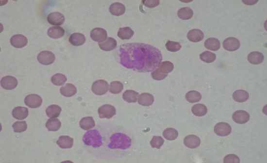
SCHISTOCYTE: In peripheral blood (×600) A fragmented red blood cell that appears in the blood in a variety of bizarre shapes, from small triangular forms to round cells with irregular surfaces. Schistocytes are found in patients with hemolytic anemias, severe burns, and several other conditions.
What is a normal schistocyte count?
The red blood cells get trapped in the fibrin strands and the sheer force of the blood flow causes the red blood cell to break. The resulting fragmented cell is called the schistocyte. A normal schistocyte count for a healthy individual is <0.5% although usual values are found to be <0.2%.
What is schistocyte fragmentation?
schistocyte [shis′təsīt] an erythrocyte cell fragment characteristic of hemolysis or cell fragmentation associated with severe burns, microangiopathic hemolytic anemias, and intravascular coagulation.
What are the characteristics of schistocytes in peripheral blood smear (ppb)?
Peripheral blood smear in patient with thrombotic thrombocytopenic purpura. Typical schistocytes are annotated. A schistocyte or schizocyte (from Greek schistos for "divided" and kytos for "hollow" or "cell") is a fragmented part of a red blood cell. Schistocytes are typically irregularly shaped, jagged, and have two pointed ends.
How do schistocytes form?
What is the name of the disease where schistocytes are present in blood?
What causes schistocytes on peripheral blood smear?
Why do schistocytes appear in hemolytic anemia?
What is a normal schistocyte count?
What is a schizocyte?
What is DIC in a coagulation cascade?
See 4 more
About this website

What conditions cause schistocytes?
Schistocytes are often seen in patients with hemolytic anemia. They are frequently a consequence of mechanical artificial heart valves, hemolytic uremic syndrome, and thrombotic thrombocytopenic purpura, among other causes.
What does the presence of schistocytes mean?
Schistocytes are split red blood cells that indicate microangiopathic hemolytic anemia. Their presence in a peripheral smear is the hallmark for diagnosing thrombotic thrombocytopenic purpura (TTP).
What does it mean if you have schistocytes in the peripheral blood?
The presence of schistocytes (fragmented red blood cells) on the peripheral blood smear suggests red blood cell injury from damaged endothelium and is a characteristic feature of microangiopathic hemolytic anemia.
What is one specific condition where the presence of schistocytes could indicate a life threatening condition?
Schistocytes can also be seen in patients with preeclampsia, malignant hypertension, renal failure, mechanical heart valves, vitamin B12 deficiency and occasionally in normal individuals. Schistocytes are seen in life threatening conditions like TTP/HUS and DIC.
What does fragmented red blood cells mean?
Fragmented red blood cells are generally the consequence of mechanical damage, usually in the context of turbulent blood flow or contact with a pathologically altered endothelium, the latter occurring most commonly in the microvasculature.
Where are schistocytes found?
Schistocytes, or “split” red cells, appear in the peripheral blood smear (>1% of total red cells)2 as an indication of the microangiopathic hemolytic anemia.
Are schistocytes seen in iron deficiency anemia?
Schistocytes were observed in patients with TMA (n=76), infection (n=20), hematologic malignancy (n=10), mechanical heart valves (n=2), renal failure (n=10), hemoglobinopathy (n=15), iron deficiency anemia (n=1), and megaloblastic anemia (n=1) and in neonates (n=11) (Table 1).
What causes red cell fragments?
The most common causes of abnormal fragmentation were malignancy with cytotoxic chemotherapy and severe iron deficiency. In two subjects, an abnormal red blood cell fragmentation pattern was the clue to a spectrin mutant in subjects with an automated blood count previously evaluated as normal.
What anemia has target cells?
There are four major circumstances in which target cells appear as the major morphologic abnormality: thalassemia, hepatic disease with jaundice, hemoglobin C disorders, and the postsplenectomy state. Lesser numbers of target cells are found in sickle cell anemia, iron deficiency, and lead intoxication.
What are the 5 symptoms of hemolytic uremic syndrome?
What are the symptoms of hemolytic uremic syndrome? Symptoms of HUS include vomiting, bloody diarrhea (loose stool/poop), stomach pain, fever, chills, and headache. As infection progresses, persons may experience fatigue, weakness, fainting, bruising, and paleness.
What are the signs and symptoms of TTP?
SymptomsBleeding into the skin or mucus membranes.Confusion.Fatigue, weakness.Fever.Headache.Pale skin color or yellowish skin color.Shortness of breath.Fast heart rate (over 100 beats per minute)
How do I report schistocytes?
Schistocytes should be identified and counted on a peripheral blood smear using optical microscopy. The blood smear should be spread, air-dried, fixed, and stained according to standard procedures with panoptical stains, as reported by ICSH (1984) and confirmed by international studies (Barnes et al., 2005).
Are schistocytes seen in iron deficiency anemia?
Schistocytes were observed in patients with TMA (n=76), infection (n=20), hematologic malignancy (n=10), mechanical heart valves (n=2), renal failure (n=10), hemoglobinopathy (n=15), iron deficiency anemia (n=1), and megaloblastic anemia (n=1) and in neonates (n=11) (Table 1).
What causes megaloblastic anemia?
There are many causes of megaloblastic anemia, but the most common source in children occurs from a vitamin deficiency of folic acid or vitamin B-12. Other sources of megaloblastic anemia include the following: Digestive diseases — Certain diseases of the lower digestive tract can lead to megaloblastic anemia.
How many schistocytes are significant per HPF?
Two (2) schistocytes per HPF correlated with 1% schistocytes on the linear plot. At UCMC, policy had been to report 2-8 schistocytes per HPF as present and >8 per HPF as increased. These findings indicated that the threshold for reporting increased schistocytes should be lowered from >8 per HPF to >2 per HPF.
What anemia has target cells?
There are four major circumstances in which target cells appear as the major morphologic abnormality: thalassemia, hepatic disease with jaundice, hemoglobin C disorders, and the postsplenectomy state. Lesser numbers of target cells are found in sickle cell anemia, iron deficiency, and lead intoxication.
The Clinical Significance of Schistocytes: A Prospective Evaluation of ...
The presence of ≥1% schistocytes on a peripheral blood smear (PBS) is an important criterion for the diagnosis of thrombotic microangiopathy (TMA). The reporting of schistocytes has been standardized by the International Council for Standardization ...
Schistocytes on the Peripheral Blood Smear - Mayo Clinic Proceedings
The presence of schistocytes (fragmented red blood cells) on the peripheral blood smear suggests red blood cell injury from damaged endothelium and is a characteristic feature of microangiopathic hemolytic anemia. Microangiopathic hemolytic anemia is an infrequent cause of Coombs-negative intravascular hemolytic anemia, and its causes include thrombotic thrombocytopenic purpura/hemolytic ...
Schistocytes are circulating red blood cell fragments seen in TTP
Schistocytes or schizocytes are defined as circulating red blood cell fragments. Detection of schistocytes is an important clue for the diagnosis of thrombotic microangiopathy (TMA), which includes both thrombotic thrombocytopenic purpura (TTP) and hemolytic-uremic syndrome (HUS).
Poikilocytosis: Symptoms, Treatment, Outlook, and More - Healthline
Poikilocytosis is the term for having abnormally shaped red blood cells (RBCs) in your blood. We'll explain the different types and what they mean for you.
Schistocytes – A Laboratory Guide to Clinical Hematology
Images show peripheral blood smears with schistocytes present (indicated by the arrow). From MLS Collection, University of Alberta. Image 1: 100x oil immersion.
Evaluation of Normal Reference Range of Schistocytes and Burr Cells in ...
Abstract. Introduction:Schistocytes are split red blood cells that indicate microangiopathic hemolytic anemia. Their presence in a peripheral smear is the hallm
What is a fragmented red cell?
A fragmented red cell which arises from either an intrinsic increase in cell fragility or due to intravascular rugosities that traumatise cells; schistocytes are a nonspecific finding seen in hemolysis, trauma, prosthetic heart valves, megaloblastic and microangiopathic anaemioccidianhemolytic uremic syndrome, TTP.
What is a schistocyte?
SCHISTOCYTE: In peripheral blood (×600) A fragmented red blood cell that appears in the blood in a variety of bizarre shapes, from small triangular forms to round cells with irregular surfaces. Schistocytes are found in patients with hemolytic anemias, severe burns, and several other conditions.
What is the name of the syndrome that shows a littoral cell angioma of the sple?
Littoral cell angioma of the spleen revealed by a Kasabach-Merritt syndrome in a 22-month-old child
What is MAHA in blood?
MAHA results from intravascular fragmentation of red blood cells, which produces schistocyteson peripheral blood smear.
What is the diagnosis of TTP?
The diagnosis of TTP was distinguished from haemolytic anaemia related to her CTD by means of the profuse presence of schistocyteson peripheral smear, normal complement levels and negative Coombs test.
What is abnormal shape of red blood cells?
A red blood cell having an abnormal shape as a result of fragmentation that occurs as the cell flows through damaged small vessels.
What is a helmet cell?
a fragment of an erythrocyte, commonly observed in the blood in hemolytic anemia; called also helmet cell and schizocyte.
What is a schistocyte?
Schistocytes. Schistocytes or schizocytes are defined as circulating red blood cell fragments. Detection of schistocytes is an important clue for the diagnosis of thrombotic microangiopathy (TMA), which includes both thrombotic thrombocytopenic purpura (TTP) and hemolytic-uremic syndrome (HUS).
What are the morphological criteria for schistocytes?
The morphological criteria required for identification of schistocytes includes cells smaller than intact red blood cells and have shapes with sharp angles and straight borders, small crescents, helmet cells, keratocytes, and microspherocytes.
What is the schistocyte threshold for TMA?
For diagnosis of transplant associated TMA, schistocyte threshold of 4% is recommended together with thrombocytopenia, increased lactate dehydrogenase, decreased hemoglobin concentration, and decreased haptoglobin.
What causes red blood cell fragmentation?
Non-TMA causes of red blood cell fragmentation include red blood cell membrane defects, thalassemia, meg aloblastic anemia, primary myelofibrosis, and thermal injury . The red blood cell fragments in these cases show high variability in shapes and marked aniso-poikilocytosis which are not specific for the diagnosis of TMA.
Is a schistocyte count clinically meaningful?
A schistocyte count should be considered clinically meaningful if schistocytes represent the main morphological red blood cell abnormality in the smear.
How many spicules are there in an erythrocyte?
An erythrocyte with 10 to 30 spicules distributed over the surface of the cell, as seen in heart disease, stomach cancer, kidney disease, and dehydration. Synonym: echinocyte
What is the function of B lymphocytes?
1. A lymphocyte that synthesizes and secretes antibodies. B lymphocytes originate and differentiate in the bone marrow and then populate the spleen, lymph nodes, and other lymphoid tissues. When exposed to an antigen, a B cell divides to form (a) plasma cells, which produce antigen-specific antibodies, and (b) a lesser number of memory B cells, which can quickly differentiate into plasma cells upon a second exposure to the original antigen. Antibody production is a key part of the humoral immune response of adaptive immunity. The humoral immune response is effective against bacteria, viruses, and other pathogens, and provides the rationale for vaccination. Synonym: B lymphocyteSee: ; T cell;
What are the cells that are found in the blood stream?
Any cell normally found circulating in the blood stream. Blood cells include red cells and white cells; red cells generally remain inside blood vessels, while white cells can also more into the tissues outside the blood vessel walls.
What is the red blood cell?
A cell in the blood of vertebrates that transports oxygen and carbon dioxide to and from the tissues. In mammals, the red blood cell is disk-shaped and biconcave, contains hemoglobin, and lacks a nucleus. Also called erythrocyte, red cell, red corpuscle.
Which cell breaks down antigens and displays their fragments on surface receptors next to major histocompatibility?
A cell that breaks down antigens and displays their fragments on surface receptors next to major histocompatibility complex molecules. This presentation is necessary for some T lymphocytes that are unable to recognize soluble antigens. Macrophages are the primary antigen-presenting cells, but B cells and dendritic cells also can act as APCs.
Where are adult stem cells found?
Adult stem cells are found in many tissues, such as bone marrow, brain, retina, skin, intestines, liver, testis, and pancreas.
What is a schistocyte in a PBS?
The presence of ≥1% schistocytes on a peripheral blood smear (PBS) is an important criterion for the diagnosis of thrombotic microangiopathy (TMA). The reporting of schistocytes has been standardized by the International Council for Standardization in Hematology (ICSH). Despite the availability of guidelines, however, the assessment of schistocytes remains subjective. More recently, the automated fragmented red cell (FRC) parameter has been evaluated. However, local studies are not available.
What is the mean value of schistocytes in TMA?
The schistocyte counts were normally distributed in the TMA and non-TMA groups with mean (±SD) values of 3.44±1.84% and 1.11±0.83%, respectively (p<0.0001). In addition, the mean values for hemoglobin and platelet count were significantly lower in the TMA group (p<0.049 and p<0.001 respectively). The red cell distribution width (RDW) was significantly higher in the TMA group, whereas there was no difference for LDH (Table 2).
How do schistocytes form?
This occurs when there is damage to the blood vessel and a clot begins to form . The formation of the fibrin strands in the vessels occurs as part of the clot formation process. The red blood cells get trapped in the fibrin strands and the sheer force of the blood flow causes the red blood cell to break. The resulting fragmented cell is called the schistocyte.
What is the name of the disease where schistocytes are present in blood?
Excessive schistocytes present in blood can be a sign of microangiopathic hemolytic anemia (MAHA).
What causes schistocytes on peripheral blood smear?
The causes of MAHA can be disseminated intravascular coagulation, thrombotic thrombocytopenic purpura, hemolytic-uremic syndrome, HELLP syndrome, malfunctioning cardiac valves etc.
Why do schistocytes appear in hemolytic anemia?
Schistocytes are often seen in patients with hemolytic anemia. They are frequently a consequence of mechanical artificial heart valves and hemolytic uremic syndrome, thrombotic thrombocytopenic purpura, among other causes.
What is a normal schistocyte count?
A normal schistocyte count for a healthy individual is <0.5% although usual values are found to be <0.2%. A schistocyte count of >1% is most often found in thrombotic thrombocytopenic purpura, although they are more often seen within the range of 3–10% for this condition. A schistocyte count of <1% but greater than the normal value is suggestive of disseminated intravascular coagulation, but is not an absolute diagnosis. The standard for a schistocyte count is a microscopic examination of a peripheral blood smear.
What is a schizocyte?
A schistocyte or schizocyte (from Greek schistos for "divided" and kytos for "hollow" or "cell") is a fragmented part of a red blood cell. Schistocytes are typically irregularly shaped, jagged, and have two pointed ends.
What is DIC in a coagulation cascade?
Disseminated intravascular coagulation or DIC is caused by a systemic response to a specific condition including sepsis and severe infection, malignancy, obstetric complications, massive tissue injury, or systemic diseases. Disseminated intravascular coagulation is an activation of the coagulation cascade which is usually a result of an increased exposure to tissue factor. The activation of the cascade leads to thrombi formation which causes an accumulation of excess fibrin formation in the intravascular circulation. The excess fibrin strands cause mechanical damage to the red blood cells resulting in schistocyte formation and also thrombocytopenia and consumption of clotting factors. Schistocyte values between .5% and 1% are usually suggestive of DIC.
