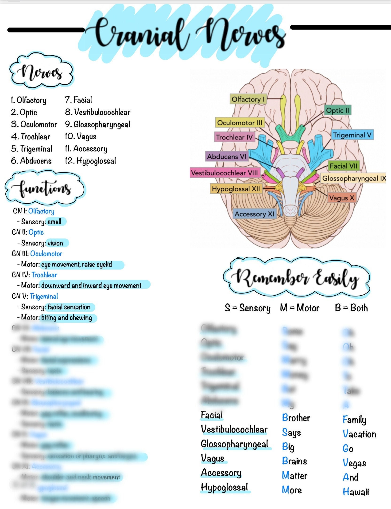
Eighth cranial nerve: The eighth cranial nerve is the vestibulocochlear nerve. The vestibulocochlear nerve is responsible for the sense of hearing and it is also pertinent to balance, to the body position sense.
What does eighth cranial nerve mean?
The vestibulocochlear nerve, also known as cranial nerve eight (CN VIII), consists of the vestibular and cochlear nerves. Each nerve has distinct nuclei within the brainstem. The vestibular nerve is primarily responsible for maintaining body balance and eye movements, while the cochlear nerve is responsible for hearing. NCBI Skip to main content
What cranial nerve is damaged?
The vestibulocochlear nerve, CN VIII, controls hearing and balance. Loss or a decrease of hearing may indicate nerve damage. Motor Function Nerves. Cranial nerves associated primarily with motor function are IV, VI, XI and XII. CN IV is the trochlear nerve, which controls inward and downward eye movement. Damage can cause double vision.
How to conduct a cranial nerve examination?
To assess the corneal reflex:
- Clearly explain what the procedure will involve to the patient and gain consent to proceed.
- Gently touch the edge of the cornea using a wisp of cotton wool.
- In healthy individuals, you should observe both direct and consensual blinking. The absence of a blinking response suggests pathology involving either the trigeminal or facial nerve.
Which cranial nerves are not purely sensory?
Trochlear nerve is basically a cranial nerve of motor functionality that exercises control over the superior oblique eye muscle. This makes it a motor nerve as opposed to the sensory functionality of the rest indulging in vision, smell and hearing respectively. Therefore, option A is the most appropriate.

What happens if the 8th cranial nerve is damaged?
CN VIII pathology can result from direct trauma, congenital malformations, tumor formation, infection, and vascular injury. Presenting symptoms include vertigo, nystagmus, tinnitus, and sensorineural hearing loss.
Does cranial nerve 8 affect balance?
Abstract. The vestibulocochlear nerve (8th cranial nerve) is a sensory nerve. It is made up of two nerves, the cochlear, which transmits sound and the vestibular which controls balance.
How do you assess the 8th cranial nerve?
8th Cranial nerveHearing is first tested in each ear by whispering something while occluding the opposite ear. ... Vestibular function can be evaluated by testing for nystagmus. ... If patients have acute vertigo during the examination, nystagmus is usually apparent during inspection.More items...
What cranial nerve causes vertigo?
The vestibulocochlear nerve sends balance and head position information from the inner ear (see left box) to the brain. When the nerve becomes swollen (right box), the brain can't interpret the information correctly. This results in a person experiencing such symptoms as dizziness and vertigo.
What happens if the vestibulocochlear nerve is damaged?
The vestibular nerve communicates messages about head position and motion from your inner ear to your brain. When this nerve is damaged, these messages become jumbled and inaccurate, confusing your brain and producing the dizziness, nausea and movement issues.
What are the symptoms of nerve damage in the ear?
Symptoms can include:mild to severe hearing loss.sounds fading in and out.difficulty understanding spoken words (speech perception)normal hearing but with poor speech perception.worsened speech perception in noisy environments.
Can MRI show cranial nerve damage?
Nerve damage can usually be diagnosed based on a neurological examination and can be correlated by MRI scan findings. The MRI scan images are obtained with a magnetic field and radio waves. No harmful ionizing radiation is used.
Can cranial nerve damage be repaired?
Treatment. If a cranial nerve is completely cut in two, it cannot be repaired. However, if it is stretched or bruised but the nerve remains intact, it can recover. This takes time and can cause a variety of unpleasant symptoms including tingling and pain.
Is cranial nerve 8 sensory or motor?
sensory nervesCranial nerves I, II, and VIII are pure sensory nerves. Cranial nerves III, IV, VI, XI, and XII are pure motor nerves.
Why do I lose balance when I walk?
Losing your balance while walking, or feeling imbalanced, can result from: Vestibular problems. Abnormalities in your inner ear can cause a sensation of a floating or heavy head and unsteadiness in the dark. Nerve damage to your legs (peripheral neuropathy).
What vitamin deficiency can cause dizziness?
Low Vitamin B12 Levels Can Cause Dizziness “Vitamin B12 deficiency is easy to detect and treat, but is an often overlooked cause of dizziness,” he notes. Ask your doctor about having a simple blood test to check your B12 levels if you're having dizzy spells.
What neurological disorders cause balance problems?
Illnesses like multiple sclerosis, Parkinson's disease, and cervical spondylosis slowly damage the way your nervous system talks to your brain, which can affect your balance.
Which cranial nerve is concerned with the maintenance of balance?
The vestibulocochlear nerve or auditory vestibular nerve, also known as the eighth cranial nerve, cranial nerve VIII, or simply CN VIII, is a cranial nerve that transmits sound and equilibrium (balance) information from the inner ear to the brain.
Which nerve relates to hearing equilibrium and balance?
vestibulocochlear nervesCranial nerves The vestibular nerve handles balance and equilibrium, while the cochlear nerve is responsible for hearing. The vestibulocochlear nerves originate in the monitoring receptors of the internal ear—the vestibule and cochlea.
What do the 7th and 8th cranial nerves do?
The 8th nerve, also known as the vestibulocochlear nerve, mainly transmits information from the hearing organ (cochlea) and the vestiular organ to the brain. The 8th nerve traverses the internal auditory canal, which also contains the 7th nerve, the nervous intermedius, and the labyrinthine artery.
Is cranial nerve 8 sensory or motor?
sensory nervesCranial nerves I, II, and VIII are pure sensory nerves. Cranial nerves III, IV, VI, XI, and XII are pure motor nerves.
Overview
A number of cranial nerves send electrical signals between your brain and different parts of your neck, head and torso. These signals help you smell, taste, hear and move your facial muscles.
Function
Your cranial nerves play a role in controlling your sensations and motor skills.
Anatomy
Two of your cranial nerve pairs originate in your cerebrum. The cerebrum is the largest portion of your brain that sits above your brainstem. These two pairs of cranial nerves include:
Conditions and Disorders
Some conditions or injuries can damage parts of the brain where cranial nerves are located. In some cases, a condition may damage only one cranial nerve. Trauma or surgery could injure or sever a nerve.
Care
You can keep your brain, cranial nerves and entire nervous system healthier with a few lifestyle changes. You can:
What are the cranial nerves?
Overview of the Cranial Nerves. Twelve pairs of nerves—the cranial nerves—lead directly from the brain to various parts of the head, neck, and trunk. Some of the cranial nerves are involved in the special senses (such as seeing, hearing, and taste), and others control muscles in the face or regulate glands.
How to test cranial nerve function?
When doctors suspect a cranial nerve disorder, they ask the person detailed questions about the symptoms. They also test the function of the cranial nerves by asking the person to do simple tasks, such as to follow a moving target with the eyes.
How do you know if you have cranial nerve disorders?
Symptoms. Symptoms of cranial nerve disorders depend on which nerves are damaged and how they were damaged. Cranial nerve disorders can affect smell, taste, vision, sensation in the face, facial expression, hearing, balance, speech, swallowing, and muscles of the neck. For example, vision may be affected in various ways:
What nerve causes facial pain?
If the 8th cranial nerve (auditory or vestibulocochlear nerve) is damaged or malfunctions, people may have problems hearing and/or have vertigo —a feeling that they, their environment, or both are spinning. Cranial nerve disorders can also cause various kinds of facial or head pain.
What nerves control the movement of the eye?
The muscles are controlled by the following cranial nerves: 3rd cranial nerve. 4th cranial nerve. 6th cranial nerve.
What nerves are damaged in the eye?
If any of the three cranial nerves that control eye movement (3rd, 4th, or 6th cranial nerve) is damaged, people cannot move their eyes normally. Symptoms include double vision when looking in certain directions.
How many pairs of nerves are there in the brain?
Twelve pairs of nerves—the cranial nerves—lead directly from the brain to various parts of the head, neck, and trunk. Some of the cranial nerves are involved in the special senses (such as seeing, hearing, and taste), and others control muscles in the face or regulate glands. The nerves are named and numbered (according to their location, ...
What is the function of cranial nerves?
Cranial nerves function to relay various types of information to and from the body. Some of the nerves are motor nerves, and they move muscles. Others are sensory nerves; they carry information from the body to the brain. Some cranial nerves are a combination of motor and sensory nerves.
Why are cranial nerves important?
The cranial nerves have several functions critical for day-to-day life , so they are an important focus for physicians as well as patients affected by disorders of cranial nerve function.
How to treat cranial nerve problems?
Some treatments for cranial nerve problems involve surgery. Of course, this is risky and should be used as a last resort. Some cranial nerve problems, like tumors, may be successfully treated with radiation. The focused beam of radiation can help to shrink or eliminate a tumor that is affecting the cranial nerve.
What nerve does not enter the olfactory bulb?
Originally thought to support the function of smell, it is now known that the terminal nerve does not enter the olfactory bulb and does not function in smelling things.
Which nerves are responsible for causing the iris to constrict?
First, the oculomotor nerve transmits signals that allow the eyes to move in every direction not controlled by other cranial nerves. Second, the oculomotor nerve carries parasympathetic fibers to the iris, causing the iris to constrict when you're in bright light.
Where are the cranial nerves located?
The cranial nerves are all located on the underside of your brain inside your skull. They come in pairs, one on each side of the brain, and are numbered in Roman numerals I through XII. These are often labeled as CN I, CN II, and so on. The first two cranial nerves, the olfactory nerve, and the optic nerve arise from the cerebrum, and the remaining ten nerves originate in the brain stem. The nerves then travel from their origin to various body parts in your head, face, mouth, and — in some cases — in the periphery of the body.
What is the CN II?
The Optic Nerve (CN II) The optic nerve transmits electrical signals from the retina of your eye to the brain, which transforms these signals into an image of what we see in the world around us. Disorders of the optic nerve, such as optic neuritis, can lead to visual disturbances, double vision, and blindness. 1 .
What are the cranial nerves?
Overview of the Cranial Nerves. Twelve pairs of nerves—the cranial nerves—lead directly from the brain to various parts of the head, neck, and trunk. Some of the cranial nerves are involved in the special senses (such as seeing, hearing, and taste), and others control muscles in the face or regulate glands.
How to test cranial nerve function?
When doctors suspect a cranial nerve disorder, they ask the person detailed questions about the symptoms. They also test the function of the cranial nerves by asking the person to do simple tasks, such as to follow a moving target with the eyes.
How do you know if you have cranial nerve disorders?
Symptoms. Symptoms of cranial nerve disorders depend on which nerves are damaged and how they were damaged. Cranial nerve disorders can affect smell, taste, vision, sensation in the face, facial expression, hearing, balance, speech, swallowing, and muscles of the neck. For example, vision may be affected in various ways:
What nerve causes facial pain?
If the 8th cranial nerve (auditory or vestibulocochlear nerve) is damaged or malfunctions, people may have problems hearing and/or have vertigo —a feeling that they, their environment, or both are spinning. Cranial nerve disorders can also cause various kinds of facial or head pain.
What nerves control the movement of the eye?
The muscles are controlled by the following cranial nerves: 3rd cranial nerve. 4th cranial nerve. 6th cranial nerve.
What nerves are damaged in the eye?
If any of the three cranial nerves that control eye movement (3rd, 4th, or 6th cranial nerve) is damaged, people cannot move their eyes normally. Symptoms include double vision when looking in certain directions.
How many pairs of nerves are there in the brain?
Twelve pairs of nerves—the cranial nerves—lead directly from the brain to various parts of the head, neck, and trunk. Some of the cranial nerves are involved in the special senses (such as seeing, hearing, and taste), and others control muscles in the face or regulate glands. The nerves are named and numbered (according to their location, ...
What are the functions of the cranial nerves?
Each has a different function for sense or movement. The functions of the cranial nerves are sensory, motor, or both: Sensory cranial nerves help a person to see, smell, and hear. Motor cranial nerves help control muscle movements in the head and neck.
What nerve helps the body sense changes in the position of the head with regard to gravity?
The vestibular nerve helps the body sense changes in the position of the head with regard to gravity. The body uses this information to maintain balance.
How many cranial nerves are there?
The twelve cranial nerves are a group of nerves that start in the brain and provide motor and sensory functions to the head and neck. Each cranial nerve has its unique anatomical characteristics and functions. Doctors can identify neurological or psychiatric disorders by testing cranial nerve functions. Last medically reviewed on October 10, 2019.
Which nerve provides movement to most of the muscles that move the eyeball and upper eyelid, known as extraocular?
The oculomotor nerve provides movement to most of the muscles that move the eyeball and upper eyelid, known as extraocular muscles.
Which nerve is involved in eye movement?
The trochlear nerve is also involved in eye movement.
Which nerves help control muscle movements in the head and neck?
Motor cranial nerves help control muscle movements in the head and neck.
Where do olfactory receptors travel?
When a person inhales fragrant molecules, olfactory receptors within the nasal passage send the impulses to the cranial cavity, which then travel to the olfactory bulb. Specialized olfactory neurons and nerve fibers meet with other nerves, which pass into the olfactory tract. The olfactory tract then travels to the frontal lobe and other areas ...
What is the eighth paired cranial nerve?
The vestibulocochlear nerve is the eighth paired cranial nerve. It is comprised of two parts – vestibular fibres and cochlear fibres. Both have a purely sensory function. In this article, we will consider the anatomical course, special sensory functions and clinical relevance of this nerve.
Which nerve is responsible for hearing and balance?
The vestibulocochlear nerve is unusual in that it primarily consists of bipolar neurones. It is responsible for the special senses of hearing (via the cochlear nerve), and balance (via the vestibular nerve).
What is the clinical significance of labyrinthitis?
Clinical Relevance: Labyrinthitis. Labyrinthitis refers to inflammation of the membranous labyrinth, resulting in damage to the vestibular and cochlear branches of the vestibulocochlear nerve. The symptoms are similar to vestibular neuritis, but also include indicators of cochlear nerve damage:
What is the clinical relevance of vestibular neuritis?
Clinical Relevance: Vestibular Neuritis. Vestibular neuritis refers to inflammation of the vestibular branch of the vestibulocochlear nerve. The aetiology of this condition is not fully understood, but some cases are thought to be due to reactivation of the herpes simplex virus.
What is vestibular neuritis?
Vestibular neuritis refers to inflammation of the vestibular branch of the vestibulocochlear nerve. The aetiology of this condition is not fully understood, but some cases are thought to be due to reactivation of the herpes simplex virus.
Where does vestibulocochlear nerve come from?
Both sets of fibres combine in the pons to form the vestibulocochlear nerve. The nerve emerges from the brain at the cerebellopontine angle and exits the cranium via the internal acoustic meatus of the temporal bone.
Where does the vestibular nerve originate?
Anatomical Course. The vestibular and cochlear portions of the vestibulocochlear nerve are functionally discrete, and so originate from different nuclei in the brain: Vestibular component - arises from the vestibular nuclei complex in the pons and medulla. Cochlear component - arises from the ventral and dorsal cochlear nuclei, ...
