What is the function of the corticospinal?
The corticospinal tract, AKA, the pyramidal tract, is the major neuronal pathway providing voluntary motor function. This tract connects the cortex to the spinal cord to enable movement of the distal extremities.
What type of information does the corticospinal tracts carry?
The corticospinal tract carries motor signals from the primary motor cortex in the brain, down the spinal cord, to the muscles of the trunk and limbs. Thus, this tract is involved in the voluntary movement of muscles of the body.
What muscles does the corticospinal tract control?
The lateral corticospinal tract sends fibers predominantly to the extremity muscles, and the cortical innervation is contralateral, in other words, the left motor cortex controls the right extremities. The anterior corticospinal tract sends fibers mainly to the trunk or axial muscles.
What are the functions of the corticospinal and Corticobulbar tracts?
The corticobulbar tract conducts impulses from the brain to the cranial nerves. These nerves control the muscles of the face and neck and are involved in facial expression, mastication, swallowing, and other motor functions. The corticospinal tract conducts impulses from the brain to the spinal cord.
Does the corticospinal tract control movement?
The corticospinal tract controls primary motor activity for the somatic motor system from the neck to the feet. It is the major spinal pathway involved in voluntary movements. The tract begins in the primary motor cortex, where the soma of pyramidal neurons are located within cortical layer V.
What is the corticospinal tract quizlet?
corticospinal tract pathway. major descending motor pathway carrying voluntary impulses from the brain to your skeletal muscles, innervates all muscles controlled by spinal nerves. corticospinal tract originates in. cerebral cortex, pre-central gyrus. You just studied 29 terms!
What happens if anterior corticospinal tract is damaged?
If the corticospinal tract is interrupted in the cerebrum, voluntary movement of the limbs is limited on the contralateral side of the body. By contrast, a lesion below the decussation impairs voluntary movement on the same, or ipsilateral, side.
Is corticospinal sensory or motor?
It is generally assumed that the main function of the corticospinal tract (CST) is to convey motor commands to bulbar or spinal motoneurons. Yet the CST has also been shown to modulate sensory signals at their entry point in the spinal cord through primary afferent depolarization (PAD).
What causes damage to the corticospinal tract?
Damage to the corticospinal tract can be caused by stroke and by spinal cord injury, and commonly results in a flaccid paralysis of the affected side.
What does the corticospinal tract travel through?
0:072:01Tract. The corticospinal tract is a major tract that carries movement related information from theMoreTract. The corticospinal tract is a major tract that carries movement related information from the motor cortex. To the spinal cord. The neurons that travel in the corticospinal tract are called upper
Where is corticospinal tract?
The corticospinal tract is a white matter motor pathway starting at the cerebral cortex that terminates on lower motor neurons and interneurons in the spinal cord, controlling movements of the limbs and trunk.
What does anterior corticospinal tract control?
In contrast to the lateral corticospinal tract which controls the movement of the limbs, the anterior corticospinal tract controls the movements of axial muscles (of the trunk). A few of its fibers pass to the lateral column of the same side and to the gray matter at the base of the posterior grey column.
What tracts carry motor information?
Descending tracts carry motor information, like instructions to move the arm, from the brain down the spinal cord to the body.
What tract carries somatosensory information?
The somatosensory system consists of the two main paired pathways that take somatosensory information up to the brain: the medial lemniscal or posterior pathway, and the spinothalamic or anterolateral pathway.
What does the corticospinal tract travel through?
0:072:01Tract. The corticospinal tract is a major tract that carries movement related information from theMoreTract. The corticospinal tract is a major tract that carries movement related information from the motor cortex. To the spinal cord. The neurons that travel in the corticospinal tract are called upper
What does anterior corticospinal tract control?
In contrast to the lateral corticospinal tract which controls the movement of the limbs, the anterior corticospinal tract controls the movements of axial muscles (of the trunk). A few of its fibers pass to the lateral column of the same side and to the gray matter at the base of the posterior grey column.
How does the corticospinal tract develop?
As growth continues, the corticospinal tract axons will reach the lower part of the cervical spinal cord by 24 weeks gestation. [3][7] After birth, the corticospinal tract continues to develop. The tract is then refined, and motor control develops. The research proposes that refinement of the corticospinal tract happens through the elimination of transient termination and growth within the gray matter of the spinal cord.; this is followed by developing control of the corticospinal tract’s role in voluntary motor function.[2] The tract continues development through puberty, which is when the gender differences in white matter emerge. Studies have shown that androgens play a role in axonal development through the proliferation of neural cell bodies and the prevention of cell death following axonal injury. As a result, the development of white matter in males and females diverges during adolescence. [8]
How much of the corticospinal tract decussates to the contralateral side?
As the corticospinal tract continues to travel down into the medulla, 75 to 90% of the fibers will decussate to the contralateral side via the pyramidal decussation. [5][3]The 5 to 15% of fibers that do not decussate within the pyramidal decussation make up the anterior corticospinal tract. This tract extends into the spinal cord, ...
Why is the corticospinal tract important?
Clinically, the corticospinal tract is important in ischemic infarcts, rehabilitation, and various neurodegenerative disorders. The corticospinal tract, AKA, the pyramidal tract, is the major neuronal pathway providing ...
What is the correlation between neuromyelitis optica and MS?
Damage to the corticospinal tract has correlations with neuromyelitis optica (NMO) and multiple sclerosis (MS). Both autoimmune diseases involve an inflammatory process that causes extensive damage to neurologic structures involved in the corticospinal tract resulting in extensive neurologic disability, including optic neuritis and transverse myelitis. [11][12]
What is the compromise of the corticospinal tract?
Compromise of the corticospinal tract during development presents may present as a tract that is completely absent, hypoplastic, or malformed. Disorders with the absence of corticospinal tracts include anencephaly, where there is a failure of the rostral neural tube to close; congenital aqueduct stenosis with a narrowing of the cerebral aqueduct; and microcephaly, which is a defect in proliferation. Underdeveloped corticospinal tracts present in lissencephaly, a defect in migration leading to absent gyration, Walker-Warburg syndrome, migration deficiencies yielding cerebro-ocular dysplasia with muscular atrophy; holoprosencephaly, and the failure of the brain hemispheres to separate. Corticospinal tract malformations usually involve diffuse brain malformation and are most often associated with an abnormal trajectory of the pathway.[3] These pathologies present with a range of problems, including the lack of motor control due to the involvement of the corticospinal tract.
Where do the corticospinal fibers go?
After leaving the brainstem and entering the spinal cord, the fibers run down through the anterior and lateral corticospinal tract. When they get to their target level, the fibers of the anterior corticospinal tract decussate through the anterior white commissure before synapsing to a neuron in the anterior horn of the gray matter. The lateral corticospinal tract fibers have previously decussated at the level of the pyramid and synapse at a neuron on the anterior horn when they get to the appropriate level. These neurons, known as anterior horn cells, then project to the limbs and axial muscles to provide voluntary motor function. [3]
Where does the corticospinal tract originate?
The corticospinal tract originates primarily from the frontoparietal cortices, including the primary motor cortex, secondary motor area, and somatosensory cortex.[2] The corticospinal tracts then come together to form bundles, which travel through the internal capsule and cerebral peduncles. The bundles then travel down to the brainstem.[3] As the tract reaches the pons, the bundles take on a more compact structure and continue to condense as they descend. As a result, the neural structure of the corticospinal tract takes up more surface area in the upper pons than in the lower pons.[4] As the corticospinal tract continues to travel down into the medulla, 75 to 90% of the fibers will decussate to the contralateral side via the pyramidal decussation. [5][3]The 5 to 15% of fibers that do not decussate within the pyramidal decussation make up the anterior corticospinal tract. This tract extends into the spinal cord, but only travels down to the levels of the lower thoracic cord. Various collaterals also exist for the corticospinal tract, with the aberrant pyramidal tract being the most representative. The aberrant pyramidal tract separates from the corticospinal tract within the midbrain and pons, then descends through the medial lemniscus.[6] This collateral pathway may provide an alternative motor pathway in the case of a cerebral infarct, which will be a topic of discussion below. [2]
What is the role of nerve cells in the spinal cord?
The nerve cells of the corticospinal tract play a very important role in motor function execution in the body, an example of this being them sending out several collateral fibres that move downwards to the spinal cord , making a connection in a variety of locations such as the basal ganglia, thalamus, several sensory nuclei, and so forth. Furthermore, corticospinal tract fibres discontinue in multiple locations throughout the spinal cord, which also includes the posterior horn (that is usually a part of the process that handles sensory input that is received).
What is the corticospinal tract?
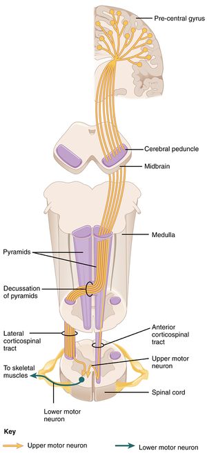
The Corticospinal Tract (also shortly referred to as CST), further recognized as the Pyramidal Tract, is a network of axons that connects the spinal cord to the cerebral cortex. It includes many structural and functional components of the neuromuscular system as it runs downwards towards the spinal cord, such as the decussation of pyramids, motor neurons, the cerebral peduncles, precentral gyrus, and skeletal muscle cells which are the target tissues that receive the motor information sent from the higher parts of the brain. In humans, it demonstrates actual command of motor skills and is most simply in command of perfect, digital movement patterns. In case of injury, side effects will be exhibited on the contralateral part of the body when there is only a unilateral malformation of the right or left corticospinal tract.
What percentage of the corticospinal tract does not decussate?
The remaining ten percent of corticospinal tract fibres do not decussate; instead, they do proceed to move down into the ipsilateral spinal cord; and this particular segment of the corticospinal tract is recognized as the anterior corticospinal tract.
What is the pyramidal trunk?
The Corticospinal Tract (also shortly referred to as CST), further recognized as the Pyramidal Tract, is a network of axons that connects the spinal cord to the cerebral cortex.
What is the function of motor nerve cells?
Motor nerve cells are the brain and spinal cord cells that help the body to move, talk, gulp down food and drinks, and inhale and exhale by transmitting signals from the brain to the muscles cells of the body that performs these tasks . The nerve fibres of motor neurons are of the maximum length in the body, with a singular axon stretching from the bottom surface of the spinal cord to the feet.
Where does the sloping spinal tract stem from?
It is a component of the sloping spinal tract system, which stems from the cortex or brainstem (Figure 1).
Which nerve cells move in the corticospinal tract?
Upper motor neurons are the nerve cells that move in the corticospinal tract; they recombine on lower motor neurons in the spinal cord, which communicate directly with skeletal muscle to cause muscle activity. The figure below gives a visual concept of the corticospinal tract and the components of the nervous system it incorporates, which are briefly explained below.
What is the corticospinal tract?
The Corticospinal Tract: Through the Spinal Cord and Peripheral Nervous System. The Lateral Corticospinal Tract. The Ventral Corticospinal Tract. Summary of the Role of the Corticospinal Tract. References. The corticospinal tract, also known as the pyramidal tract, is one of the descending spinal tracts necessary for the passing ...
How Does the Corticospinal Tract Communicate With the Rest of the Nervous System?
The corticospinal tract maintains connections with multiple regions of the cerebrum, primarily the motor cortex. The motor cortex is recognized to have three main components, the primary motor cortex, premotor cortex, and the supplementary motor area – each of these maintain their own unique connections and methods of communication with the corticospinal tract.
What are the two sub-tracts of the corticospinal tract?
Just like many other major nerve tracts, the corticospinal tract can be divided into two sub-tracts: the lateral corticospinal tract and the ventral (anterior) corticospinal tract.
What is the term for the extension of the nerve fibers that travel out to the peripheral nervous system?
As the nerve fiber travels out of the central nervous system, they transition into what is known as the “ lower motor neuron .” The lower motor neurons are the extension of the nerve fibers that travel out to the peripheral nervous system in order to innervate the skeletal muscles.
What are the fibers that connect the spinal cord to the cranial nerves?
These nerves are not called cortico spinal nerves, however, they are instead called “cortico nuclear fibers” since they innervate cranial nerve nuclei.
Why is proprioceptive information important?
With proprioceptive information, your brain is able to develop a more accurate motor plan since it will be aware of the original positioning of your body. For instance, if you wanted to scratch the itch on your eyebrow, but your arm was buried under a blanket, your motor plan would be to free your arm from the blanket, lift your arm, and scratch.
Which spinal tract is responsible for the transmission of information from the central nervous system to the peripheral nervous system?
The corticospinal tract , also known as the pyramidal tract, is one of the descending spinal tracts necessary for the passing of information from the central nervous system to the peripheral nervous system, particularly to musculature of the axial region of the body (the trunk) and distal regions (limbs and fingers/toes).
Where does the corticospinal tract originate?
The corticospinal tract originates in several parts of the brain , including not just the motor areas, but also the primary somatosensory cortex and premotor areas. Most of the neurons originate in the primary motor cortex (precentral gyrus, Brodmann area 4) or the premotor frontal areas.
Where do the corticospinal and corticobulbar tracts pass?
Then both tracts pass through the brain stem, from the pons and then to the medulla. The corticospinal tract, along with the corticobulbar tract, form two pyramids on either side of the medulla of the brainstem—and give their name as pyramidal tracts.
How many neurons are in the corticospinal tract?
There are more than one million neurons in the corticospinal tract, and they become myelinated usually in the first two years of life. The corticospinal tract is one of the pyramidal tracts, the other being the corticobulbar tract .
What is the function of the pyramidal tract?
Function. The primary purpose of the corticospinal tract is for voluntary motor control of the body and limbs. However, connections to the somatosensory cortex suggest that the pyramidal tracts are also responsible for modulating sensory information from the body. Because some of the connections cross the midline at the level ...
What is the fastest conduction rate of any signal from the brain to the spinal cord?
These cells are notable because of their rapid conduction rate, over 70m/sec, the fastest conduction of any signals from the brain to the spinal cord. There are two divisions of the corticospinal tract, the lateral corticospinal tract and the anterior corticospinal tract.
Where do the anterior corticospinal tract neurons stay?
The anterior corticospinal tract neurons, the remaining 10%, stay ipsilateral in the spinal cord but decussate at the level of the spinal nerve in which they exit, and control the trunk, shoulder and neck muscles.
Which white matter pathway is the corticospinal tract?
Pyramidal white matter motor pathway. Corticospinal pathway. The corticospinal tract is a white matter motor pathway starting at the cerebral cortex that terminates on lower motor neurons and interneurons in the spinal cord, controlling movements of the limbs and trunk.
What is the lateral corticospinal tract?
The lateral corticospinal tract can suffer damage in a variety of ways. The most common types of injury are central cord syndrome, Brown-Sequard syndrome, and anterior spinal cord syndrome. The corticospinal tract controls primary motor activity for the somatic motor system from the neck to the feet. It is the major spinal pathway involved in ...
Which side of the brain controls the right side of the spinal cord?
This crossover causes the left side of the brain to control the right side of the spinal cord and the right side of the brain to control the left side of the spin al cord. A small number of axons remain on the ipsilateral side to form the anterior corticospinal tract.
What are the most common types of spinal cord injuries?
The most common types of injury include central cord syndrome, Brown-Sequard syndrome, and anterior spinal cord syndrome. Improved patient outcome measures may be facilitated by prompt diagnosis and management of spinal cord trauma.
How to evaluate spinal cord injury?
For any spinal cord lesion, the extent of trauma needs to be evaluated. If the injury involves the cervical region, cervical immobilization should be performed during the initial evaluation to prevent additional injury to the cord. An exam should include all primary functions of the spinal cord (i.e., motor, primary touch, proprioception, autonomic function, and pain, temperature, and light touch). Assessment of sensory function for primary touch as well as pain and light touch can be performed by touching a patient at various dermatome regions of the body with a blunt or sharp object. To assess corticospinal tract function, examine muscle tone and spasticity for extensors and flexors of the arms and legs. Test for motor strength and function by having the patient move different groups of muscles with and without resistance — test for proprioception with a finger to nose test, rapid alternating movements test, or Romberg test. If the patient is ambulatory, examine their gate for motor ability and coordination.[13] If the injury occurs in the lower lumbar region, the bowel and bladder can be affected.[8] In these cases, the rectal tone can be assessed to determine the severity of autonomic compromise.
What are the bilateral deficits in spinal cord syndrome?
Patients with anterior spinal cord syndrome have bilateral deficits to the corticospinal tracts and spinothalamic tracts. They experience bilateral paralysis and paresis below the site of the lesion.[10] They also experience bilateral pain/temperature and light touch deficits due to the injury of the spinothalamic tract.[12] Sacral sparing of the spinothalamic tract occurs due to a dual blood supply by the posterior spinal arteries. The posterior spinal arteries wrap around the peripheral aspect of the cord. This secondary supply enables the full functionality of the peripheral spinothalamic tract, which transmits pain, temperature, and light touch for the feet.
Where do axons move?
Axons of both anterior and lateral corticospinal tracts move into the gray matter of the ventral horn to synapse onto lower motor neurons. These lower motor neurons exit the spinal cord to contract muscle.[1] .
Which tract of the body is responsible for motor control?
While the anterior corticospinal tract assists with axial muscle motor control, the lateral corticospinal tract is the primary pathway for motor information to the body. Injuries to the lateral corticospinal tract results in ipsilateral paralysis (inability to move), paresis (decreased motor strength), and hypertonia (increased tone) ...
What is the function of the lateral corticospinal tract?
The primary responsibility of the lateral corticospinal tract is to control the voluntary movement of contralateral limbs.[1] The origination of the Lateral corticospinal tract is in the primary motor cortex which lies in the precentral gyrus. When a stimulus is engaged, the cell body of the lateral corticospinal tract (in the primary motor cortex, the upper motor neuron) will send an impulse through the tract that will eventually travel to the anterior horn of the spinal cord from where it will transmit the impulse via lower motor neurons into the muscle fibers. This pathway can be scrutinized into greater detail.
What diseases affect the lateral corticospinal tract?
This includes strokes, poliomyelitis, spinal muscular atrophy, amyotrophic lateral sclerosis, vitamin B12 deficiency, Friedreich ataxia, and Brown-Sequard syndrome.
What is the defining feature of whether there will be ipsilateral or contralateral deficits?
Due to the pyramidal decussation of the lateral corticospinal tract in the caudal medulla, damage rostral or caudal to this decussation will be the defining feature of whether there will be ipsilateral or contralateral deficits. For example, if there is a lesion in the precentral gyrus of the left cerebral cortex, the patient will exhibit upper motor neuron signs with damage to the right side of the body. Contrarily, if there is spinal cord damage on the left side (below the pyramidal decussation), motor deficits will be present on the left side of the body. If there is spinal cord damage at the level of the anterior horn, then lower motor neuron signs will be present with ipsilateral deficits.
What are the pathways that connect the brain and spinal cord?
There are also pathways connecting different areas of the brain. These pathways are called white matter tracts in the central nervous system (CNS). These are all nerve fibers covered with myelin sheaths which are derived from oligodendrocytes. The corticospinal tract belongs to one of the most important descending tracts of the CNS. It contains fibers from the upper motor neurons to synapse on the lower motor neurons. Upper motor neurons (UMN) can be described as the nerve fibers responsible for the communication between the brain to the spinal cord. Lower motor neurons (LMN) are the nerve fibers responsible for the communication between the spinal cord to muscle.
What is spinal muscular atrophy?
Spinal muscular atrophy, also known as Werdnig-Hoffman disease in infants and Kugelberg-Welander disease in juveniles, is a congenital degeneration of the anterior horns of the spinal cord. Since this disease results in symmetric degeneration of the anterior horns, it results in symmetric weakness with lower motor neuron signs. Infants are characteristically described as "floppy babies" with associated tongue fasciculations. This disease carries an autosomal recessive inheritance with a mutation in the SMN1gene. [4]
What is the term for damage to the upper motor neurons in the cerebral cortex?
Wallerian degeneration of the corticospinal tract can often be visualized in computed tomogram (CT) or magnetic resonance imaging ( MRI) of the brain (see below).
Where does the impulse go when moving a leg?
When a motor act is planned and initiated by the premotor and supplemental motor cortex to the primary motor cortex in the precentral gyrus to move the leg, an impulse generated from the primary motor cortex will be conducted through the lateral corticospinal tract ipsilaterally through the corona radiata. It passes through the posterior limb of the internal capsule, through the cerebral peduncle and basis pontis, decussates at the caudal medulla (pyramidal decussation), and then continues to descend contralaterally into the spinal cord. Once that impulse reaches the cell body in the anterior horn (lower motor neuron) of the spinal cord, the motor fibers from the lower motor neurons will leave the spinal cord, proceed through the spinal nerve root, plexus, peripheral nerve, and finally to the neuromuscular junction where the impulse is transmitted to the muscle fibers resulting in contraction of that limb muscles. Damage to any of these structures may cause motor deficits.
What is the function of the lateral corticospinal tract?
Function. The lateral corticospinal tract is responsible for the voluntary movement of the contralateral upper and lower limbs. The upper motor neurons of the LCST, the giant pyramidal cells of Betz, preserve a somatotopic organization, called the motor homunculus.
Where do corticospinal fibers decussate?
In the anterior aspect of the lower medulla, the majority of corticospinal fibers decussate ( pyramidal decussation ). The crossed fibers form the lateral corticospinal tract while the uncrossed fibers enter the anterior corticospinal tract. The former is responsible for providing voluntary motor information to the muscles of the limbs while the latter supplies the axial muscles of the trunk. Both tracts run along the spinal cord, synapsing with lower motor neurons in the anterior gray horn on the same side. The lower motor neurons leave the spinal cord through the ventral root and form peripheral nerves which innervate the musculature of the body.
What is the difference between the corticobulbar and corticospinal?
Decussation of the corticospinal tract occurs at the junction of the medulla oblongata and spinal cord while the corticobulbar tracts decussate above each relevant cranial nerve nuclei. Thus lower motor neurons of the musculature of the body receive motor input mostly from the contralateral hemisphere, the lower motor nuclei of cranial nerves receive bilateral innervation. With regards to the corticospinal tract this means that injury above the pyramidal decussation leads to contralateral motor deficits. Whereas damage below the pyramidal decussation will result in ipsilateral motor deficits.
What is the corticobulbar tract?
Corticobulbar tract. Definition: motor pathway from the motor cortex of the brain to the motor nuclei of cranial nerves within the brainstem. Function: responsible for voluntary movement of the muscles of the face (CN. VII), head and neck (CN.
Which part of the LCST is responsible for innervation of the cervical region?
This somatotopic organization is preserved all along the corticospinal tracts, whereby the more medial part of the LCST is responsible for innervation of the cervical region and the lateral part of the tract sends efferent output to the lower thoracic, lumbar and sacral regions, respectively.
Which tract is responsible for innervating the muscles of the face, head and neck, as well as the muscles involved?
Therefore the corticobulbar tract is responsible for innervating the muscles of the face, head and neck, as well as the muscles involved in swallowing, phonation and facial expression. This article will describe the anatomy and function of the corticobulbar and corticospinal tracts. Key facts about the corticobulbar and corticospinal tracts.
Which tract provides voluntary control of muscular movements?
Corticospinal and corticobulbar tract (Tractus corticospinalis et corticobulbaris) The pyramidal tract provides voluntary control of muscular movements. It consists of two distinct pathways, the corticobulbar tract and the corticospinal tract.