The deep fibular nerve innervates the muscles in the anterior compartment of the leg, including:
- Tibialis anterior
- Extensor hallucis longus
- Extensor digitorum longus
- Fibularis tertius
Is fibular nerve same as peroneal nerve?
The deep branch of the fibular nerve, also known as the deep fibular (peroneal) nerve, arises from the bifurcation of the common fibular nerve as well. This nerve runs through the interosseous membrane to enter the extensor (anterior) compartment of the leg for which it provides innervation.
What muscles does the common peroneal nerve innervate?
function and location The peroneal nerve, from the ventral division, travels to the anterior surface of the leg and innervates the tibialis anterior, the fibularis muscles, and extensor muscles that elevate the foot and fan the toes.
Which nerve innervates the supinator?
Supinator muscle
- Origin and insertion. The wide supinator muscle consists of superficial and deep layers. ...
- Relations. Supinator is found deep in the forearm, superficial only to the parts of the radius and ulna over which the muscle lies.
- Innervation. Supinator is innervated by the posterior interosseous nerve (C7, C8), a branch of the radial nerve. ...
- Blood supply
- Function. ...
What nerve innervates the peroneal muscles?
The deep peroneal nerve sends off motor branches to several muscles in the calf, including: 1
- Tibialis anterior
- Extensor hallucis longus
- Extensor digitorum longus
- Fibularis tertius

Where does the deep fibular nerve terminate?
The deep fibular nerve terminates in the dorsum of the foot as a cutaneous nerve. This innervates the webbed space of skin between the great toe (hallux) and the second toe.
Why is the deep fibular nerve compressed?
There are two main reasons why the deep fibular nerve could be compressed. The first is that the anterior leg muscles have been excessively used and so are compressing the nerve within the anterior compartment. The patient will experience pain in the anterior leg.
What causes the deep fibular nerve to become entrapped?
This causes paralysis of the muscles in the anterior compartment of the leg, and so a patient loses the ability to dorsiflex the foot.
What is the function of the nerve roots?
Motor function: Innervates the muscles in the anterior compartment of the leg, as well as some of the intrinsic muscles of the foot. Sensory function: Supplies the triangular region of skin between the 1st and 2nd toes.
What is the deep peroneal nerve?
The deep fibular nerve (deep peroneal nerve) is a nerve of the leg. It is one of the terminal branches of the common fibular nerve. In this article, we shall look at the anatomy of the deep fibular nerve – its anatomical course, motor and sensory functions, and any clinical relevance.
Which nerve descends first, the tibialis anterior or the extensor hallucis longus?
During its descent, the deep fibular nerve is initially lateral, then anterior and finally medial to the anterior tibial artery.
Which nerve innervates the muscles in the anterior compartment of the leg?
The deep fibular nerve innervates the muscles in the anterior compartment of the leg, including: These muscles are responsible for dorsiflexion of the foot at the ankle joint. During the gait cycle for walking, dorsiflexion is required: When a person strikes their heel on the floor in the stance phase.
What is the deep fibular nerve?
Deep Fibular (Peroneal) Nerve. The deep fibular nerve was very recently known as the anterior tibial nerve. It begins at the bifurcation of the common fibular nerve, between the fibula and the proximal part of the fibularis longus. It passes obliquely forward, deep to the extensor digitorum longus, to the front of the interosseous membrane.
Where is the deep fibular nerve located?
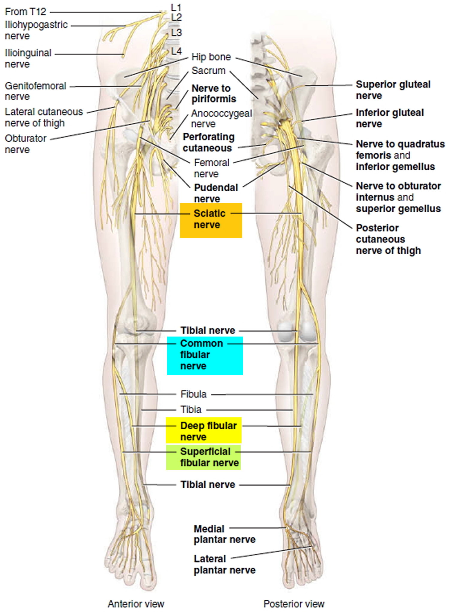
The deep fibular nerve begins in the lateral compartment deep to the fibularis longus as one of the two divisions of the common fibular nerve, which was previously called the deep peroneal nerve. It continues around the fibular neck, piercing the anterior intermuscular septum, and runs obliquely forward between the extensor digitorum longus and tibialis anterior on the interosseous membrane, where it lies with the anterior tibial artery ( Figure 46.5 ). It descends with the artery and lies first lateral, then anterior, and finally lateral to it at the ankle, where it divides into its terminal branches.
What muscles does the tibialis anterior have?
In the leg, it supplies the four muscles of the anterior compartment: tibialis anterior, extensor digitorum longus, extensor hallucis longus, and fibularis tertius. It gives off an articular branch above the inferior extensor retinaculum to supply the ankle joint.
What are the four muscles of the anterior compartment?
The four muscles of the anterior compartment are the tibialis anterior, extensor digitorum longus, extensor hallucis longus, and fibularis tertius. As a group, these muscles originate at the proximal tibia, at the adjacent fibula, and at the interosseous membrane. All four muscles are innervated by the deep fibular nerve, and all four perform dorsiflexion as one of their primary actions.
What nerve divides into two dorsal digital nerves?
Before dividing, it gives off an interosseous branch that supplies the first metatarsophalangeal joint. The deep fibular nerve may end as three terminal branches instead of two.
What is the lateral terminal branch of the ankle?
It is usually divided into medial and lateral branches. It also gives an articular branch to the ankle joint. The lateral terminal branch crosses the ankle deep to the extensor digitorum brevis and supplies the extensor digitorum brevis.
How many terminal branches does the deep fibular nerve have?
The deep fibular nerve may end as three terminal branches instead of two. A number of variations in the digital distribution of the nerve have been reported. It may supply the medial side of the great toe, adjacent sides of the second and third toes, or the lateral 3.5 toes. The nerve may have no digital branches at all.
Where does the fibular nerve go?
The nerve then pierces the fascia of the leg to enter the anterior compartment of the leg. The common fibular nerve wraps around the fibular neck. Then, the nerve terminates by dividing into two terminal branches, the superficial and deep fibular (peroneal) nerves, underneath the fibularis longus muscle.
Where is the common fibular nerve located?
Common fibular (peroneal) nerve. The common fibular (peroneal) nerve (L4-S2) is a short, thin nerve located in the posterior compartment of the lower extremity . It arises as a terminal branch of the sciatic nerve around the apex of the popliteal fossa . The common fibular (peroneal) nerve courses inferolaterally through the popliteal fossa, ...
What is the superficial branch of the peroneal nerve?
The superficial branch of the common peroneal nerve, also known as the superficial fibular (peroneal) nerve, arises at the bifurcation of the common peroneal nerve, between the fibula and the proximal part of fibularis longus muscle. This nerve then descends deep to the fibularis longus muscle.
What nerve divides the anterior compartment of the leg?
When it reaches the anterior compartment of the leg (underneath the fibularis longus muscle) , the nerve divides into the superficial fibular (peroneal) nerve and deep fibular (peroneal) nerve. The superficial fibular (peroneal) nerve supplies the muscles of the lateral compartment of the leg and provides sensation to the anterolateral aspect ...
Why is the fibula nerve so close to the neck?
Injuries to the common fibular nerve are not uncommon due to the fact that this nerve is relatively unprotected. Its close proximity to the neck of the fibula makes it vulnerable to damage when the bone gets injured. The nerve can also be damaged due to compression caused by swelling or inflammation in the fibular compartment of the leg.
What is the root value of the common fibular nerve?
The common fibular (peroneal) nerve (root value L4-S2) is the smaller of two terminal branches of the sciatic nerve, the other being the tibial nerve . The common fibular (peroneal) nerve contains mainly fibers derived from the posterior division of the sacral plexus .
Which nerve provides sensory supply to the skin of the lower anterolateral aspect of the leg and most of the do?
The common fibular nerve provides sensory supply for the skin of the lower anterolateral aspect of the leg and most of the dorsum of foot. More specifically, the superficial branch provides sensory supply to the anterolateral aspect of the leg, extending from midway down the leg to the majority of the dorsal aspect of the foot and toes.
Which nerve innervates the extensor hallucis brevis?
After its bifurcation past the ankle joint, the lateral branch of the deep peroneal nerve innervates the extensor digitorum brevis and the extensor hallucis brevis, while the medial branch goes on to provide cutaneous innervation to the webbing between the first and second digits.
Which nerve passes through the tarsus, beneath the extensor digitorum brevis,?
Before it divides it gives off to the first space an interosseous branch which supplies the metatarsophalangeal joint of the great toe and sends a filament to the first Interosseous dorsalis muscle. Lateral terminal branch - This nerve passes across the tarsus, beneath the extensor digitorum brevis, supplying latter.
What nerve supplies the extensor muscles?
In the leg, the deep peroneal nerve supplies muscular branches to the anterior compartment of extensor muscles in the leg which include the tibialis anterior, extensor digitorum longus, peroneus tertius, and extensor hallucis longus (propius), and an articular branch to the ankle-joint. After its bifurcation past the ankle joint, the lateral branch of the deep peroneal nerve innervates the extensor digitorum brevis and the extensor hallucis brevis, while the medial branch goes on to provide cutaneous innervation to the webbing between the first and second digits.
What nerve is the right lower extremity?
Deep peroneal nerve. Nerves of the right lower extremity Posterior view. The deep peroneal nerve begins at the bifurcation of the common peroneal nerve between the fibula and upper part of the peroneus longus, passes infero-medially, deep to extensor digitorum longus, to the anterior surface of the interosseous membrane, ...
What nerve is on the lateral side of the leg?
Lateral side of the leg. Deep peroneal nerve is the nerve of the anterior compartment of the leg and the dorsum of the foot. It is one of the terminal branches of the common peroneal nerve. It corresponds to the posterior interosseus nerve of the forearm. It begins at the lateral side of the fibula bone, and then enters ...
Where does the deep peroneal nerve begin?
Anatomical terms of neuroanatomy. The deep peroneal nerve begins at the bifurcation of the common peroneal nerve between the fibula and upper part of the peroneus longus, passes infero-medially, deep to extensor digitorum longus, to the anterior surface of the interosseous membrane, and comes into relation with the anterior tibial artery above ...
Which nerve terminates in the ankle?
Close to the ankle joint, deep peroneal nerve terminates by dividing into medial and lateral terminal branches. Medial terminal branch - This nerve accompanies the dorsalis pedis artery along the dorsum of the foot, and, at the first interosseous space, divides into two dorsal digital nerves which supply the adjacent sides ...
Which nerve is responsible for pulling the foot back?
By innervating the tibialis anterior, extensor hallucis longus, extensor digitorum longus, and fibularis tertius, the deep peroneal nerve is responsible for pulling the foot back—the opposite motion of pointing the toes. This motion, which is called dorsiflexion, is important for walking. Dorsiflexion is required both when your heel strikes ...
Which nerve is located in the back of the leg?
Tibial nerve. Common peroneal nerve. The tibial nerve continues down the back of the leg while the common peroneal nerve wraps around the outside of your knee to get to the front of the calf. Just below the knee, the common peroneal nerve separates into two terminal branches: 1 . Superficial peroneal nerve.
What is the deep peroneal nerve?
Rehabilitation. The deep peroneal nerve, also called the deep fibular nerve, is a peripheral nerve of the calf. It's a terminal branch of the common peroneal nerve, which is a branch of the sciatic nerve. The deep peroneal nerve contains both motor and sensory fibers.
Which branch of the deep peroneal nerve connects to the extensor hallucis brevis?
The medial branch, which is a cutaneous (of the skin) nerve. The lateral and medial are the terminal branches of the deep peroneal nerve.
Which nerve sends off motor branches to several muscles in the calf?
The deep peroneal nerve sends off motor branches to several muscles in the calf, including: 1 . It also sends a branch to the ankle joint, then puts off two branches into the foot: The lateral branch, which connects to the extensor digitorum brevis and extensor hallucis brevis muscles.
Which nerve provides motor function to muscles?
The upper portion of the deep peroneal nerve provides motor function to muscles, while the lower portion provides both motor and sensory function to portions of the foot. 1
What test measures how fast electrical signals move through the nerves?
Nerve conduction tests , which measure how fast electrical signals move through the nerves
What nerves are involved in peroneal nerve injury?
What is a peroneal nerve injury? A peroneal nerve injury affects a major nerve in your leg called the fibular or common peroneal nerve. This nerve starts in the back part of your knee and allows you to feel the outsides of the lower legs, the tops of the feet, and the skin between the big toe and second toe. It also controls some of the muscles in ...
What is a peroneal nerve injury?
A peroneal nerve injury affects a major nerve in your leg called the fibular or common peroneal nerve. This nerve starts in the back part of your knee and allows you to feel the outsides of the lower legs, the tops of the feet, and the skin between the big toe and second toe. It also controls some of the muscles in the leg and the foot.
What causes a peroneal nerve to hurt?
Causes of a peroneal nerve injury. The common peroneal nerve runs very close to the surface of your skin just below the knee, which is why it is so easy to injure. It can become injured in the following ways: A cut through the nerve (such as by trauma or during an orthopedic surgery).
Why does one foot feel numb?
Numbness and tingling in one foot can be commonly caused by nerve damage that can lead to sciatica, tarsal tunnel syndrome, or a fibular nerve injury. Read below for more information on causes of numbness in one foot and how to find relief. Lower Leg. Tingling Lower Leg.
Why do my feet go numb?
Numbness in the feet can be caused from trauma from an injury or nerve damage to any part of the leg that may affect your feet. Other causes of feet numbness include restless leg syndrome, sciatica, or tarsal tunnel syndrome. Read below for more information on causes and treatment options.
Can a broken peroneal nerve be repaired?
You can also injure it by wearing high heels for long periods of time. Treatment depends on how severe your injury is, but can include surgery to repair or replace the damaged nerve.
Can you wear high heel shoes with fibular nerve entrapment?
Over-the-counter pain medications. You may have to avoid wearing high heel shoes and crossing your legs. If you have a deep fibular nerve entrapment injury: Change your shoes from high heels or tight-fitting shoes to supportive shoes with a well-padded tongue. Try different shoe lacing configurations.