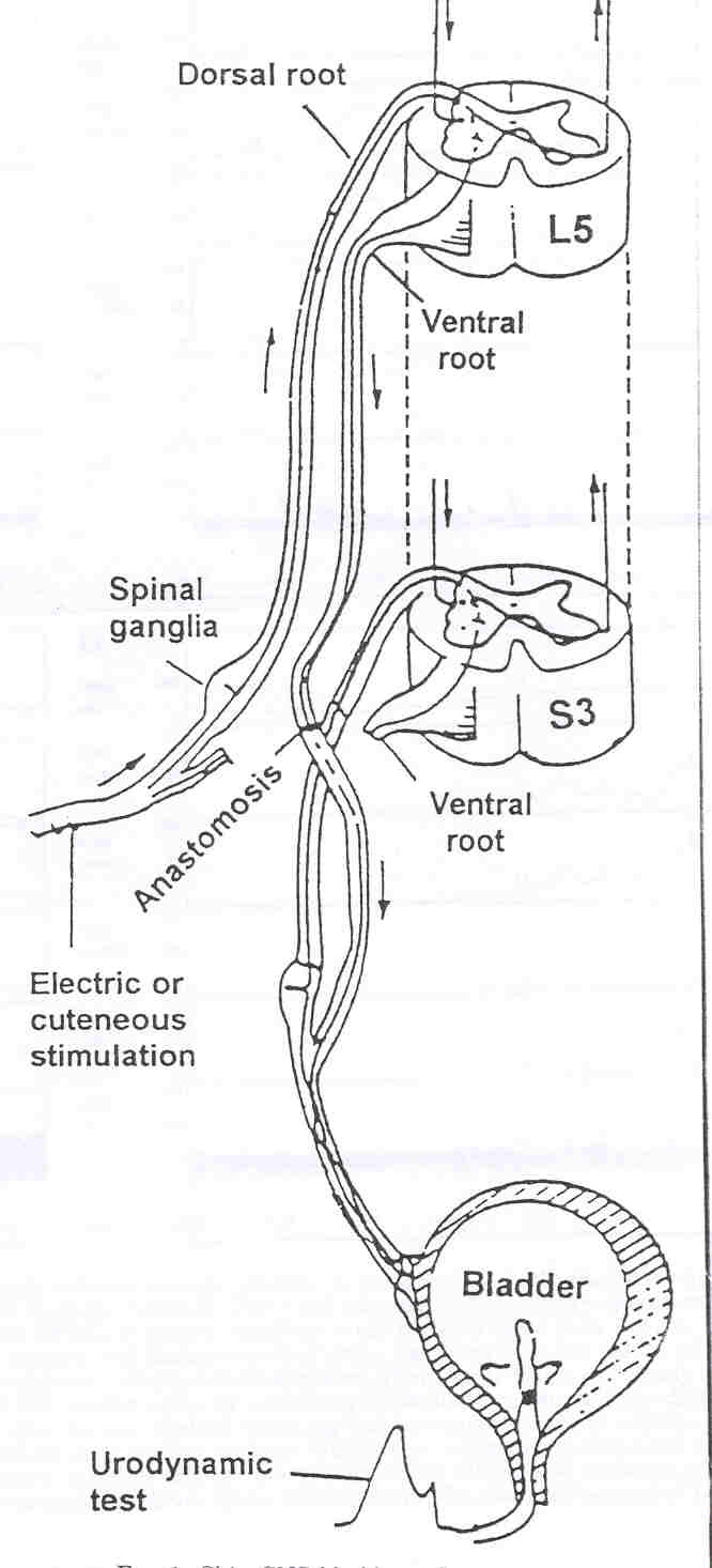
The lumbar nerve roots are pairs of nerves that exit from the spinal cord, below each vertebra in the lumbar spine. These include L1, L2, L3, L4, and L5, and are named for the vertebra above them. These nerve roots are made up of a front or anterior root that controls motor movements, and a back or posterior root that controls sensory feelings.
What does the L1 L2 L3 and L4 nerve do?
L1 spinal nerve provides sensation to the groin and genital regions and may contribute to the movement of the hip muscles. L2, L3, and L4 spinal nerves provide sensation to the front part of the thigh and inner side of the lower leg. These nerves also control movements of the hip and knee muscles.
What is the function of L1 vertebra?
L1 (1st Lumbar Vertebra) By: Tim Barclay, PhD. Last Updated: Oct 24, 2017. The L1 vertebra (1st lumbar vertebra) is the smallest and most superior of the lumbar vertebrae. As the first vertebra in the lumbar region, the L1 vertebra bears the weight of the upper body and acts as a transition between the thoracic and lumbar vertebrae.
What is the dermatomal distribution of the L1 and L2 spinal nerve?
The dermatomal distribution of the L1 spinal nerve is located in the groin and the upper part of the buttock. The distribution of the L2 spinal nerve is located in the outside thigh.
What are the symptoms of L1 nerve damage?
Consequently, what are the symptoms of l1 nerve damage? This damage is caused by compression of the nerve roots which exit the spine, levels L1- S4. The compression can result in tingling, radiating pain, numbness, paraesthesia and occasional shooting pain.

What are the symptoms of L1 nerve damage?
What Are the Symptoms of an L1 Injury? The L1 vertebra is the topmost section of the lumbar spinal column. This section of the spine contains a portion of the spinal cord. Injuries to the L1 spine can affect hip flexion, cause paraplegia, loss of bowel/bladder control, and/or numbness in the legs.
What nerves are affected by L1?
A limited description of the specific lumbar spinal nerves includes: L1 innervates the abdominal internal obliques via the ilioinguinal nerve; L2-4 innervates iliopsoas, a hip flexor, and other muscles via the femoral nerve; L2-4 innervates adductor longus, a hip adductor, and other muscles via the obturator nerve; L5 ...
Where does L1 cause pain?
L1 or L2 symptoms include pain in lower back and groin area and/or pain that radiate to upper front and inside of thigh. L3 or L4 symptoms include pain in lower back and /or pain that radiates to the quadriceps in the front of the thigh.
What are the symptoms of L1 nerve root compression?
Definition/Description This damage is caused by compression of the nerve roots which exit the spine, levels L1- S4. The compression can result in tingling, radiating pain, numbness, paraesthesia, and occasional shooting pain.
What type of paralysis would occur at L1?
Lumbar level injuries result in paralysis or weakness of the legs (paraplegia).
What nerve is between L1 and L2?
There are five lumbar nerve pairs. The first of these nerve roots exits between L1 and L2. There are five sacral nerve pairs.
Where is the L1 nerve root?
Your lumbar vertebrae, known as L1 to L5, are the largest of your entire spine. Your lumbar spine is located below your 12 chest (thoracic) vertebra and above the five fused bones that make up your triangular-shaped sacrum bone.
What nerves are affected by T12 and L1?
T12-L1 Pinched Nerve: The T12 spinal nerves are responsible for the abdominal muscles and the skin over the buttocks. A pinched nerve at this level may cause pain into the buttocks or over the abdomen. Localized symptoms of pinched nerve in the thoracic spine may include pain or stiffness of the midback.
What does narrowing of L1 and L2 mean?
In this study, disk space narrowing at level L1/L2 appeared to be associated with pain in the hip region, especially in men. The strength of the associations increased for participants with chronic hip pain and in those without radiological signs of hip osteoarthritis.
How do you fix S1 nerve compression?
Common injection treatments for L5-S1 include:Lumbar epidural steroid injections. Steroids injected directly into the spinal epidural space can help decrease inflammation and reduce the sensitivity of nerve fibers to pain, generating fewer pain signals. ... Radiofrequency ablation.
Can a pinched nerve affect one side of your body?
Nerves also stimulate certain muscles and organs so they function properly. For nerves that serve the skin and musculoskeletal system, the symptoms of a pinched nerve affect the nerve's normal function. A pinched nerve generally affects only one side of the body. Its effects can range from mild to severe.
Is nerve root compression serious?
Nerve root compression that is severe enough to cause weakness in the arms or legs requires prompt diagnosis and surgical treatment because compression leads to death of the nerve cells and can permanently affect the function of the sensory and motor nerves downstream from the point of compression.
What is the L1 vertebral arch?
The vertebral arch is a thin bony ring attached to the posterior of the vertebral body. In the L1 vertebra, it is a bit smaller than the vertebral body, but is much thicker and stronger than the arches of the cervical and thoracic vertebrae above it.
What is the intervertebral disc?
Intervertebral discs - made of gel surrounded by strong, rubbery fibrocartilage - lie between the bodies of the L1 vertebra and its neighboring vertebrae. The intervertebral discs support the spinal column, absorb shock force and body weight, and provide flexibility to the lower back.
What is the body of L1?
Like the other lumbar vertebrae, L1 has a large, roughly cylindrical region of bone known as the body, or centrum, which makes up most of its mass. The bodies of lumbar vertebrae are much wider than they are deep, convex on their anterior surface and concave on their posterior surface. « Back Show on Map ». Anatomy Term.
Which process extends vertically from the vertebral arch?
Many muscles that flex, extend, rotate and stabilize the lumbar spine attach to the spinous process. Finally, a pair of articular processes extends vertically from the vertebral arch, with one connecting to the T12 vertebra above and the other to the L2 vertebra below.
Where does the spinous process extend?
The thin, rectangular spinous process extends posteriorly from the vertebral arch toward the skin of the back. In the L1 vertebra, it points more inferiorly than it does in any other lumbar vertebra, making it somewhat resemble the spinous processes of the neighboring thoracic vertebrae. Many muscles that flex, extend, ...
Which vertebra is the smallest?
The L1 vertebra (1st lumbar vertebra) is the smallest and most superior of the lumbar vertebrae. As the first vertebra in the lumbar region, the L1 vertebra bears the weight of the upper body and acts as a transition between the thoracic and lumbar vertebrae.
How many pairs of lumbar spinal nerves are there?
There are 5 pairs of lumbar spinal nerves that progressively increase in size from L1 to L5. These nerves exit the intervertebral foramina below the corresponding vertebra. For example, the L4 nerve exits beneath the L4 vertebra through the L4-L5 foramen. These nerves course down from the lower back and merge with other nerves to form ...
What are the two nerves that branch off from the right and left sides of the spinal cord?
Lumbar Spinal Nerves. Two spinal nerves branch off from the right and left sides of the spinal cord or the cauda equina at each spinal segment. These spinal nerves are formed by 2 types of fibers—sensory fibers that send messages to the brain (feeling pain when the leg is hurt) and motor fibers that receive messages from the brain ...
What nerve controls the hip, knee, foot, and toe?
The L5 spinal nerve controls hip, knee, foot, and toe movements. Read more about Spinal Cord and Spinal Nerve Roots. advertisement.
What type of nerve sends messages to the brain?
These spinal nerves are formed by 2 types of fibers—sensory fibers that send messages to the brain (feeling pain when the leg is hurt) and motor fibers that receive messages from the brain (lifting the leg to get out of a car).
Where does the spinal nerve travel?
The spinal nerve travels a short distance inside the intervertebral foramen, after which it branches off into several nerves that innervate different parts of the body. Doctors may sometimes refer to the part of the spinal nerve exiting the intervertebral foramen as the nerve root or use the terms nerve root and spinal nerve interchangeably.
Which spinal nerves innervate the lower limbs?
The 5 pairs of lumbar spinal nerves innervate the lower limbs. While innervation can vary among individuals, some common patterns include 2: L1 spinal nerve provides sensation to the groin and genital regions and may contribute to the movement of the hip muscles. L2, L3, and L4 spinal nerves provide sensation to the front part ...
Which nerves carry sensory information to the brain?
The dorsal root fibers carry sensory information from the dermatome to the brain. A myotome is a group of muscles controlled by the ventral root fibers of a spinal nerve. The ventral root fibers carry motor signals from the brain to the myotome. When a spinal nerve gets irritated or compressed, sensory and/or motor deficits may occur in ...
