What does each wave of an ECG represent?
What does each wave of an ECG represent? There are three main components to an ECG: the P wave, which represents the depolarization of the atria; the QRS complex, which represents the depolarization of the ventricles; and the T wave, which represents the repolarization of the ventricles. Click to see full answer.
What is an S wave?
S waves are a type of transverse wave. What this means is that the oscillations of an S wave’s particles are perpendicular to the wave propagation’s direction. The main restoring force is because of the shear stress. Therefore, the propagation of S waves cannot take place in liquids with very low or zero viscosity.
What is the normal speed of an ECG?
A standard ECG is recorded at 25mm/sec and with a frequency cut off of no lower than 150Hz in adults, and 250Hz in children. On the standard ECG paper, with standard calibration, the squares represent: The standard calibration signal will look like this: This will be present at the beginning or end of all four rows of the trace, and shows:
What wave comes after the T wave of an ECG?
U wave on ECG occurs after the T wave and is usually seen in the mid precordial leads. In hypokalemia, T wave becomes flattened and U wave becomes prominent (or apparently so because of near absence of T waves). Important conditions associated with U waves are systemic hypertension, aortic and mitral regurgitation and coronary artery disease [1].

What does a large S wave indicate?
A prominent S-wave in lead I is typically present in cases of congenital heart disease, valvular heart disease, and cor pulmonale that cause right ventricular enlargement and fibrosis.
What is R wave and S wave in ECG?
So now it is possible to break down the QRS complex into 3 distinct waves: Q wave representing septal depolarisation. R wave representing ventricular depolarisation. S wave representing depolarisation of the Purkinje fibres.
What happens at the S wave?
An S wave, or shear wave, is a seismic body wave that shakes the ground back and forth perpendicular to the direction the wave is moving.
What is an abnormal S wave?
An S wave of less than 0.3 mV in lead V1 is considered abnormally small. If the amplitude of the entire QRS complex is less than 1.0 mV in each of the precordial leads, the voltage is considered abnormally low.
Why is R wave positive?
The R wave is very positive because early ventricular depolarization is largely directed toward this lead. The S wave is also present because the terminal depolarization of the upper wall of the left ventricle is directed away from aVF.
Why are Q and S waves negative?
By definition, a Q wave on the electrocardiogram (ECG) is an initially negative deflection of the QRS complex. Technically, a Q wave indicates that the net direction of early ventricular depolarization (QRS) electrical forces projects toward the negative pole of the lead axis in question.
Which phrases describe S waves?
This is Expert Verified Answer. Answer: The phrase that best describes S waves is that they behave like a ripple.
What is difference between P and S waves?
P waves can travel through liquid and solids and gases, while S waves only travel through solids. Scientists use this information to help them determine the structure of Earth. For example, if an earthquake occurs on one side of Earth, seismometers around the globe can measure the resulting S and P waves.
What are the characteristics of S waves?
S-wave Motion S Wave—secondary body waves that oscillate the ground perpendicular to the direction of wave travel. They travel about 1.7 times slower than P waves. Because liquids will not sustain shear stresses, S waves will not travel through liquids like water, molten rock, or the Earth's outer core.
What does AFIB look like on an ECG?
This means an ECG showing atrial fibrillation will have no visible P waves and an irregularly irregular QRS complex. The ventricular rate is frequently fast, unless the patient is on AV nodal blocking drugs such as beta-blockers or non-dihydropyridine calcium channel blockers.
What are the most common ECG abnormalities?
Electrocardiographic abnormalities include first-degree heart block, right and left bundle branch block, premature atrial and ventricular contractions, nonspecific T-wave changes, and evidence of ventricular hypertrophy.
What causes deep S wave?
Left ventricular hypertrophy causes increased R-wave amplitudes in V4–V6 and deeper S-waves in V1–V3.
Where is the S wave in ECG?
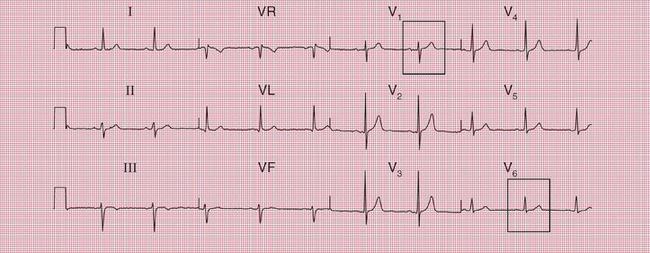
The S wave is the first downward deflection of the QRS complex that occurs after the R wave. However, a S wave may not be present in all ECG leads in a given patient. In the normal ECG, there is a large S wave in V1 that progressively becomes smaller, to the point that almost no S wave is present in V6.
What does a high R wave mean?
Tall R waves in V1 can be caused by abnormal electrical conduction (RBBB or left-sided VT, which slowly spreads across the right ventricle, or a left-sided accessory pathway), loss of posterior myocardium (old or acute posterior MI) or chronic anterior hypertrophy (HCM), chronic or acute RV strain (RVH, PE), congenital ...
What happens to the R wave and S wave as you go from V1 V6?
The R wave in the precordial leads steadily increases in amplitude from lead V1 to V6, with a corresponding decrease in S wave depth, culminating in a predominantly positive complex in V6. Thus, the QRS complex gradually changes from being predominantly negative in lead V1 to being predominantly positive in lead V6.
Is the R wave always positive?
The convention is that the Q wave is always negative and that the R wave is the first positive wave of the complex. If the QRS complex only includes an upward (positive) deflection, then it is an R wave. The S wave is the first negative deflection after an R wave.
1. What is the full form of ecg?
ECG stands for electrocardiograph.
2. How many different types of wave are there in ecg?
There are mainly three types of ECG peak:The P-wave represents the depolarisation of the atria, which leads to the contraction of both the atria.Th...
3. How is ecg helpful ?
ECG (electrocardiogram) measures or records the electrical activity of our heart at rest and also it provides information about our heart rate and...
4. Why is ecg done?
ECG is done because of following reasons:To detect any heart related issues.To detect whether you have had any heart attack.To find out if any medi...
5. Explain different types of ecg?
There are mainly three different types of ECG is performed:1. Resting ECG: when your body is in resting state.under this type machine examine your...
6. Define some differences between electrocardiogram and echocardiogram.
Both electrogram and electrocardiogram and echocardiogram is used to monitor the heart pulse rate. The major difference which differentiate these t...
7. How do you prepare for an electrocardiogram?
In order to prepare yourself for an electrocardiogram you need to follow some of the major steps which are described as below:Strictly avoid oily o...
8. What happens during an echocardiogram?
this particular test also is used to measure the heart pulses. Echocardiogram is a graphical representation of the outlines of the heart's moment....
9. Are you awake during a transesophageal echocardiogram?
The following steps will be carried out in order to make the process easier:While carrying out echocardiogram, a person is given a sedative which w...
10. What is the P wave on electrocardiogram?
In order to represent the electrical depolarization of the atria of the heart, P waves aur PR segment is used. This is an integral part of an elect...
Is the presence of the S wave clinically significant?
The presence or absence of the S wave does not bear major clinical significance. Rarely is the morphology of the S wave discussed.
Is there a S wave in V1?
In the normal ECG, there is a large S wave in V1 that progressively becomes smaller, to the point that almost no S wave is present in V6. A large slurred S wave is seen in leads I and V6 in the setting of a right bundle branch block. The presence or absence of the S wave does not bear major clinical significance.
Is the S wave present in lead I?
Rarely is the morphology of the S wave discussed. In the setting of a pulmonary embolism, a large S wave may be present in lead I — part of the S1Q3T3 pattern seen in this disease state.
What does an EKG tell you?
A lot!: For a cardiologist, an EKG can give information about the rate, the rhythm, whether there are signs of a congenital abnormality, signs of a heart attach or blocked artery, the thickness of the heart muscle, previous heart attack, and more. In experienced hands, an EKG can tell an incredible amount of information.
Is Q wave on EKG normal?
Probably nothing: Q waves of certain width and height on an EKG has been associated with an old heart attack. This hallmark is not very specific and if its boderline and no clinical history or no real cardiac risk factors , its probably a normal variant.
Is persistent S wave abnormal?
Ecg: Persistent S wave in the lateral precordium doesn't have any particular evil important, it is often a normal finding. Occasionally it can be seen with pathologic conditions but there are generally other associated findings.
Is a small S wave in a lead normal?
Depends: A small S wave in these leads is usually normal. A prominent S-wave may be seen in patients with right sided overload conditions, such as pulmonary disease. A large S wave in V1 and lead I when the QRS is wide is seen in right bundle branch block (RBBB). In rare cases, a person with dextrocardia may be diagnosed by ECG with one feature being a persistent s-wave in V5-V6. Review with your doctor.
What does ECG stand for?
ECG. ECG stands for electrocardiograph. It gives a graphical representation of the electrical activity of the heart during a cardiac cycle which helps to further detect the abnormalities and help us to measure the functioning of the heart. To obtain a standard ECG graph, a patient is connected to the machine with three electrical leads, ...
What are the different types of waves produced by an electrocardiogram?
Different Types of Waves Produced By Electrocardiogram. 1. The P-Wave. It represents the electrical excitation (or depolarisation) of the atria, which leads to the contraction of both the atria.
How do ECG electrodes work?
These electrodes are connected to the ECG machine with the help of wires that help in detecting the electrical impulses occurring at each heartbeat. These electrodes can detect every minute form of changes happening in heart muscles and draw every depolarising pattern of heartbeat on a graph.
What is resting ECG?
Resting ECG: when your body is in resting state.under this type machine examine your heart beat during resting condition. 2. Ambulatory ECG: This type of ECG is conducted for 24 hours. The heart’s electrical impulses are measured by a device called the Holter Monitor.
What is the peak of the ECG?
There are mainly three types of ECG peak: The P-wave represents the depolarisation of the atria, which leads to the contraction of both the atria. The QRS complex represents the depolarisation of the ventricles, which leads to the ventricular contraction.
What is the name of the test that measures the heart's electrical impulses?
Ambulatory ECG: This type of ECG is conducted for 24 hours. The heart’s electrical impulses are measured by a device called the Holter Monitor. Cardiac Stress Test: This test is used to measure ECG when you are on an exercise bike.
What are the two determinants of QRS voltage?
Two determinants of QRS voltages are: Size of the ventricular chambers (i.e., the larger the chamber, the larger the voltage) Nearness of chest electrodes to ventricular chamber (the closer, the larger the voltage) 3. T-Wave.
What is the basic of ECG?
Basics of ECG- Interpretation of waves and intervals. A normal ECG is electrical representation of a normal heart beat or sinus rhythm. The cardiac action potential causing deporalization and repolarization of various cardiac tissues gives a pattern of rhythic change is Electrocardiograph which can be used to diagnose different diseases of the CVS.
What is normal ECG?
A normal ECG is electrical representation of a normal heart beat or sinus rhythm. The cardiac action potential causing deporalization and repolarization of various cardiac tissues gives a pattern of rhythic change is Electrocardiograph which can be used to diagnose different diseases of the CVS.
Where does the P wave originate?
A normal P wave originates from the Sinoatrial Node , SA node. It represents atrial depolarization.Normal P wave has a
What is the J point in a QRS?
The J point is the the junction between the termination of the QRS complex and the beginning of the ST segment. Abnormal J (slurring elevation) is seen in Hypothermia -” Osborne Wave.”
What is the most obvious part of the ECG?
QRS Complex. The QRS complex is the main spike seen in the standard ECG. It is the most obvious part of the ECG, which is clearly visible. The QRS complex represents the depolarization of ventricles. It shows the beginning of systole and ventricular contraction.
What does the P wave represent?
P wave- It represents the depolarization of atria and represents atrial contraction. It precedes the QRS complex
What does a widened QRS complex mean?
The widened or prolonged QRS complex indicates the bundle branch block or hyperkalemia.
