How long does a torn ATFL take to heal?
How long does a torn ankle ligament take to recover? Part of the recovery time may include physical therapy, stretching or exercise programs. A torn ligament can take anywhere from two weeks to several months to heal, depending on other factors of the injury like the person’s age and where the injury occurred.
Does ATFL tear require surgery?
Why an ATFL Rupture does not need surgery. A lot of people panic when they read “ruptured” on a scan or a report. A lot of the time, it’s a cause for concern. In the case of an ATFL rupture, not so much. The ATFL is one of three ligaments making up the lateral ankle complex, along with the PTFL and CFL. The role of the ATFL is to help stop the ankle rolling, particularly when the foot is pointing down.
What are the functions of annular ligament in femur?
annular ligament It runs around the radialhead form the anterior and the posterior margin o the radial notch, to approximate the radial head to he radial notch and enclose the radial circumference. It encircles 80% o the radial head and functions to maintain the relationship between the head o the radius and the humerus and ulna.
What causes anterior cruciate ligament injury?
The anterior cruciate ligament can be injured in several ways :
- Changing direction rapidly
- Stopping suddenly
- Slowing down while running
- Landing from a jump incorrectly Direct contact or collision, such as a football tackle
What is the function of the Talofibular ligament?
The anterior talofibular ligament (ATFL) is part of the lateral collateral ligament complex of the ankle. Its role is to stabilize the talus. It is also the weakest of the lateral collateral ankle ligaments.
Can you walk with a torn anterior talofibular ligament?
Small tears of the ATFL will cause pain, tenderness, and swelling, but walking is usually still possible. Larger ATFL tears will cause greater pain, swelling and bruising, and you may have difficulty walking.
How important is the anterior talofibular ligament?
The anterior talofibular ligament is an important structure in lateral ankle instability, particularly in a plantarflexed foot, but it otherwise adds little physical support and probably acts more for proprioception.
How long does it take for a torn anterior talofibular ligament to heal?
ligament. Mild tenderness and swelling around the ankle, typically recovers in 5-14 days. Partial tearing of anterior talofibular ligament and some tearing of the calcaneofibular ligament. Moderate tenderness and swelling around the ankle, typically will take 2-3 weeks to recover.
Does a tear of the anterior talofibular ligament require surgery?
Clinical Management Of ATFL Rupture The concern would be that they develop chronic instability in the ankle or an anterior ankle impingement long term, which would require surgery. This can occur in patients who either present to physiotherapy late, or are non- compliant with their rehab.
How do you treat a torn Talofibular ligament?
Rehabilitation Program Initial treatment of all grades of lateral ankle sprains consists of rest, ice, compression, and elevation (RICE), as well as nonsteroidal anti-inflammatory drugs (NSAIDs). Ice should be applied to the injured ankle for approximately 20 minutes, 3-4 times per day.
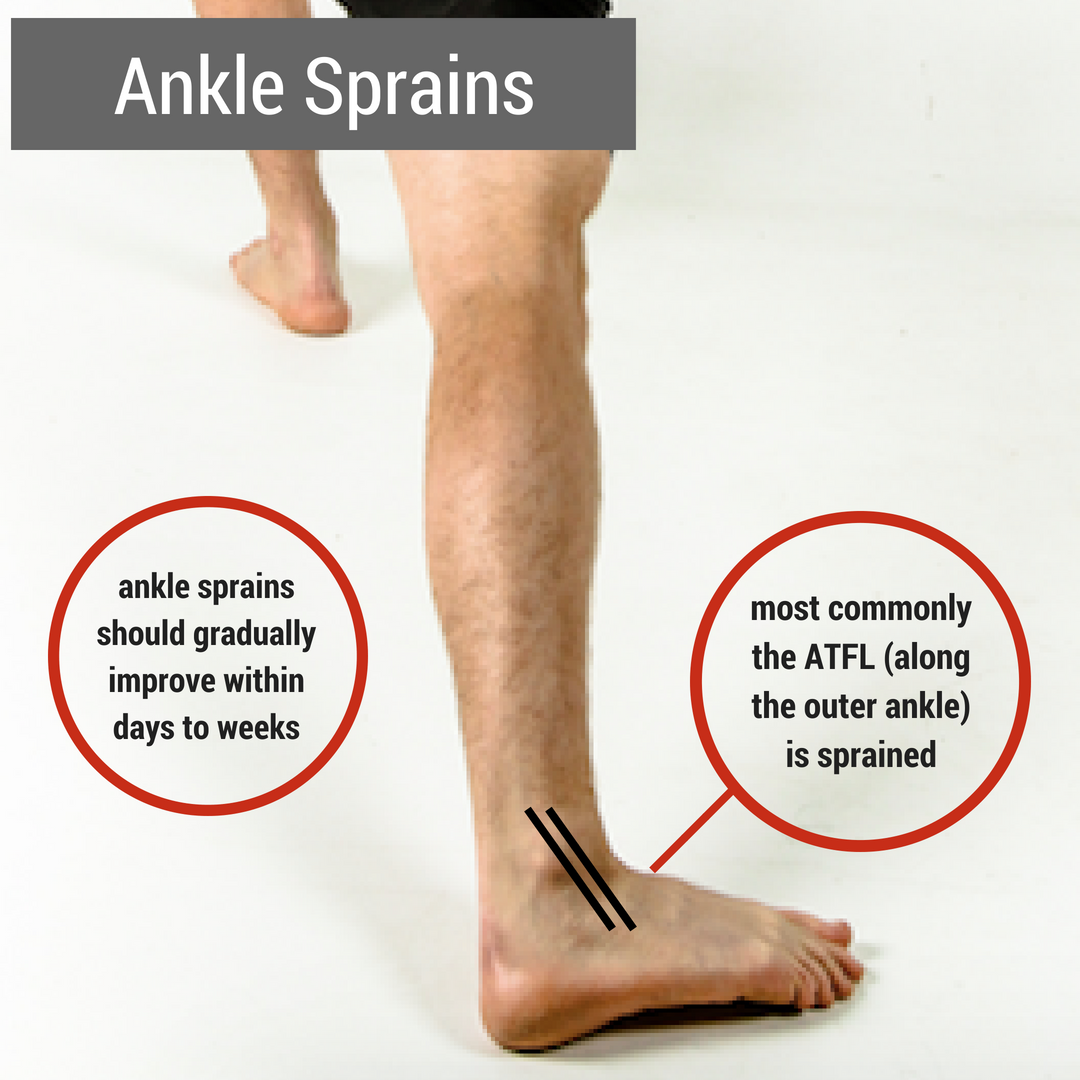
Why is ATFL ligament most commonly injured?
Due to its low ultimate load and the anatomical positions of origins and insertions, the ATFL is most commonly injured in a lateral ankle sprain [30]. The subtalar joint is formed by the articulation between the bottom of the talus and the calcaneus [18].
How do you know if you tore your anterior talofibular ligament?
Anterior talofibular ligament tear diagnosis Bony tenderness or deformity. Suspicion of a fracture or syndesmotic injury. Severe pain or swelling that makes the physical examination unreliable. Inability to walk.
Is an ATFL tear serious?
The ATFL is one of four major ligaments in the ankle that provides stability and flexibility. According to Dr. Van Boerum a sprain of the ATFL rarely requires surgery; however, if he continues to injure the ankle long term, he may need surgery down the road for chronic instability, but this is not very common.
Is walking good for ligament injury?
The short answer is yes. After the pain and swelling subsides and if there is no other injury to your knee, you may be able to walk in straight lines, go up and down stairs and even potentially jog in a straight line.
Which are the 3 most commonly injured ankle ligaments?
The three ligaments that compose the lateral complex are the anterior talofibular (ATFL), the calcaneofibular (CFL), and posterior talofibular (PTFL) and they tend to be injured in this order with the anterior talofibular ligament being injured most commonly.
What helps ligaments heal faster?
5 Treatment Solutions for Your Ligament InjuryRest. The generally accepted wisdom on how to initially treat a ligament injury can be summed up in one acronym: RICE, which stands for Rest, Ice, Compression, and Elevation. ... Reduce Swelling. ... Ligament Injections. ... PRP Therapy. ... Balance Training.
How do you know if you tore your anterior talofibular ligament?
Anterior talofibular ligament tear diagnosis Bony tenderness or deformity. Suspicion of a fracture or syndesmotic injury. Severe pain or swelling that makes the physical examination unreliable. Inability to walk.
Is an ATFL tear serious?
The ATFL is one of four major ligaments in the ankle that provides stability and flexibility. According to Dr. Van Boerum a sprain of the ATFL rarely requires surgery; however, if he continues to injure the ankle long term, he may need surgery down the road for chronic instability, but this is not very common.
What movement does the anterior talofibular ligament prevent?
It is one of the lateral ligaments of the ankle and prevents the foot from sliding forward in relation to the shin. It is the most commonly injured ligament in a sprained ankle—from an inversion injury—and will allow a positive anterior drawer test of the ankle if completely torn.
How do you test for ATFL?
Perform the squeeze test just above the anterior tibiofibular ligament. Squeeze the bones together firmly and slowly, hold and then quickly release. If there is pain upon release at the area of the anterior tibiofibular ligament, then a sprain of that ligament is highly suspected.
How long does it take for talofibular ligament to heal?
In its mildest form, a strain to the anterior talofibular ligament will mend itself in three to four days. Last medically reviewed on January 19, 2018.
What ligament absorbs the most impact when the foot is planted unnaturally?
Because of its lateral position in the ankle, the anterior talofibular ligament absorbs most of the negative impact when the foot is planted unnaturally or when the ankle twists in an awkward way. The sprains to this joint that occur from it being stretched beyond its means are typically mild. However, if the ligament becomes slightly ...
What is the ligament that connects the talus to the fibula?
Originating from the fibular malleolus — an area at the end of the calf bone (fibula) — the anterior talofibular ligament connects the talus (ankle) bone to the anterior (front) fibula. It measures 2 millimeters thick, 10-12 millimeters wide, and about 20 millimeters in length. It, along with other ligaments and bones, maintains stability in the ankle joint, protecting it from force.
How thick is the ankle ligament?
It measures 2 millimeters thick, 10-12 millimeters wide, and about 20 millimeters in length. It, along with other ligaments and bones, maintains stability in the ankle joint, protecting it from force. When a ligament in the ankle becomes bruised, stretched, or torn, a “sprain injury” occurs, restricting the motion of the ankle. ...
Which ligament is the weakest in the ankle?
The weakest and most commonly injured ligament in the ankle is the anterior talofibular ligament. This is a lateral ligament, which means it consists of a band of connective tissue and is located on the outside of the ankle. It is near the posterior talofibular ligament.
What is the purpose of talar tilt test?
Talar Tilt Test: This test is primarily performed to determine the integrity of the calcaneofibular ligament (CFL), however, can also give valuable information about the ATFL. The test is performed with the ankle held in neutral position while the talus is tilted into adduction and abduction. Repeat the test with the foot in plantar flexion to evaluate the integrity of the ATFL. A positive test result is 5° to 10° of increased inversion as compared with the non-injured ankle, indicating a CFL injury.
What is the most common injury in college athletics?
Ankle ligament sprains were also reported to be the most common injury for college athletics in the United States. The anterior talofibular ligament is the most commonly injured ligament in the ankle.
How to test for anterior drawer?
The Anterior drawer tests the integrity of the ATFL and the anterior joint capsule. A positive test result is when there is greater than 5 mm of anterior motion of the STJ as compared with the non-injured ankle. An audible clunk may also be present during test. Due to increased pain and swelling acutely, the anterior drawer test has been found to have a markedly increased sensitivity when performed 4 to 5 days after injury.
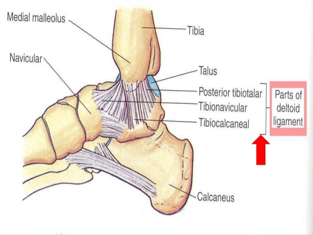
What is the ATFL?
The Anterior Talo-Fibular Ligament (ATFL) is one of three ligaments that make up to Lateral Collateral Ligament of the ankle. The ATFL is a short ligament that widens slightly from top to bottom.
What is swelling distal to the lateral malleolus of the ankle that may extend to the foot if?
Swelling observed distal to the lateral malleolus of the ankle that may extend to the foot if the lateral capsule is torn.
Where does the anterior talofibular ligament originate?
The anterior talofibular ligament originates from the anterior edge of the lateral malleolus of the fibula and attaches to the neck of the talus, in front of the lateral malleolar facet.
What does stress radiograph reveal?
Stress radiographs may reveal excessive anterior translation of the talus or inversion of the talus.
ATFL Injuries
The Anterior Talofibular ligament (ATFL) is an intracapsular ankle ligament and part of the lateral collateral ligament complex. The ATFL Provides proprioceptive sense and stability to the foot and ankle.
Treatment
Treatment for most of the sprain in an ankle ligament differs based on the severity or grade of the sprain or tear.
What is the function of the tibiofibular ligament?
One of the primary functions of the tibiofibular ligament is to make up the back ‘wall’ area of the recipient socket for the talar trochlea of the ankle. The trochlea is a fibrous structure that is shaped similarly to a pulley.
Which ligament is located on the rear of the tibiofibular syndesmosis?
The posterior tibiofibular ligament is a fibrous band of connective tissue that travels horizontally over the rear surface of the tibiofibular syndesmosis, which is a meeting area of the fibula (calf bone) and the tibia (shinbone) that is composed of the interosseus membrane and both the interosseous and anterior ligaments.
Is the posterior inferior ligament flat?
It is significantly smaller in size than the lateral malleolus’ anterior ligament, which is flat and triangular in shape. Another common name for the ligament is the posterior inferior ligament. Last medically reviewed on April 14, 2015.
What happens when you twist your ankle?
When all the body's weight is placed on the lateral ankle ligaments, the anterior tibiofibular ligament stretches or tears resulting in a high sprain of the ankle. Sometimes, pieces of bone may also be torn off with the ligament. In some cases, this twisting force on the ankle can cause other damage. The bones around the ankle may become broken, ...
What is the ligament that holds the fibula and tibia together?
The anterior tibiofibular ligament is often referred to as the high ankle ligament. This ligament slants downward, distally and laterally, between the margins of the fibula and tibia. It’s a point just above the ankle joint where these two bones meet, and the ligament holds the fibula and tibia bones together (syndesmosis).
How to tell if you have a sprained ankle or foot?
The more severe sprains of the ankle and foot are syndesmosis injuries, and they may also cause many problems for people trying to return to normal activity. When you sprain your ankle, you will feel acute and localized pain on the side of the injured ankle. Mild and moderate syndesmosis sprains can feel like a typical sprained ankle at first. Symptoms include swelling and pain on the outside of the ankleand may be accompanied by discoloration and bruising of the skin. The pain radiates upward along the side of the lower leg, and the ankle may feel unstable and weak.
What is squeeze test?
Any pain associated with this squeeze test is the marker of a syndesmosis ligament injury. If a syndesmosis injury is suspected, X-rays are then used to determine its severity, and an enlarged gap between the tibia and fibula will confirm the diagnosis.
How long does it take to recover from a syndesmosis injury?
Recovering from even mild injuries of this type can take as long as six to seven weeks.
What is anterior tibiofibular injury?
Anterior Tibiofibular Ligament Injury. An ankle syndesmosis injury (a severe form of ankle sprain), involves damage to supportive ligaments in the ankle. This type of injury is also known as a high ankle sprain because it occurs in the ligaments above the ankle joint.
How to diagnose anterior tibiofibular ligament injury?
The diagnosis of anterior tibiofibular ligament injury is usually done by examining the ankle. The physician moves the ankle in different positions to check the functionality of the ligament around the ankle. The ligament is stressed by holding the lower leg still, while turning the ankle outward. Another test is performed by holding ...
What is the weakest ligament in the lateral collateral complex of the ankle?
Approximately two-thirds of ankle sprains tend to be isolated injuries to the anterior talofibular ligament (ATFL), the weakest ligament in the lateral collateral complex of the ankle. There is general agreement that avulsion is more common at the fibular than the talar end of the ligament 2.
What is the most common ligament injury?
Anterior talofibular ligament injury is the most common of the ligament injuries that can occur as part of the lateral ligament complex injuries 2. The injuries can comprise either soft tissue tears, avulsion fractures or both.
Which ligament has high sensitivity and specificity rates?
Has been shown to have high sensitivity and specificity rates for chronic anterior talofibular ligament tears 6.
What does MRI show on a ligament?
MRI may show detachment, discontinuity, thickening, thinning, contour irregularity of the ligament, a bright rim sign 5 or an associated bony avulsion. Heterogeneity with increased intraligamentous signal intensity on fat-suppressed T2-weighted or intermediate-weighted images is indicative of intrasubstance edema or hemorrhage.
Where is the ligament located?
the ligament appears as a thin, straight, low signal intensity band extending from the talus to the fibular malleolus
Which approach do orthopedists favor?
Many orthopedists favor a functional approach based on clinical examination: