Vascular Tunic
- Iris The iris is the anterior most portion of the vascular tunic and functions as a moveable diaphragm between the anterior and posterior chambers. ...
- Ciliary Body The ciliary body, like the iris, contains both neurectodermal and mesodermal tissue. ...
- Choroid The choroid is the third component of the uvea. ...
What are the 3 parts of the vascular tunic?
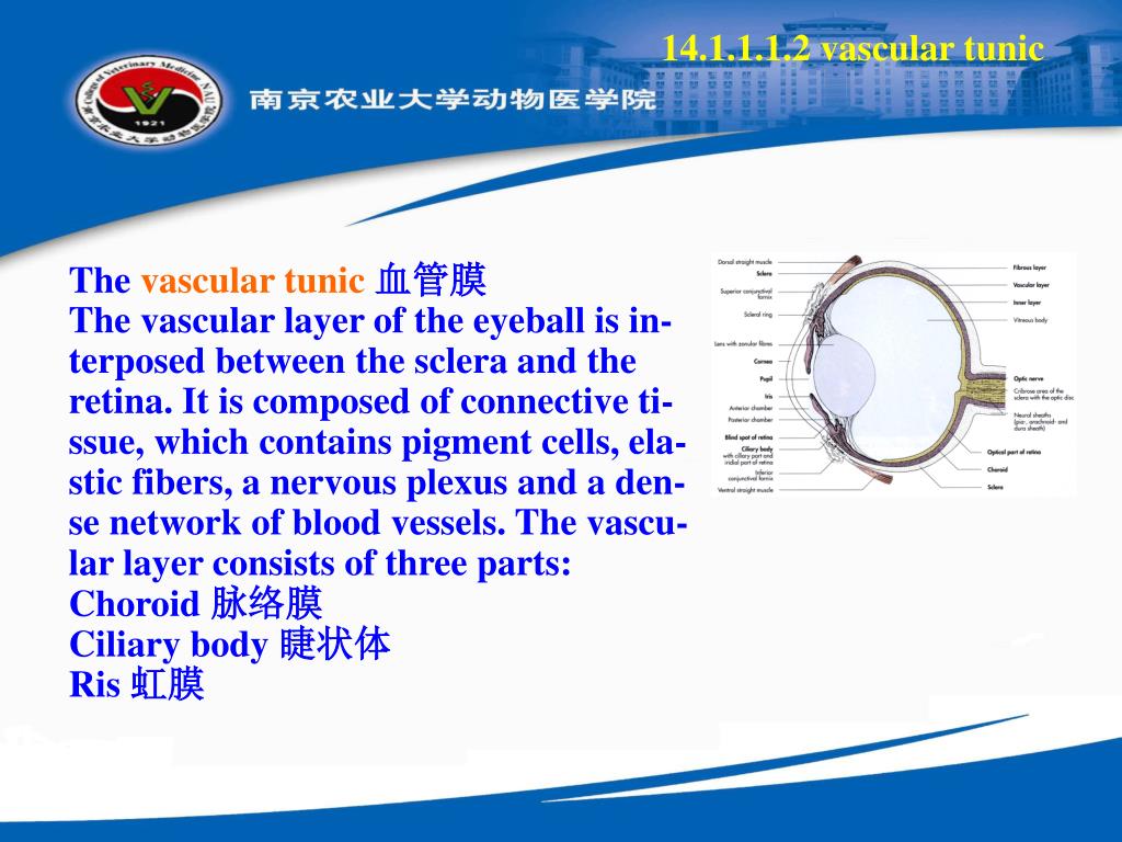
Vascular Tunic The vascular tunic is comprised of three distinct regions, (1) the iris, (2) the ciliary body, and (3) the choroid. The vascular tunic is mesodermal in origin and is situated between the outer fibrous tunic and the inner nervous tunic. The vascular tunic is also refered to as the uvea.
What is the vascular tunic of the eye made of?
—The vascular tunic of the eye is formed from behind forward by the choroid, the ciliary body, and the iris. The choroid invests the posterior five-sixths of the bulb, and extends as far forward as the ora serrata of the retina.
What are the 3 tunics of the eye?
The Tunics of the Eye F IG. 869– Horizontal section of the eyeball. From without inward the three tunics are: (1) A fibrous tunic, (Fig. 869) consisting of the sclera behind and the cornea in front; (2) a vascular pigmented tunic, comprising, from behind forward, the choroid, ciliary body, and iris; and (3) a nervous tunic, the retina.
What is the function of the vascular tunic?
The vascular tunic is also refered to as the uvea. The iris is the anterior most portion of the vascular tunic and functions as a moveable diaphragm between the anterior and posterior chambers.

What is the vascular tunic made up of?
The vascular tunic is comprised of three distinct regions, (1) the iris, (2) the ciliary body, and (3) the choroid. The vascular tunic is mesodermal in origin and is situated between the outer fibrous tunic and the inner nervous tunic. The vascular tunic is also refered to as the uvea.
What is part of the nervous tunic?
Nervous tunic: The inner nervous tunic is the retina. The retina consists of an outer pigmented epithelium covered by nervous tissue (the neural layer) on the inside. The dark color of the pigmented epithelium absorbs light (as with the choroid) and stores vitamin A used by photoreceptor cells in the neural layer.
What are the 3 tunics of the eye and what is the function of each?
Answer and Explanation: 1) Fibrous tunic : It forms an outer layer that consists of the sclera and the cornea. Sclera : Its function is to support the shape of an eye, protect the internal delicate structures. Cornea : Its function is to refracts the incoming light and protection of anterior part of the eye.
What are the two parts of the first tunic?
From without inward the three tunics are: (1) A fibrous tunic, (Fig. 869) consisting of the sclera behind and the cornea in front; (2) a vascular pigmented tunic, comprising, from behind forward, the choroid, ciliary body, and iris; and (3) a nervous tunic, the retina.
What is the vascular layer of the eye?
choroidThe vascular layer of the eye, also known as the uvea or uveal tract, consists of the three layers that are continuous with each other. From posterior to anterior, these are the choroid, ciliary body and iris.
Is the lens part of the vascular tunic?
Answer: e. All of the above are part of the vascular tunic (uvea). The anterior portion of the uvea contains the iris and ciliary body, while the posterior portion contains the choroid. The lens is found between the anterior and posterior segments of the uvea.
What are the 3 main layers of the eye?
Eye Anatomy and FunctionThe outer layer of the eyeball is a tough, white, opaque membrane called the sclera (the white of the eye). ... The middle layer is the choroid. ... The inner layer is the retina, which lines the back two-thirds of the eyeball.
What are the 3 coats of the eye?
The wall of the eyeouter layer – made up of the sclera and cornea (called the fibrous tunic)middle layer – made up of the uvea (called the vascular tunic)inner layer – made up of the retina (called the neural tunic)
What are the three tunics of the eye quizlet?
What are the three layers of the eye? The sclera, the choroid layer, and the retina.
What are the types of tunics?
Contents1.1 Indian tunic.1.2 Celtic tunic.1.3 Greek tunic.1.4 Roman tunic.1.5 Germanic tunic.
What tunic means?
Definition of tunic 1a : a simple slip-on garment made with or without sleeves and usually knee-length or longer, belted at the waist, and worn as an under or outer garment by men and women of ancient Greece and Rome. b : surcoat. 2a : a hip-length or longer blouse or jacket. b : a short overskirt.
Which is not one of the tunics or layers of the eye?
The eyeball has three layers: the outer fibrous tunic, the middle vascular tunic, and the inner sensory tunic. The part of the fibrous tunic shown here is the sclera (top arrow bar). Just below it, but not labeled, is the choroid (part of the vascular tunic).
What part of eye is retina?
Retina: The retina is the light-sensitive tissue at the back of the eye. The retina converts light into electrical impulses that are sent to the brain through the optic nerve. Vitreous gel: The vitreous gel is a transparent, colorless mass that fills the rear two-thirds of the eyeball, between the lens and the retina.
What's a sclera?
Listen to pronunciation. (SKLAYR-uh) The white layer of the eye that covers most of the outside of the eyeball.
What is optic nerve head?
The optic nerve head (ONH) is the structure in the posterior ocular fundus that allows the exit of the retinal ganglion cell axons and the entry and exit of the retinal blood vessels.
What is the white part of the eye made of?
collagen fibersThe sclera is the white outer coating of the eye. It is tough, fibrous tissue that extends from the cornea (the clear front section of the eye) to the optic nerve at the back of the eye. The sclera gives the eyeball its white color. The cornea and sclera are made of the same type of collagen fibers.
What is the vascular tunic?
872, 873, 874). —The vascular tunic of the eye is formed from behind forward by the choroid, the ciliary body, and the iris.
What is the fibrous tunic?
The Fibrous Tunic (tunica fibrosa oculi). —The sclera and cornea (Fig. 869) form the fibrous tunic of the bulb of the eye; the sclera is opaque, and constitutes the posterior five-sixths of the tunic; the cornea is transparent, and forms the anterior sixth.
What is the arteria centralis retino?
The arteria centralis retinæ (Fig. 879) and its accompanying vein pierce the optic nerve, and enter the bulb of the eye through the porus opticus. The artery immediately bifurcates into an upper and a lower branch, and each of these again divides into a medial or nasal and a lateral or temporal branch, which at first run between the hyaloid membrane and the nervous layer; but they soon enter the latter, and pass forward, dividing dichotomously. From these branches a minute capillary plexus is given off, which does not extend beyond the inner nuclear layer. The macula receives two small branches (superior and inferior macular arteries) from the temporal branches and small twigs directly from the central artery; these do not, however, reach as far as the fovea centralis, which has no bloodvessels. The branches of the arteria centralis retinæ do not anastomose with each other—in other words they are terminal arteries. In the fetus, a small vessel, the arteria hyaloidea, passes forward as a continuation of the arteria centralis retinæ through the vitreous humor to the posterior surface of the capsule of the lens.
What are the three tunics?
From without inward the three tunics are: (1) A fibrous tunic, (Fig. 869) consisting of the sclera behind and the cornea in front; (2) a vascular pigmented tunic, comprising, from behind forward, the choroid, ciliary body, and iris; and (3) a nervous tunic, the retina.
What is the epithelium of the cornea?
The corneal epithelium ( epithelium corneæ anterior layer) covers the front of the cornea and consists of several layers of cells. The cells of the deepest layer are columnar; then follow two or three layers of polyhedral cells, the majority of which are prickle cells similar to those found in the stratum mucosum of the cuticle. Lastly, there are three or four layers of squamous cells, with flattened nuclei.
Is the cornea a vessel or nerve?
Vessels and Nerves. —The cornea is a non-vascular structure; the capillary vessels ending in loops at its circumference are derived from the anterior ciliary arteries. Lymphatic vessels have not yet been demonstrated in it, but are represented by the channels in which the bundles of nerves run; these channels are lined by an endothelium. The nerves are numerous and are derived from the ciliary nerves. Around the periphery of the cornea they form an annular plexus, from which fibers enter the substantia propria. They lose their medullary sheaths and ramify throughout its substance in a delicate net-work, and their terminal filaments form a firm and closer plexus on the surface of the cornea proper, beneath the epithelium. This is termed the subepithelial plexus, and from it fibrils are given off which ramify between the epithelial cells, forming an intraepithelial plexus.
