However, toxic change in neutrophils do not reflect a “toxic effect” of bacteria on neutrophils but are morphologic abnormalities acquired during maturation under conditions that intensely stimulate neutrophil production and shorten the maturation time in the bone marrow
Bone marrow
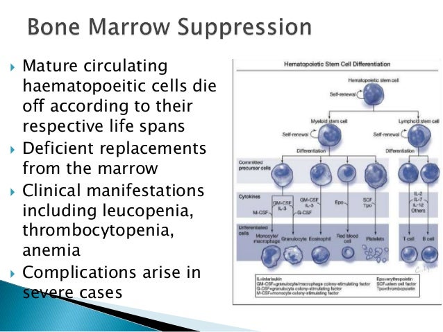
Bone marrow is a semi-solid tissue which may be found within the spongy or cancellous portions of bones. In birds and mammals, bone marrow is the primary site of new blood cell production or hematopoiesis. It is composed of hematopoietic cells, marrow adipose tissue, and supportive stromal cells. In adult humans, bone marrow is primarily located in the ribs, vertebrae, sternum, and bone…
What do azurophilic granules in neutrophils indicate?
In many infections or toxic stimulations, neutrophils respond with large dark blue intracytoplasmic azurophilic granules . These toxic granules may be unmasked in metamyelocytes, bands, and segmented neutrophils in such instances. Their presence indicates active phagocytosis with an increase in lysosomal activity.
What is toxic granulation in neutrophils?
Toxic granulation is the accumulation of big, dark granules in segmented neutrophils (or, sometimes, in earlier neutrophil precursors). It is likely due to the demand placed on the marrow to get those neutrophils out in the circulation as soon as possible in order to fight the infection.
What are toxic morphological changes in neutrophils?
Toxic morphological changes are seen in neutrophils. A left shift with an increase in immature granulocytes typically accompanies toxic changes. In order to report toxic changes, typically two out of the three features should be seen in the majority of neutrophils: 1. Toxic Granulation:1-3
What does it mean when your neutrophils are high?
Neutrophils show abnormal granules. Look for a neutrophil leukocytosis or leukopenia. The blood film may suggest bacterial infection with toxic granulation of neutrophils (increased band forms) even when the white cell count is normal. Leukopenia is a poor prognostic sign.

What do toxic neutrophils indicate?
Toxic neutrophils exhibit a variety of nuclear and cytoplasmic abnormalities in Romanowsky-stained blood smears, and are associated with inflammation and infection.
What causes toxic granulation of neutrophils?
Toxic granulation is seen in cases of severe infection, as a result of denatured proteins in rheumatoid arthritis or, less frequently, as a result of autophagocytosis. Infection is the most frequent cause of toxic granulation. This phenomenon may be seen in cells which also contain Döhle bodies and/or vacuoles.
What are the main features of toxicity in the neutrophil?
In order to report toxic changes, typically two out of the three features should be seen in the majority of neutrophils:Toxic Granulation: 1-3 Dark blue-black peroxidase positive granules that appear in the cytoplasm of the neutrophil. ... Toxic Vacuolation: 1-3 ... Dohle Bodies: 1-3
What does toxic granulation present mean?
'Toxic granulation' is the term used to describe an increase in staining density and possibly the number of granules that occurs regularly with bacterial infection and often with other causes of inflammation (Fig.
What does toxic changes present mean?
TOXIC CHANGES in neutrophils are morphologic abnormalities acquired during maturation under conditions that intensely stimulate neutrophil production and shorten the maturation time in marrow. On the standard hemogram, their presence is reported under the WBC morphology section.
Is Toxic Vacuolation serious?
Clinical significance Toxic vacuolation is associated with sepsis, particularly when accompanied by toxic granulation.
What is an inflammatory Leukogram?
Inflammatory leukogram (cat) Inflammatory leukogram (dog) The blood neutrophil count with inflammation represents the balance between tissue demand and bone marrow supply. The leukogram pattern can be variable depending on the source and severity of the inflammation.
What is a left shift in neutrophils?
Left shift describes when immature neutrophils are released from the bone marrow due to an outpouring of cells, typically due to infection. • In any acute inflammation, an increase in neutrophils is often seen. Increases may be seen after a heart attack (or other infarct) and necrosis. •
Why do neutrophils increase in bacterial infections?
Under infectious or inflammatory conditions, neutrophil granulopoiesis can be increased, typically termed “emergency granulopoiesis”, in order to restore homeostasis in the bone marrow after recruitment of neutrophils to peripheral sites (1).
Is toxic granulation sepsis?
Toxic granulation is often found in patients with bacterial infection and sepsis, although the finding is nonspecific. Patients being treated with chemotherapy or granulocyte colony stimulating factor, a cytokine drug, may also exhibit toxic granulation.
What granules are in neutrophils?
Neutrophils contain at least four different types of granules: (1) primary granules, also known as azurophilic granules; (2) secondary granules, also known as specific granules; (3) tertiary granules; and (4) secretory vesicles (Figure 1).
What does Vacuolated neutrophils present mean?
The presence of vacuolated polymorphonuclear neutrophils in blood smears of patients suffering from infection appears to be associated with massive bacterial growth and to constitute a very early symptom of rapidly life-threatening septicaemia.
What are the three main findings of toxic changes?
These clues are called toxic changes and they encompass three main findings: toxic granulation (as seen above), Dohle bodies (also present above – look closely), and cytoplasmic vacuolization. If you see any of these changes, you can be quite certain that the patient has an infection.
What is toxic granulation?
Toxic granulation is the accumulation of big, dark granules in segmented neutrophils (or, sometimes, in earlier neutrophil precursors). It is likely due to the demand placed on the marrow to get those neutrophils out in the circulation as soon as possible in order to fight the infection.
What are toxic changes in neutrophils?
Toxic morphological changes are seen in neutrophils. A left shift with an increase in immature granulocytes typically accompanies toxic changes. In order to report toxic changes, typically two out of the three features should be seen in the majority of neutrophils:
What does it mean when you have a left shift on a blood smear?
A Left shift refers to the increase presence of immature bands and myeloid precusors. Reaction to infection, inflammation, stress, and granulocyte colony-stimulating factor therapy.
Why is thinking about neutrophils important?
Thinking about the function of neutrophils makes understanding an increase in the number easier to understand. Mechanisms that can increase the number of these white blood cells include:
What is the role of neutrophils in the body?
They are our "first responders" playing the role of the first line of defense against infectious organisms that enter our bodies.
What is the normal neutrophil count?
A normal (absolute) neutrophil count is between 2500 and 7500 neutrophils per microliter of blood. 2 The neutrophil count may be high with infections, due to increased production in the bone marrow as with leukemia, or due to physical or emotional stress.
Why are there immature neutrophils in blood?
Immature neutrophils may be found on a blood smear if the body is stressed and there is a great need for more neutrophils. When this occurs, an increased number of immature neutrophils can make their way to the blood from the bone marrow before reaching maturity.
Why does bone marrow stop producing white blood cells?
The bone marrow may slow down or cease to produce white blood cells, for example, when the bone marrow is injured as with chemotherapy, or a vitamin deficiency is present which causes inadequate production.
Why do neutrophils live in the blood?
These neutrophils may become "demarginated" and circulate in the bloodstream due to stress, infections, and sometimes exercise. The release of neutrophils along the blood vessels into the bloodstream is one reason why the white blood cell count can sometimes rise rapidly (it takes longer for new neutrophils to be produced or released from the bone marrow).
How long do neutrophils live?
Neutrophils have a very short lifespan, living on average only 8 hours, 4 but our bodies produce roughly 100 billion of these cells each day. After being released from the bone marrow, around half of these cells are present along the lining of blood vessels and the other half are found in tissues of the body.
What does a blood film on a neutrophil mean?
Full blood count and blood film. Look for a neutrophil leukocytosis or leukopenia. The blood film may suggest bacterial infection with toxic granulation of neutrophils (increased band forms) even when the white cell count is normal. Leukopenia is a poor prognostic sign.
Which type of neutrophilia has a variable but usually limited left shift?
Variable but usually limited left shift typical in nonneoplastic neutrophilia
What is toxic granulation?
Toxic granulation is the term used to describe an increase in staining density and possibly number of granules that occurs regularly with bacterial infection and often with other causes of inflammation ( Fig. 5.75). It can also be a feature of administration of granulocyte colony-stimulating factor. Fractionally larger, coarser granules may be seen ...
How to tell if you have a thrombocytosis?
Laboratory assessment shows evidence of significant inflammation in the first few days of the disease. The white blood count is frequently elevated and shifted to the left, often with toxic granulations; there may be a mild anemia. The platelet count is often elevated, but in atypical cases the evolution to a characteristic thrombocytosis with platelet counts greater than 700 000 may be delayed into the second or third week of the illness. Sedimentation rates and CRP values are elevated. The initial evaluation should also include an electrocardiogram and echocardiography. Coronary artery dilatations but not true aneurysms are found early on.
Can hyperglycemia occur in severe sepsis?
Hypoglycemia or hyperglycemia may occur in severe sepsis. In patients who are diabetic close monitoring and normalization of blood glucose are essential.
Can GM-CSF cause myelodysplastic syndrome?
With all types of cytokine treatments, the marrow may show an increase in immature cells and cellularity, which can mimic neoplastic conditions. The effects of G-CSF or GM-CSF can mimic acute myeloid leukemia (AML), but occasion ally myelodysplastic syndromes or myeloproliferative neoplasms can be simulated. The single most important factor in dealing with these situations is obtaining an appropriate clinical and treatment history; this can resolve most difficulties. GM-CSF and G-CSF will cause increases in immature cells in the peripheral blood. These cells typically include left-shifted myeloid elements in a variety of stages of maturation. In particular, myeloid blasts can occasionally be seen. Neutrophils and precursors usually display toxic granulation (Fig. 6.19 ). In the marrow, there will usually be an increase in the M:E ratio associated with left-shifted granulopoiesis with numerous promyelocytes and myelocytes. Promyelocytes may be increased to a degree to mimic acute promyelocytic leukemia. Again, toxic changes are characteristically present early in therapy. A slight increase in blasts may be seen. Concurrent flow cytometry on G-CSF-treated samples may show some dyspoietic changes in maturation and antigen expression and may mimic a myeloid stem cell disorder. With GM-CSF, in addition to the effect on granulopoiesis, there are often increased monocytes, monocytic precursors, and variably increased numbers of eosinophils. On more than one occasion changes of growth factor (G-CSF or GM-CSF) therapy have been misinterpreted as acute leukemia or myeloproliferative neoplasms. Presence of only modestly elevated blast counts, a spectrum of maturation in the granulocytic lineage (albeit concentrated in the early forms), toxic granulation, and a good history allow one to avoid mistaking growth factor effects for a myeloid malignancy. Patience will also clarify the matter because it is possible to follow the maturation of the granulocytic cells over the course of a few days or weeks. G-CSF and GM-CSF effects are also reviewed in Chapter 14 on acute myeloid leukemias.
