What is perivascular space in the brain?
Perivascular space. The brain pia mater is reflected from the surface of the brain onto the surface of blood vessels in the subarachnoid space. In the brain, perivascular cuffs are regions of leukocyte aggregation in the perivascular spaces, usually found in patients with viral encephalitis .
What is found in the anterior surface of the midbrain?
Tegmentum: This anterior surface of the midbrain contains numerous structures including the reticular formation, the periaqueductal gray (PAG) matter, certain cranial nerve nuclei, sensory and motor nerve pathways (the corticospinal and spinothalamic tract), the red nucleus, the substantia nigra, and the ventral tegmental area (VTA).
What are perivascular cuffs in the brain?
In the brain, perivascular cuffs are regions of leukocyte aggregation in the perivascular spaces, usually found in patients with viral encephalitis. Perivascular spaces vary in dimension according to the type of blood vessel.
What is the blood supply to the midbrain?
The midbrain receives blood supply from the basilar artery and its branches, including the posterior cerebral artery and the superior cerebellar artery. There are also two cranial nerves present in the midbrain: The trochlear nerve (cranial nerve IV). The midbrain is a complex region of your brainstem that serves many functions.

What fluid is in the midbrain?
Cerebrospinal fluid (CSF) is a clear, colorless body fluid found within the tissue that surrounds the brain and spinal cord of all vertebrates. The cerebrospinal fluid circulates in the subarachnoid space around the brain and spinal cord, and in the ventricles of the brain.
What are the fluid-filled spaces in the brain called?
Also called CSF. Cerebrospinal fluid (CSF, shown in blue) is made by tissue that lines the ventricles (hollow spaces) in the brain. It flows in and around the brain and spinal cord to help cushion them from injury and provide nutrients.
What is the fluid-filled space located in the hypothalamus?
The hypothalamus lies below the hypothalamic sulcus separating it from the thalamus above. Like the thalamus, a thin vertical space filled with CSF called the 3rd ventricle is positioned midline between the two halves of the hypothalamus thalamus.
What is the ventricular space in the midbrain?
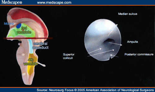
The third ventricle is continuous caudally with the cerebral aqueduct, which runs though the midbrain. At its caudal end, the aqueduct opens into the fourth ventricle, a larger space in the dorsal pons and medulla. The fourth ventricle narrows caudally to form the central canal of the spinal cord.
Where is cerebrospinal fluid found in the brain?
CSF is secreted by the CPs located within the ventricles of the brain, with the two lateral ventricles being the primary producers. CSF flows throughout the ventricular system unidirectionally in a rostral to caudal manner.
Where is CSF fluid formed?
CSF formation. Most CSF is formed in the cerebral ventricles. Possible sites of origin include the choroid plexus, the ependyma, and the parenchyma[2]. Anatomically, choroid plexus tissue is floating in the cerebrospinal fluid of the lateral, third, and fourth ventricles.
What is the foramen of Monro?
The foramen of Monro is a short conduit between the paired lateral ventricles and the third ventricle of the brain. This deep structure becomes clinically significant when obstructed and leads to obstructive (non-communicating) hydrocephalus.
What is arachnoid space?
The subarachnoid space consists of the cerebrospinal fluid (CSF), major blood vessels, and cisterns. The cisterns are enlarged pockets of CSF created due to the separation of the arachnoid mater from the pia mater based on the anatomy of the brain and spinal cord surface.
What is in cerebrospinal fluid?
Your brain and spinal cord have a surrounding protective layer of cerebrospinal fluid (CSF). CSF contains nutrients that your brain can use. The CSF layer also supports and cushions your brain and spinal cord from sudden movements. The effect is similar to putting a grape inside a jar.
What is foramen of Luschka?
Results: The foramina of Luschka are paired apertures located in the lateral recesses of the fourth ventricle, within the posterior cranial fossa. The foramina of Luschka are of importance clinically as their blockage can disrupt the flow of cerebrospinal fluid leading to the development of hydrocephalus.
Where is the choroid plexus located?
the brainThe choroid plexus resides in the innermost layer of the meninges (pia mater) which is in close contact with the cerebral cortex and spinal cord. It is a highly organized tissue that lines all the ventricles of the brain except the frontal/occipital horn of the lateral ventricles and the cerebral aqueduct.
What is the function of the ventricular system of the brain?
The ventricular system produces, transports, and excretes CSF, which coats the central nervous system. Each ventricle contains choroid plexus, which makes the circulating CSF.
What are the names of the 4 ventricles of the brain?
The fourth ventricle is one of the four connected fluid-filled cavities within the human brain. These cavities, known collectively as the ventricular system, consist of the left and right lateral ventricles, the third ventricle, and the fourth ventricle.
What ventricle is associated with the brainstem?
The fourth ventricleAnatomy. The fourth ventricle is a broad tent-shaped cerebrospinal fluid (CSF) cavity located behind the brain stem and in front of the cerebellum in the center of the posterior fossa (Fig. 31-1). CSF enters through the cerebral aqueduct, which opens into the fourth ventricle at its rostral end.
What do the ventricles of the brain control?
Aside from cerebrospinal fluid, your brain ventricles are hollow. Their sole function is to produce and secrete cerebrospinal fluid to protect and maintain your central nervous system.
What is the midbrain?
The mesencephalon , or midbrain, is divided into two major areas in chondrichthyans: the dorsal tectum mesencephali (comprised of the optic tectum and the torus semicircularis) and the ventral tegmentum mesencephali, also referred to as the tegmentum (Hofmann, 1999 ). This brain area is similarly organized across the majority of vertebrates and is well developed across cartilaginous fishes ( Smeets et al., 1983; Smeets, 1998; Lisney and Collin, 2006; Lisney et al., 2007; Yopak and Lisney, 2012; Yopak, 2012b ).
Where is the floor of the mesencephalon?
Examine the brain in ventral view ( Figure 7.76 ). The floor of the mesencephalon is formed by the cerebral peduncles, slightly elevated regions lateral and posterior to the mammillary bodies. The relatively wide and flattened oculomotor nerve arises from the surface of each cerebral peduncle.
What is the mesencephalon of a dolphin?
As in other mammals, the mesencephalon of dolphins is a narrow central part of the brainstem ( Figs. 6.6 and 6.9 b). It is composed of three macroscopic levels: (1) the tectum (SC) dorsally, (2) the intermediate tegmentum (t), which includes the anterior (mesencephalic) part of the reticular formation, and (3) the crus cerebri (cc) ventrally (see also Fig. 6.27 ). Between the tectum and tegmentum is situated the mesencephalic part of the ventricular system (mesencephalic aqueduct; see Fig. 6.9 b), which in dolphins is narrow and low in the rostral part and much higher and wider in the caudal part.
What is the tegmentum of a dolphin?
Tegmentum. In the mesencephalon of dolphins, the anterior most part of the tegmentum, also called pretectum, is well developed. In a transverse section at the level of the posterior commissure ( Figs. 6.26c and 6.27c) lie two tegmental nuclei, the appearance of which is unique among mammals, perhaps with the exception of the elephant ( Cozzi et al., 2001; Oelschläger et al., 2010 ). The two nuclei are called “elliptic nucleus” (E) and situated at the anterior end of and within the central gray, rostral and dorsal to the oculomotor nuclear complex and near the nucleus ruber ( Figs. 6.9, 6.26, and 6.27 ). The elliptic nucleus seems to represent a strongly hypertrophied nucleus of Darkschewitsch ( Fig. 6.48, D) that, during ontogeny, tightly associates with the adjacent nucleus interstitialis Cajal (I). Both nuclei are very large in comparison with those in the human, particularly the nucleus of Darkschewitsch. In terrestrial mammals, the interstitial nucleus is engaged in oculomotor function, that is, in vertical eye (and head) movements but also sends fiber bundles into the spinal cord ( Barone and Bortolami, 2004 ). The nucleus of Darkschewitsch receives fiber systems from the cerebellum, vestibular nuclei, pretectal nuclei, and the motor cortex. Projections of the nucleus of Darkschewitsch run directly to the spinal cord and indirectly via the medial tegmental tract (mtt) to the inferior olivary complex (medial accessory nucleus) and from there to the cerebellum ( Voogd et al., 1998 ). Strong cortical projections, including such from the auditory cortex, run along the pyramidal tract through the crus cerebri to the pons and are also redirected to the cerebellar cortex. Acoustic stimuli, however, were also reported to be conveyed via the inferior colliculus and the superior colliculus and further to the paramedian pontine reticular formation ( Benninghoff and Drenckhahn, 2004 ).
What are the two tecti in a mesencephalon?
Tectum. The roof of the mesencephalon is characterized by the lamina tecti (quadrigeminal plate), which comprises two rostral and two caudal colliculi (superior and inferior colliculi of primates). h The superior colliculi of dolphins seem to be well developed, as other centers of the visual pathway are. The inferior colliculi, however, are much larger in comparison, in accordance with the overall impressive size of the auditory system. In histological sections as well as in MR scans, the dolphin superior colliculus exhibits a pattern of faint lines that is known from other mammals. In terrestrial mammals, the superior layers represent a sensory apparatus that gets its input from the retina and visual cortex, whereas the deeper layers comprise an integration apparatus for the processing of multimodal input ( Benninghoff and Drenckhahn, 2004 ). In dolphins, the rostral and caudal colliculi so far have not been investigated systematically.
Where is the tegmentum located?
The tegmentum, located ventral to the tectum and rostral to the medulla oblongata, receives projections from most brain centers and is highly conserved across vertebrates ( Striedter, 2005 ). Compared to the optic tectum, which has been the subject of significant study across most vertebrate classes, few comparative studies have examined the allometry and relative size of the tegmentum ( Platel and Delfini, 1986; Kotrschal and Junger, 1988; Boire and Baron, 1994; Striedter, 2005; Yopak and Lisney, 2012 ), cytoarchitecture, and pathways ( Smeets et al., 1983; Morita and Finger, 1985; Gonzalez et al., 1999; de Arriba and Pombal, 2007; Demski, 2012 ), although it is interconnected with the tectum and is related, in part, to visual processing. The lateral portion of the tegmentum receives input from the spinal cord ( Hayle, 1973) and tectum ( Boord and Northcutt, 1982; Smeets et al., 1983) and the lateral mesencephalic complex is the site for termination of secondary electrosensory, mechanosensory, and auditory fibers ( Boord and Northcutt, 1982; Corwin and Northcutt, 1982 ). Cell groups in this area have been identified, including the midbrain reticular formation, which plays an important role in controlled locomotion in elasmobranchs ( Demski, 1977; Droge and Leonard, 1983a,b ), as observed in mammals ( Mori et al., 1980) and teleosts ( Kashin et al., 1974 ).
Where do spinothalamic nuclei end?
In mammals, spinothalamic, dorsal columnar, and trigeminal nucleus projections terminate in the mesencephalon (intercollicular area) and in four thalamic areas including: ventral thalamus (zona incerta); the ventrobasal and posterior thalamic nuclei; a perigeniculate area at the ventromedial edge of the medial geniculate; and the intralaminar nuclei, particularly the central lateral nucleus ( Giesler et al., 1981; 1988; Cliffer et al., 1991; Willis and Coggeshall, 1991; LeDoux et al., 1987 ).
Which space drains fluid from neuronal cells?
The spaces ultimately drain fluid from neuronal cell bodies to the cervical lymph nodes. In particular, the “tide hypothesis” suggests that the cardiac contraction creates and maintains pressure waves to modulate the flow to and from the subarachnoid space and the perivascular space.
Where is a dilated perivascular space located?
Dilated perivascular spaces are categorized into three types: Type 1 are located on the lenticulostriate arteries projecting into the basal ganglia. Type 2 are located in the cortex following the path of the medullary arteries. Type 3 are located in the midbrain.
What is the cause of a dilated space in the perivascular space?
CADASIL syndrome (cerebral autosomal dominant arteriopathy with subcortical infarcts and leukoencephalopathy syndrome) is a hereditary stroke condition due to a Notch 3 gene mutation on Chromosome 19. Studies have noted that in comparison to family members lacking the affected haplotype that leads to the condition, an increased number of dilated spaces is observed in individuals with CADASIL. These perivascular spaces are localized primarily in the putamen and temporal subcortical white matter and they appear to correlate with age of the individual with the condition rather than severity of the disease itself.
What are the perivascular spaces of the subarachnoid space?
Perivascular spaces surrounding arteries in the cerebral cortex and the basal ganglia are separated from the subpial space by one or two layers of leptomeninges, respectively, as well as the pia mater. By virtue of the leptomeningeal cell layer, the perivascular spaces belonging to the subarachnoid space are continuous with those of the subpial space. The direct communication between the perivascular spaces of the subarachnoid space and the subpial space is unique to the brain’s arteries, as no leptomeningeal layers surround the brain’s veins. Use of the scanning electron microscope has determined that the spaces surrounding blood vessels in the subarachnoid space are not continuous with the subarachnoid space because of the presence of pia mater cells joined by desmosomes.
What is the significance of perivascular spaces?
The clinical significance of perivascular spaces comes primarily from their tendency to dilate . The importance of dilation is hypothesized to be based on changes in shape rather than size. Enlarged spaces have been observed most commonly in the basal ganglia, specifically on the lenticulostriate arteries. They have also been observed along the paramedial mesencephalothalamic artery and the substantia nigra in the mesencephalon, the brain region below the insula, the dentate nucleus in the cerebellum, and the corpus callosum, as well as the brain region directly above it, the cingulate gyrus. Upon the clinical application of MRI, it was shown in several studies that perivascular space dilation and lacunar strokes are the most commonly observed histological correlates of signaling abnormalities.
What is the median eminence?
The median eminence, a brain structure at the base of the hypothalamus, contains capillaries with wide perivascular spaces. In humans, perivascular spaces surround arteries and veins can usually be seen as areas of dilatation on MRI images.
What is the perivascular space?
A perivascular space, also known as a Virchow–Robin space, is a fluid-filled space surrounding certain blood vessels in several organs, including the brain, potentially having an immunological function, but more broadly a dispersive role for neural and blood-derived messengers.
What are the three divisions of the brain?
The three divisions of the brain that make up the brainstem are the: A. medulla oblongata, thalamus, and pons. B. medulla oblongata, thalamus, and midbrain. C. medulla oblongata, midbrain, and pons. D. medulla oblongata, pons, and cerebellum. D.
Do cerebral functions have a typical location?
Many cerebral functions have a typical location. This fact is known as:
What is perivascular space?
Typically, these so-called perivascular spaces help clear waste and toxins from the brain and might be linked with changes in the aging brain, researchers say. "Dilated perivascular spaces, which are a common MRI finding, especially in the elderly, are not just an incidental finding," said study author Dr.
How many people have enlarged perivascular spaces?
Of the 31 people with enlarged perivascular spaces in both areas of the brain that the researchers looked at, 39% developed dementia. The people with severe enlargement of perivascular spaces in both areas of the brain were also more likely to have greater decline four years later on their overall scores of cognition than people with mild ...
Which is more likely to develop dementia: perivascular space or perivascular space?
They found that those with the most enlarged perivascular spaces were nearly three times more likely to develop dementia than those with fewer or no enlarged spaces.
Does Finney think enlarged spaces are a diagnostic tool?
Finney doesn't think that these enlarged spaces will be a diagnostic tool, and instead, "It's really going to be a marker of risk. It's not going to tell you if you have dementia, it's not going to tell you if you're going to get it.