See more
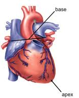
Mar 18, 2022 · The apex of the heart is lowest tip of the organ that points downward at the base, forming what almost looks like a rounded point. “Apex” is a Latin word meaning “summit” or “peak,” and this part of the heart can be thought of in those terms, too. It’s formed mostly by the left ventricle and extends pretty far out to the left in most people.

What is the apex of the heart attached to?
The inferior tip of the heart, known as the apex, rests just superior to the diaphragm. The base of the heart is located along the body's midline with the apex pointing toward the left side.Jul 30, 2020
What is the apex of a heart?
In the anatomical position, the apex of the heart is the confluence of the inferior and left borders. It is a projection inferiorly, anteriorly and to the left of the left ventricle. Anterolaterally, it is covered by the anterior surface of the mediastinal pleura.
What form the base of the heart?
The base of tbe heart (basis cordis), directed upward, backward, and to the right, is separated from the fifth, sixth, seventh, and eighth thoracic vertebræ by the esophagus, aorta, and thoracic duct. It is formed mainly by the left atrium, and, to a small extent, by the back part of the right atrium.
What end of the heart is the apex?
The base of the heart is located at the level of the third costal cartilage, as seen in Figure 1. The inferior tip of the heart, the apex, lies just to the left of the sternum between the junction of the fourth and fifth ribs near their articulation with the costal cartilages.
Where is the apex of the heart located quizlet?
The apex of the heart is the inferior tip of the heart and points toward the left side.
How do you find the apex of the heart?
The normal apex beat can be palpated in the precordium left 5th intercostal space, half-inch medial to the left midclavicular line and 3–4 inches left of left border of sternum.
Is the base of the heart superior to the apex?
The base of the heart is probably better termed its posterior surface. It is not the most inferior surface of the organ but rather the most superior. It assumed the term because it is thought to resemble the base of the pyramid or cone which extends obliquely to the left to the apex of the heart.
What structure is posterior to the heart?
Toward the front of the body. The sternum lies anterior to the heart. Towards the back of the body. The heart lies posterior the sternum.
What forms the right border of the heart?
The right border of the heart (right margin of heart) is a long border on the surface of the heart, and is formed by the right atrium. The atrial portion is rounded and almost vertical; it is situated behind the third, fourth, and fifth right costal cartilages about 1.25 cm. from the margin of the sternum.
Why is apex of heart at bottom?
The heart weighs about 300 g and is located within the mediastinum; it is cone-shaped and tilted forward and to the left. Because of rotation during fetal development, the apex of the heart (tip of the cone) is at its bottom and lies left of the midline.
How is the base of the heart different from the apex of the heart quizlet?
Distinguish between the base of the heart and the apex of the heart. The great vessels are located on the base, and the apex ends at a point. Name the three layers of the heart wall, and indicate the layer that is also called the visceral pericardium.
What are the chambers of the heart?
A typical heart has two upper and two lower chambers. The upper chambers, the right and left atria, receive incoming blood. The lower chambers, the more muscular right and left ventricles, pump blood out of the heart. The heart valves, which keep blood flowing in the right direction, are gates at the chamber openings.
What is the apex of the heart?
After removal of the left thoracic wall, the blunt apex of the heart, which is formed by the left ventricle, can be observed between the left cranial and caudal lung lobes as the longitudinal axis of the heart presents a deviation of approximately 45 degrees toward the left.
Where is the heart located in the thoracic cavity?
The Heart Is Located in the Center of the Thoracic Cavity. The heart is located in the middle of the thoracic cavity, oriented obliquely, with the apex of the heart pointing down and to the left, as shown in Figures 5.4.1 and 5.4.2.
What is Situs inversus?
Situs inversus is a rare congenital abnormality in which the normal location of the thoracic (and abdominal) organs is reversed. The aortic arch, left ventricle, and cardiac apex are all on the right side. It may be associated with Kartagener’s syndrome (see p. 218). View chapter Purchase book.
How are the right and left ventricles separated?
The right and left ventricles are already separated to a great extent by the muscular interventricular septum arising from the apex of the heart. Through much of development there is a physiological communication between the ventricles at the tip of the septum allowing free mixing of right and left ventricular blood as it travels through a common outflow tract. As the outflow septates to give rise to separate aortic and pulmonary artery/ductus arteriosus flow, the ventricles complete septation. Both septation of the outflow and the ventricles is dependent on fusion of cardiac cushions at what was the inner curvature of the heart, an area known as the atrioventricular canal (Fig. 2.4 ). The factors that are required for formation of these cushions have been studied in detail (see comprehensively review in Ref. 78). Signals from the myocardium, including TGFβ family members, VEGF, and Notch, result in a transformation of cells in the adjacent myocardium to transform into mesenchyme and migrate into the cardiac jelly in between (Fig. 2.5). The deposition of an appropriate hyaluronan and proteoglycan-rich extracellular matrix has been shown to be essential for the normal development of the cushions as in the absence of these ECM proteins the cushions do not form.79,80 Continued migration of cells from the endocardium, together with rapid proliferation, results in the formation of primitive valve-like structures that allow only unidirectional flow of blood through the heart. Ventricular septation itself is completed by the fusion of the interventricular muscular septum with the atrioventricular cushions and the proximal outflow cushions. The tissues derived from the cushions become fibrous tissue and are the membranous part of the interventricular septum in the formed heart. Deficiencies in this process give rise to subaortic, subpulmonary, and doubly committed membranous VSDs, depending on whether they sit under the aorta, pulmonary artery, or between both (Fig. 2.4 ). Given, the number of structures that must fuse in order to separate the atria from the ventricles, and each into left and right sides, it is not surprising that malformations in this area are relatively common. The most severe situation is an AVSD, where mixing of left and right atrial flows is complicated with mixing of ventricular flows and is very common in Down syndrome (see below).
How many chambers does the heart have?
The heart consists of four chambers: two smaller atria at the top (the base) and two larger ventricles at the apex. A band of fibrous tissue separates the atria from the ventricles and seats the four cardiac valves. A muscular septum separates the right from left atrium and the right from left ventricle.
Where is the tricuspid valve located?
The tricuspid, mitral, aortic, and pulmonary valves are all grouped in this connective tissue ring set in an oblique plane beneath the sternum, at right angles to the major axis of the heart. The apex of the heart taps against the chest wall, causing the apex beat in the fifth left intercostal space.
How many lung lobes are there in a rat?
There are five lung lobes in the rat: one left lobe and four on the right (cranial, middle, accessory, and caudal lobes). The middle lobe lies in contact with the diaphragm and apex of the heart and is notched to accommodate the caudal vena cava. For this reason, the middle lobe is sometimes referred to as the postcaval lobe. Bronchial branching follows a monopodial pattern in rats where each main intrapulmonary longitudinal airway has much smaller side branches (Monteiro, 2014 ). The respiratory bronchioles are relatively short and rudimentary ( Boorman, 1990 ), resulting in terminal bronchioles almost immediately connecting into alveolar ducts, each of which subdivides four or five times.
Where is the heart located?
The heart is located toward the back of the sternum and midline to the lungs. There are many parts and functions of the heart, with one of them being the apex. The apex is the lower tip of the heart and sits above the diaphragm. The apex of the heart points to the left of the body. When the heart beats, this area of the muscle touches the wall ...
What is the pericardium?
The pericardium acts as a protector of the heart. It is a thin sac that envelops the heart and holds it in place. Its protective features don't stop there. It also lubricates the heart as it beats to guard it against friction with tissue.
Which chamber of the heart is the strongest?
The Left Ventricle. This chamber of the heart was already explained, but there are a few other facts about the left ventricle. This chamber of the heart plays a factor in our blood pressure. When the pressure is being measured, it uses the fast contractions of the left ventricle. This is the strongest chamber of the heart.
What is the function of the right ventricle?
Its main function is to deliver blood, with very low pressure, to the lungs through the pulmonary artery. The deoxygenated blood is sent to the right ventricle from the right atrium.
Where does blood flow to the right ventricle?
Blood is received into the right atrium from the systemic veins and then is delivered to the right ventricle. The deoxygenated blood that flows to the right atrium comes from the rest of the body. There are two coronary arteries, one that pumps blood to the left side of the heart and the other to the right side.
What is the function of the left atrium?
The Left Atrium. Blood flows through the left atrium and then goes on to the left ventricle. The blood that pumps into the left atrium flows in from the lungs and is oxygenated blood. The Right Ventricle. The right ventricle is in the lower part of the right chamber of the heart. Its main function is to deliver blood, with very low pressure, ...
Where is the heart located?
The heart is placed within the middle mediastinum. Laterally and posteriorly it is surrounded by the lungs, while the sternum is located anteriorly. The superior border, or base of the heart, is the portion located opposite the apex where the big blood vessels enter and leave the heart. These aspects explain why the heart and great vessels project onto the middle of the thorax.
What are the surface projections of the heart?
The surface projections of the heart represent points on the thoracic wall that map out the outline and valves of the heart. These include four borders (superior, right, inferior, left) and four valves (left atrioventricular, right atrioventricular, aortic, pulmonary). The main reference points used for the surface projections of the heart are the borders of the sternum and costal cartilages, the clavicle and intercostal spaces. The latter favour sound transmission, facilitating clinical maneuvers such as percussion, auscultation and palpation to pinpoint the cardiac location.
What are the different types of heart valves?
There are four existing heart valves: 1 The left atrioventricular (mitral) valve between the left atrium and left ventricle. It projects posteriorly to the left side of the sternum, at the level of the left fourth costal cartilage. 2 The right atrioventricular (tricuspid) valve between the right atrium and right ventricle. It is located posteriorly to the right side of the sternum at the level of the right fourth costal cartilage. 3 The aortic valve between the left ventricle and aorta. One can find it at the level of the third intercostal space, posterior to the left side of the sternum. 4 The pulmonary valve between the right ventricle and pulmonary trunk, projects at the junction of the sternum and left third costal cartilage.
Why are heart valves important?
Their function is to force blood to flow in only one direction, preventing backflow (regurgitation) due to intra-arterial pressure. In that way, the direction of blood flow is always constant and unidirectional throughout the human body. Therefore, valves are extremely important! Next time you hear an abnormal sound during valve auscultation, realize the impact of the pathology on the patient's entire circulatory system..
What is the purpose of a stethoscope during auscultation?
During auscultation, clinicians use a stethoscope to listen to sounds above specific localizations of the heart, such as heart valves. Heart sounds and additional murmurs can reveal cardiovascular system diseases like valvulopathies or vascular malformations, such as a patent ductus arteriosus. Key facts about the surface projections of the heart.
What is the left border of the heart?
The left border corresponds to a line drawn from the inferior border of the second left costal cartilage to the intersection point between the fifth left intercostal space and midclavicular line. More simply, the line interconnects the left ends of the superior and inferior borders of the heart.
What are the four heart valves?
There are four existing heart valves: The left atrioventricular (mitral) valve between the left atrium and left ventricle. It projects posteriorly to the left side of the sternum, at the level of the left fourth costal cartilage. The right atrioventricular (tricuspid) valve between the right atrium and right ventricle.
