How are neural tubes formed in the brain?
There are two major ways of forming a neural tube. In primary neurulation, the cells surrounding the neural plate direct the neural plate cells to proliferate, invaginate, and pinch off from the surface to form a hollow tube.
How does the ectoderm develop into the neural tube?
The ectoderm gives rise to hair, skin, and the entire nervous system. During the course of development, the ectoderm initially develops into a structure known as the neural plate, which eventually folds into the neural tube. This process by which the neural plate becomes the neural tube is known as neurulation and is depicted in the figure below.
Where does the neural tube close during primary neurulation?
Neural tube closure begins at 4 weeks at the occipitocervical region. The hollow center of the neural tube will become the central canal of the spinal cord and ventricular system of the brain. During primary neurulation, the neural tube separates from the overlying ectoderm in a process called dysjunction.
What are the two types of neural tube development?
The neural tube develops in two ways: primary neurulation and secondary neurulation . Primary neurulation divides the ectoderm into three cell types: Primary neurulation begins after the neural plate forms. The edges of the neural plate start to thicken and lift upward, forming the neural folds.

How do neural tubes form?
In primary neurulation, the cells surrounding the neural plate direct the neural plate cells to proliferate, invaginate, and pinch off from the surface to form a hollow tube. In secondary neurulation, the neural tube arises from a solid cord of cells that sinks into the embryo ...
How does neurulation occur?
The process of neurulation begins when the underlying dorsal mesoderm (and pharyngeal endoderm in the head region) signals the ectodermal cells above it to elongate into columnar neural plate cells (Smith and Schoenwolf 1989; Keller et al. 1992). Their elongated shape distinguishes the cells of the prospective neural plate from the flatter pre-epidermal cells surrounding them. As much as 50% of the ectoderm is included in the neural plate. The neural plate is shaped by the intrinsic movements of the epidermal and neural plate regions. The neural plate lengthens along the anterior-posterior axis, narrowing itself so that subsequent bending will form a tube (instead of a spherical capsule).
What is the bending of the neural plate?
The bending of the neural plate involves the formation of hinge regionswhere the neural tube contacts surrounding tissues. In these regions, the presumptive epidermal cells adhere to the lateral edges of the neural plate and move them toward the midline (see Figure 12.3B). In birds and mammals, the cells at the midline of the neural plate are called the medial hinge point(MHP) cells. They are derived from the portion of the neural plate just anterior to Hensen's node and from the anterior midline of Hensen's node (Schoenwolf 1991a,b; Catala et al. 1996). The MHP cells become anchored to the notochord beneath them and form a hinge, which forms a furrow at the dorsal midline. The notochord induces the MHP cells to decrease their height and to become wedge-shaped (van Straaten et al. 1988; Smith and Schoenwolf 1989). The cells lateral to the MHP do not undergo such a change (Figures 12.3B,C). Shortly thereafter, two other hinge regions form furrows near the connection of the neural plate with the remainder of the ectoderm. These regions are called the dorsolateral hinge points(DLHPs), and they are anchored to the surface ectoderm of the neural folds. These cells, too, increase their height and become wedge-shaped.
What is the process of primary neurulation?
Shortly after the neural plate has formed, its edges thicken and move upward to form the neural folds, while a U-shaped neural grooveappears in the center of the plate, dividing the future right and left sides of the embryo (see Figures 12.2Cand 12.3). The neural folds migrate toward the midline of the embryo, eventually fusing to form the neural tube beneath the overlying ectoderm. The cells at the dorsalmost portion of the neural tube become the neural crestcells.
What are the three sets of cells that are formed during primary neurulation?
During primary neurulation, the original ectoderm is divided into three sets of cells: (1) the internally positioned neural tube, which will form the brain and spinal cord, (2) the externally positioned epidermis of the skin, and (3) the neural crest cells.
What are the three views of neurulation in an amphibian embryo?
Three views of neurulation in an amphibian embryo, showing early (left), middle (center), and late (right) neurulae in each case. (A) Looking down on the dorsal surface of the whole embryo. (B) Sagit-tal section through the medial plane of the embryo. (more...)
What is the role of neural plate cells in primary neurulation?
In primary neurulation, the cells surrounding the ne ural plate direct the neural plate cells to proliferate, invaginate, and pinch off from the surface to form a hollow tube.
What is the neural tube?
The neural folds pinch in towards the midline of the embryo and fuse together to form the neural tube. In secondary neurulation, the cells of the neural plate form a cord-like structure that migrates inside the embryo and hollows to form the tube. Each organism uses primary and secondary neurulation to varying degrees.
Which part of the neural tube is associated with sensation?
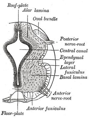
The dorsal part of the neural tube contains the alar plate, which is associated primarily with sensation. The ventral part of the neural tube contains the basal plate, which is primarily associated with motor (i.e., muscle) control.
How does Shh affect the ventral neural tube?
These transcription factors are grouped into two protein classes based on how Shh affects them. Class I is inhibited by Shh , whereas Class II is activated by Shh. These two classes of proteins then cross-regulate each other to create more defined boundaries of expression. The different combinations of expression of these transcription factors along the dorsal-ventral axis of the neural tube are responsible for creating the identity of the neuronal progenitor cells. Five molecularly distinct groups of ventral neurons form from these neuronal progenitor cells in vitro. Also, the position at which these neuronal groups are generated in vivo can be predicted by the concentration of Shh required for their induction in vitro. Studies have shown that neural progenitors can evoke different responses based on the length of exposure to Shh, with a longer exposure time resulting in more ventral cell types.
What are the neural tube patterns?
The neural tube patterns along the dorsal-ventral axis to establish defined compartments of neural progenitor cells that lead to distinct classes of neurons. According to the French flag model of morphogenesis, this patterning occurs early in development and results from the activity of several secreted signaling molecules. Sonic hedgehog (Shh) is a key player in patterning the ventral axis, while bone morphogenic proteins (BMPs) and Wnt family members play an important role in patterning the dorsal axis. Other factors shown to provide positional information to the neural progenitor cells include fibroblast growth factors (FGFs) and retinoic acid. Retinoic acid is required ventrally along with Shh to induce Pax6 and Olig2 during differentiation of motor neurons.
What is the neural groove in the embryo?
The center of the neural plate remains grounded, allowing a U-shaped neural groove to form. This neural gro ove sets the boundary between the right and left sides of the embryo. The neural folds pinch in towards the midline of the embryo and fuse together to form the neural tube.
What factors provide positional information to neural progenitor cells?
Other factors shown to provide positional information to the neural progenitor cells include fibroblast growth factors (FGFs) and retinoic acid. Retinoic acid is required ventrally along with Shh to induce Pax6 and Olig2 during differentiation of motor neurons.
What is the name of the neural tube that is open both cranially and caudally?
The mesencephalon stays as the midbrain. The rhombencephalon develops into the metencephalon (the pons and cerebellum) and the myelencephalon (the medulla oblongata ). For a short time, the neural tube is open both cranially and caudally. These openings, called neuropores, close during the fourth week in humans.
How does neural tube formation occur?
Neural tube formation begins when the area specified as neural plate by neural induction undergoes the process of convergent extension, in which it narrows and elongates. This process, first observed in amphibian embryos, is essential for initiating elevation of the neural folds in the spinal region of amniote embryos; unlike amphibians, in which the process involves the whole neural plate simultaneously, convergent extension in amniote embryos takes place in a cranial to caudal sequence as the embryo grows in length. It involves the exchange of cell neighbors within the neuroepithelial sheet and is regulated by core planar cell polarity (PCP) genes (reviewed by Tissir and Goffinet, 2013 ). Mouse mutants showing neural tube defects (NTDs) include two strains with loss of core PCP gene function, loop-tail (Vangl2), and Crash (Celsr1), in which the whole neural plate remains open (craniorachischisis) due to the failure of the cell movements of convergent extension. Core PCP genes were first discovered in Drosophila; they encode cell surface receptors such as Frizzled and cadherin family molecules and are conserved from lower invertebrates, for example, Caenorhabditis elegans, to mammals, including mice and humans. They play a number of developmental roles within the plane of epithelial sheets, and additionally have significant roles in neuronal migration, dendritic growth, and axon guidance.
When does neural tube development begin?
Neural tube development begins with the formation of the notochord around day 20 of embryogenesis. The neural tube will eventually develop into the spinal cord and brain. The surrounding mesoderm condenses to form somites.2 Somite differentiation in the early part of fetal development separates cells into the dermatome, myotome, and sclerotome cells ( Fig. 71.1 ).
What is the neurulation process?
Importantly, the process of neurulation transforms the axes of the developing CNS neuroepithelium from a planar to a tubular coordinate system . The planar (two-dimensional) neural plate is defined by rostrocaudal and mediolateral axes, whereas the tubular (three-dimensional) neural tube is defined by rostrocaudal, mediolateral, and dorsoventral axes. During this reorganization process, the lateral edge of the neural plate becomes the dorsal midline (roof plate) of the neural tube, and the medial edge (midline) of the neural plate becomes the ventral midline (floor plate) of the neural tube ( Fig. 29.5 ). The new axes become the substrate for further patterning and remain important throughout later development; however, additional axial transformations occur locally in the developing brain.
What is the hollow center of the neural tube?
The hollow center of the neural tube will become the central canal of the spinal cord and ventricular system of the brain. During primary neurulation, the neural tube separates from the overlying ectoderm in a process called dysjunction. Early dysjunction results in perineural mesenchyme access to the neural groove, ...
How are vertebrae formed?
The vertebrae and associated ligaments are formed by the development of sclerotome cells into a segmental centrum which forms cartilage and then the bony vertebral body. 2 Primary and secondary ossification centers provide growth areas to turn cartilage into bone. The secondary ossification centers in the annular epiphysis (apophyseal rings) remain open through adolescence. Scheurmann disease or kyphosis may be caused by an insult to these secondary ossification centers. Eventually, Schmorl's nodes can develop in these individuals with Scheurmann disease such that disc material herniates through the insulted vertebral endplate. Two other posterior primary ossification centers come together to form the neural arch. Rarely, particularly in the setting of poor folate intake, neural arch and tube defects (e.g. spina bifida and hemivertebrae) may arise.
Where does the neural tube close?
Neural tube closure is a key early event in brain and spinal cord development, in which the planar epithelium of the neural plate folds at the midline along the anteroposterior axis and the lateral edges of the neural plate move dorsally, contact each other, and fuse dorsally to form the neural tube (Fig. 29.5 ). Dorsal fusion first occurs in the region of the cervical spinal cord primordium, followed by separate closure events at midbrain and rostral telencephalic points. The exact location and number of dorsal fusion events vary between and within species. From each of these sites, fusion proceeds rostrally and caudally in a zipperlike mode until closure is complete (primary neurulation) from the rostral end (anterior neuropore) to the caudal (posterior neuropore). Interestingly, this mode of primary neural tube closure does not extend the full caudal length of the spinal cord but ends around the lumbosacral region. More caudal regions (mainly sacral) appear to develop by a distinct mechanism involving cavitation of the caudal eminence (tail bud) mesenchyme, known as secondary neurulation. A schematic of this process is shown in Fig. 29.6.
Which part of the brain differentiates into ependymal, mantle, and marginal layers?
Neural tube. The inferior portion of the neural tube differentiates into ependymal (central), mantle (neurons and glia), and marginal layers (axons of tract cells).2 These eventually form the future spinal cord. The neural crest develops into neurons of the peripheral nervous system.
How does the neural tube differentiate?
On the gross anatomical level, the neural tube and its lumen bulge and constrict to form the chambers of the brain and the spinal cord. At the tissue level, the cell populations within the wall of the neural tube rearrange themselves to form the different functional regions of the brain and the spinal cord. Finally, on the cellular level, the neuroepithelial cells themselves differentiate into the numerous types of nerve cells (neurons) and supportive cells (glia) present in the body.
What is the polarity of the neural tube?
The neural tube is polarized along its dorsal-ventral axis. In the spinal cord, for instance, the dorsalregion is the place where the spinal neurons receive input from sensory neurons, while the ventralregion is where the motor neurons reside. In the middle are numerous interneurons that relay information between them. The polarity of the neural tube is induced by signals coming from its immediate environment. The dorsal pattern is imposed by the epidermis, while the ventral pattern is induced by the notochord (Figure 12.13).
How does Sonic Hedgehog affect the ventral portion of the neural tube?
The importance of Sonic hedgehog in inducing and patterning the ventral portion of the neural tube can be shown experimentally. If notochord fragments are taken from one embryo and transplanted to the lateral side of a host neural tube, the host neural tube will form another set of floor plate cells at its sides (Figure 12.14B). The floor plate cells, once induced, induce the formation of motor neurons on either side of them. The same results can be obtained if the notochord fragments are replaced by pellets of cultured cells secreting Sonic hedgehog (Echelard et al. 1993; Roelink et al. 1994). Moreover, if a piece of notochord is removed from an embryo, the neural tube adjacent to the deleted region will have no floor plate cells (Placzek et al. 1990; Yamada et al. 1991, 1993).
How does the brain expand in the early embryo?
This rapid expansion is thought to be caused by positive fluid pressure exerted against the walls of the neural tube by the fluid within it. It might be expected that this fluid pressure would be dissipated by the spinal cord, but this does not appear to happen. Rather, as the neural folds close in the region between the presumptive brain and the presumptive spinal cord, the surrounding dorsal tissues push in to constrict the neural tube at the base of the brain (Figure 12.12; Schoenwolf and Desmond 1984; Desmond and Schoenwolf 1986; Desmond and Field 1992). This occlusion (which also occurs in the human embryo) effectively separates the presumptive brain region from the future spinal cord (Desmond 1982). If the fluid pressure in the anterior portion of such an occluded neural tube is experimentally removed, the chick brain enlarges at a much slower rate and contains many fewer cells than are found in normal embryos. The occluded region of the neural tube reopens after the initial rapid enlargement of the brain ventricles.
What are the three primary vesicles of the neural tube?
In this region, the neural tube balloons into three primary vesicles (Figure 12.10): forebrain (prosencephalon), midbrain (mesencephalon), and hindbrain (rhombencephalon). By the time the posterior end of the neural tube closes, secondary bulges—the optic vesicles—have extended laterally from each side of the developing forebrain.
Which transcription factors are critical in establishing the boundaries of the forebrain, midbrain, and hind?
12.4 Specifying the brain boundaries. The Pax transcription factors and the paracrine factor FGF8 are critical in establishing the boundaries of the forebrain, midbrain, and hindbrain. http://www.devbio.com/chap12/link1204.shtml
Where does the chick cerebellum come from?
12.3 Mapping the mesencephalon. The chick cerebellum has a dual origin, with most of it coming from the metencephalon, but some portions coming from the mesencephalon. Moreover, genetic abnormalities can be traced to specific brain regions by transplantations between wild-type and mutant brain regions. http://www.devbio.com/chap12/link1203.shtml
What is the neural tube?
The neural tube is the rudiment of the brain and spinal cord; its lumen gives rise to the cavities, or ventricles, of the brain and to the…. Read More. In prenatal development: Central nervous system. …and fuse, thereby creating a neural tube.
Where is the neural tube formed?
human body. The neural tube itself is formed from the ectoderm at a very early stage. Anteriorly (i.e., toward the head) it extends above the open end of the cylinder and is enlarged to form the brain. It is not in immediate contact with the epidermis, for the….
What is the term for the upper region of the neural tube to close in early embryonic development?
anencephaly. In cephalic disorder: Anencephaly. …the upper region of the neural tube to close in early embryonic development, specifically within the first month of pregnancy. (The neural tube is the primitive structure from which develops the central nervous system.) Females are more likely to be affected than males.
Where is the neural tube located in animal development?
…fuse together, and form the neural tube. In vertebrates the neural tube lies immediately above the notochord and extends beyond its anterior tip.
Which system develops the midline?
nervous system. In human nervous system: Morphological development. …the midline to form the neural tube, which will develop into the central nervous system. The neural tube detaches from the skin ectoderm and sinks beneath the surface.

Overview
In the developing chordate (including vertebrates), the neural tube is the embryonic precursor to the central nervous system, which is made up of the brain and spinal cord. The neural groove gradually deepens as the neural fold become elevated, and ultimately the folds meet and coalesce in the middle line and convert the groove into the closed neural tube. In humans, neural tube closure …
Development
The neural tube develops in two ways: primary neurulation and secondary neurulation.
Primary neurulation divides the ectoderm into three cell types:
• The internally located neural tube
• The externally located epidermis
• The neural crest cells, which develop in the region between the neural tube and epidermis but then migrate to new locations
Structure
Four neural tube subdivisions each eventually develop into distinct regions of the central nervous system by the division of neuroepithelial cells: the forebrain (prosencephalon), the midbrain (mesencephalon), the hindbrain (rhombencephalon) and the spinal cord.
• The prosencephalon further goes on to develop into the telencephalon (cerebrum) and the diencephalon (the optic vesicles and hypothalamus).
Dorsal-ventral patterning
The neural tube patterns along the dorsal-ventral axis to establish defined compartments of neural progenitor cells that lead to distinct classes of neurons. According to the French flag model of morphogenesis, this patterning occurs early in development and results from the activity of several secreted signaling molecules. Sonic hedgehog (Shh) is a key player in patterning the ventral axis, …
See also
• Neural fold
• Neural plate
• Neurulation
• Neural tube defects
• Cdx protein family
External links
• Swiss embryology (from UL, UB, and UF) iperiodembry/carnegie03
• Embryology at UNSW Notes/week3_5
• Diagram at embryology.med.unsw.edu.au
• Diagram at brainviews.com