- Tight junctions: Impermeable junctions that prevent molecules from passing through the intercellular space.
- Desmosomes: Anchoring junctions that bind adjacent cells together and help form an internal tension-reducing network of fibers.
- Gap junctions: Communicating junctions that allow ions and small molecules to pass for intercellular communication.
What is the ‘glue’ that binds cells together?
We’ve already seen the calcium-dependent cadherins involved in forming adherens junctions (desmosomes). These are essentially the ‘glue’ that binds cells together to form strong cohesive tissues and sheets of cells. Some examples of membrane proteins that enable cell-cell recognition and adhesion are illustrated on the next page.
How did cell-cell adhesion differ between the two species of sponge?
The molecules responsible for cell-cell adhesion (cell junctions) differed between the two species of sponge. d. One cell functioned as an organizer for each organism, thereby attracting only cells of the same species.
What is the function of adhesive molecules in the extracellular matrix?
Within tissues, adhesive molecules allow cells to maintain contact with one another and with structures in the extracellular matrix. One especially important class of adhesive molecules is the integrins. Integrins are more than just mechanical links, however: They also relay signals both to and from cells.
How is adhesion mediated in plant cells?
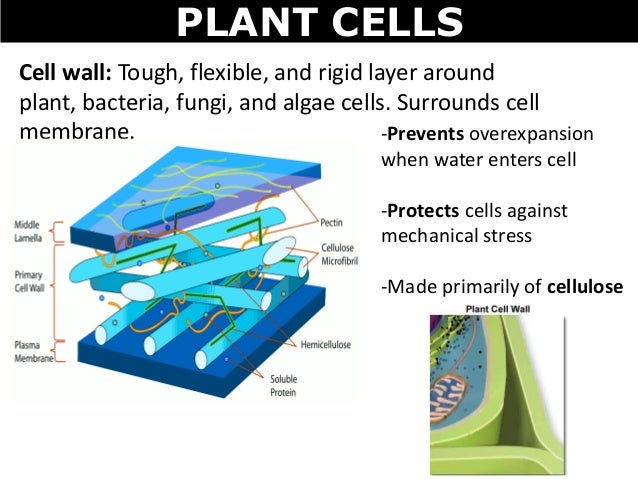
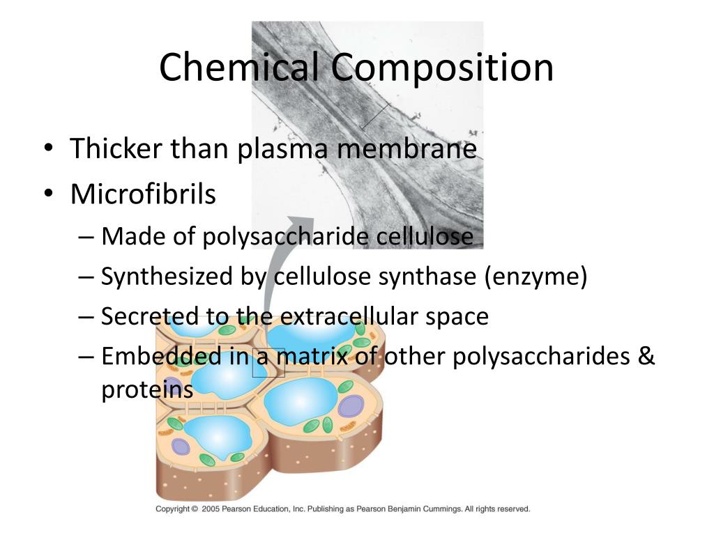
Adhesion between plant cells is mediated by their cell walls rather than by transmembrane proteins. In particular, a specialized pectin-rich region of the cell wall called the middle lamella acts as a glue to hold adjacent cells together.
What is the Würzburg researcher's interest?
What are the physical parameters that influence the membrane's fluctuation behaviour?
What is the special feature of the cell membrane of Trypanosoma?
Why is controlled adhesion important?
Where are cadherins located?
Is the process of establishing and detaching contacts dependent on physical effects?
See 1 more
About this website

What glues adjacent cell walls together?
Cellulose Fibrils The middle lamella is a layer of sticky material, also containing pectin, which serves as “glue” to hold adjacent cells together.
What attaches adjacent cells together?
Adherens junctions and desmosomes hold cells together and are formed by transmembrane adhesion proteins that belong to the cadherin family. 2. Focal adhesions and hemidesmosomes bind cells to the extracellular matrix and are formed by transmembrane adhesion proteins of the integrin family.
What bonds cells together?
Cell junctions The three main ways for cells to connect with each other are: gap junctions, tight junctions, and desmosomes. These types of junctions have different purposes, and are found in different places.
What is used for cell adhesion?
Cadherins are essential for cell–cell adhesion and cell signalling in multicellular animals and can be separated into two types: classical cadherins and non-classical cadherins.
What do desmosomes do?
Desmosomes are adhesive intercellular junctions that mechanically integrate adjacent cells by coupling adhesive interactions mediated by desmosomal cadherins to the intermediate filament cytoskeletal network.
How are cells bound together?
Cells are held together by several different complexes: tight junctions (discussed in epithelia lecture), adhering junctions, and desmosomes. These junctions consist of integral membrane proteins that contact proteins in neighboring cells and that are linked intracellularly to the cytoskeleton.
What protein holds cells together?
Adherens junctions (AJs) are protein complexes — primarily made up of cadherin — that form bonds between cells in nearly all human tissues.
Which organelle holds the cell together?
The cytoskeletonThe cytoskeleton consists of microtubules, intermediate fibers, and microfilaments, which together maintain cell shape, anchor organelles, and cause cell movement.
How cells are held together?
Cells are held together by several different complexes: tight junctions (discussed in epithelia lecture), adhering junctions, and desmosomes. These junctions consist of integral membrane proteins that contact proteins in neighboring cells and that are linked intracellularly to the cytoskeleton.
How are adjacent cells in multicellular plants held together?
Tight junctions are bands around a cell where it is 'spot welded' to an adjacent cell. No chemicals can be transferred at these points. Desmosomes come in a number of varieties from spot desmosomes to belt desmosomes. They hold cells together and anchor the internal intermediate filaments.
What is adjacent cell in biology?
The cells placed close to each other are called adjacent cells, also known as the neighbouring cells.
What holds epithelial cells together?
Epithelial cells are held together by strong anchoring (zonula adherens) junctions. The adherens junction lies below the tight junction (occluding junction). In the gap (about 15-20nm) between the two cells, there is a protein called cadherin - a cell membrane glycoprotein.
What are the proteins that bind cells together called?
Generally, proteins that interact to bind cells together are called I ntercellular C ell A dhesion M olecules ( ICAM s). These include selectins. During blood clotting, selectins on one platelet recognize and bind specific receptors on other platelets, contributing to the clot. NCAMs are another kind of ICAM, ones with sugary immunoglobulin domains ...
What is a tight junction?
Tight Junctions ( zonula occludens) are typical in sheets of epithelial cells that line the lumens of organs (e.g., intestines, lungs, etc.). Zonula refers to the fact that these structures form a band encircling an entire cell, attaching it to all surrounding cells.
How do integrins respond to fibronectin?
During development, integrins respond to fibronectin by signaling cell and tissue differentiation, complete with the formation of appropriate cell junctions. An orderly sequence of gene expression and membrane protein syntheses enable developing cells to recognize each other as different or the same.
How do cells migrate during embryogenesis?
During embryogenesis, cells migrate from a point of origin by attaching to and moving along an extracellular matrix ( ECM ), which acts as a path to the cell’s final destination . This ECM (or basal lamina) is made up of secretions from other cells…, or from the migrating cells themselves!
What is the role of cadherins in the cell membrane?
In both cases, cadherins cross cell membranes from intracellular plaque proteins, spanning the intercellular space to link adjacent cell membranes together. Plaques are in turn, connected to intermediate filaments (keratin) of the cytoskeleton, further strengthening intercellular attachments and thus, the tissue cell layer.
How do cells communicate?
17.6: How Cells are Held Together and How they Communicate. Proteins and glycoproteins on cell surfaces play a major role in how cells interact with their surroundings and with other cells. Some of the proteins in the glycocalyx of adjacent cells interact to form cell-cell junctions, while others interact with extracellular proteins ...
What are the functions of cell junctions?
A. Cell Junctions. Cell junctions serve different functions in cells and tissues. Cell junctions in healthy cells serve to bind cells tightly, to give tissues structural integrity and to allow cells in contact with one another to pass chemical information directly between them. Electron micrographs and illustrations different cell junctions are ...
What are tight junctions in the digestive tract?
For example, tight junctions between epithelial cells lining the digestive tract keep digestive enzymes and microorganisms in the intestine from leaking into the bloodstream (Note: some tight junctions may leak and allow certain ions to pass).
What is a tight junction?
Tight Junctions. In a tight junction, a series of integral protein molecules in the plasma membranes of adjacent cells fuse together , forming an impermeable junction that encircles the cell. Tight junctions help prevent molecules from passing through the extracellular space between adjacent cells.
What is the function of the gap junction?
At gap junctions the adjacent plasma membranes are very close, and the cells are connected by hollow cylinders called con nexons, composed of transmembrane proteins. The many different types of connexon proteins vary the selectivity of the gap junction channels. Ions, simple sugars, and other small molecules pass through these water-filled channels from one cell to the next.
How many junctions are there in an epithelial cell?
An epithelial cell is shown joined to adjacent cells by three common types of cell junctions. (Note: Except for epithelia, it is unlikely that a single cell will have all three junction types.)
What are the factors that bind cells together?
Typically, three factors act to bind cells together. Glycoproteins in the glycocalyx act as an adhesive. Contours in adjacent cells membranes fit together in a tight knit fashion. Special cell junctions form. Let’s take a closer look at the different types of cell junctions.
Which part of the plaque is the keratin filament?
Thicker keratin filaments extend from the cytoplasmic side of the plaque across the width of the cell to anchor to the plaque on the cell’s opposite side. In this way, desmosomes bind neighboring cells together and also contribute to a continuous internal network of strong wires.
Which junctions help form an internal tension-reducing network of fibers?
Desmosomes: Anchoring junctions that bind adjacent cells together and help form an internal tension-reducing network of fibers.
What is animal tissue?
A tissue that forms external & internal surfaces. An animal tissue consisting of sheetlike layers of tightly packed cells that line an organ, gland, duct, or body surface.
What is the region outside the plasma membrane?
In plants, the region outside the plasma membrane consisting of the porous cell walls & the extracellular air space, lamella.
What are the physical connections between two plant cells?
Physical connections between two plant cells, consisting of membrane-lined gaps in the cell walls through which the two cell's plasma membranes, cytoplasm, and smooth ER can connect directly. Similar to gap junctions. An ER tubule passes through.
What is cell attachment?
A cell-cell attachment composed of specialized proteins in the plasma membranes of adjacent animal cells. Prevents solutions from flowing between two cells.
Which cell has membrane bound organelles?
c. Eukaryotic cells have membrane-bound organelles, while prokaryotic cells do not.
What is Gaucher disease?
d. lysosomes. Gaucher disease is the most common genetic disorder affecting lipid storage in humans. The disease is caused by deficiency of an enzyme necessary for the breakdown of lipids, which leads to the accumulation of fatty material in organs of the body including the spleen, liver, kidneys, lungs, brain, and bone marrow.
Why is cell 1 the smallest?
a. Cell 1 because it has the smallest volume and will not produce as much waste as the other cells.
Do prokaryotic cells have cell walls?
a. Prokaryotic cells have cell walls, while eukaryotic cells do not .
Which cell evolved into chloroplasts?
b. endosymbiosis of a photosynthetic archaeal cell in a larger bacterial host cell to escape toxic oxygen-the anaerobic archaea evolved into chloroplasts.
Which cell has the largest surface area?
c. Cell 3 because it has the largest surface area, which will enable it to eliminate its wastes most efficiently.
Which is thicker, TEM or light microscopy?
c. Specimens visualized by TEM are much thicker than those observed by light microscopy.
Why are cadherins important?
It seems likely that cadherins are also crucial in later stages of vertebrate development, since their appearance and disappearance correlate with major morphogenetic events in which tissues segregate from one another. As the neural tubeforms and pinches off from the overlying ectoderm, for example, neural tube cells lose E-cadherinand acquire other cadherins, including N-cadherin, while the cells in the overlying ectoderm continue to express E-cadherin (Figure 19-25). Then, when the neural crest cells migrate away from the neural tube, these cadherins become scarcely detectable, and another cadherin (cadherin-7) appears that helps hold the migrating cells together as loosely associated cell groups. Finally, when the cells aggregate to form a ganglion, they re-express N-cadherin (see Figure 19-23).
Why do selectins mediate a weak adhesion?
The selectins mediate a weak adhesion because the binding of the lectindomainof the selectinto its carbohydrateligandis of low affinity. This allows the white blood cell to adhere weakly and reversibly to the endothelium, rolling along the surface of the blood vessel propelled by the flow of blood.
Which protein binds to the actin filament?
The linkage of classical cadherins to actin filaments. The cadherins are coupled indirectly to actin filaments by the anchor proteins α-catenin and β-catenin. A third intracellular protein, called p120, also binds to the cadherin cytoplasmic (more...)
How are CAMs identified?
CAMs were initially identified by making antibodies against cell-surface molecules and then testing the antibodies for their ability to inhibit cell-cell adhesion in a test tube. Those rare antibodies that inhibit the adhesion were then used to characterize and isolate the adhesion moleculerecognized by the antibodies.
What are the proteins that attach to the extracellular matrix called?
Cells adhere to each other and to the extracellular matrixthrough cell-surface proteins called cell adhesion molecules (CAMs) —a category that includes the transmembrane adhesion proteins we have already discussed. CAMs can be cell-cell adhesion moleculesor cell-matrix adhesion molecules.
How do cell motility and adhesion work together?
This may involve chemotaxisor chemorepulsion,the secretion of a soluble chemical that attracts or repels migrating cells, respectively, or pathway guidance,the laying down of adhesive or repellent molecules in the extracellular matrixor on cell surfaces to guide the migrating cells along the right paths. Then, once a migrating cell has reached its destination, it must recognize and join other cells of the appropriate type to assemble into a tissue. How this latter process occurs can be studied if cells of different embryonic tissues are artificially mingled, after which they often spontaneously sort out to restore a more normal arrangement, as we discuss next.
What is the simplest mechanism by which cells assemble to form a tissue?
The simplest mechanism by which cells assemble to form a tissue. The progeny of the founder cell are retained in the epithelium by the basal lamina and by cell-cell adhesion mechanisms, including the formation of intercellular junctions.
What are the cell-cell interactions mediated by?
In contrast to the stable cell-matrixjunctions discussed in the preceding section, the cell-cell interactions mediated by the selectins, integrins, and members of the Ig superfamily are transient adhesions in which the cytoskeletons of adjacent cells are not linked to one another. Stable adhesion junctions involving the cytoskeletons of adjacent cells are instead mediated by the cadherins. As discussed in Chapter 11, these cell-cell junctions are of two types: adherens junctionsand desmosomes, in which cadherins or related proteins(desmogleins and desmocollins) are linked to actinbundles and intermediate filaments, respectively (Figure 12.64). The role of the cadherins in linking the cytoskeletons of adjacent cells is thus analogous to that of the integrins in forming stable junctions between cells and the extracellular matrix.
What are the roles of selectins in the cell cycle?
As discussed earlier in this chapter, the selectins recognize cell surface carbohydrates (see Figure 12.14). One of their critical roles is to initiate the interactions between leukocytes and endothelial cells during the migration of leukocytes from the circulation to sites of tissue inflammation (Figure 12.62). The selectins mediate the initial adhesion of leukocytes to endothelial cells. This is followed by the formation of more stable adhesions, in which integrins on the surface of leukocytes bind to intercellular adhesion molecules (ICAMs), which are members of the Ig superfamily expressed on the surface of endothelial cells. The firmly attached leukocytes are then able to penetrate the walls of capillaries and enter the underlying tissue by migrating between endothelial cells.
How many cadherins are there in the human body?
Intriguingly, different neurons appear to express different protocadherins, suggesting that the protocadherins may play a role in the establishment of specific connections between neurons. About 50 human protocadherin genes have been identified and shown to be organized into three geneclusters. Each cluster contains multiple exons encoding the N-terminal extracellular and transmembrane protocadherin domains, but only a single set of three exons encoding the C-terminal cytoplasmic domain (Figure 12.63). The protocadherin gene clusters thus appear to consist of a variable region, encoding multiple extracellular and transmembrane domains, linked to a constant region encoding a single cytoplasmic domain. This organization of protocadherin genes strikingly resembles that of immunoglobulinand T-cell receptor genes (see Figures 5.42and 5.43), in which multiple variable region exons are joined to a single constant region exon. In the immunoglobulin and T-cell receptor genes, this occurs as a result of DNArearrangements that generate diversity in the immune system. It remains to be determined whether the variable and constant regions of protocadherins are joined at the DNA or the RNAlevel (for example, by alternative splicing) and to what extent rearrangements of protocadherin genes might contribute to the establishment of specific synaptic connections in the brain.
What are gap junctions made of?
Gap junctions are constructed of transmembrane proteinscalled connexins (Figure 12.66). Six connexins assemble to form a cylinder with an open aqueous pore in its center. Such an assembly of connexins in the plasma membraneof one cell then aligns with the connexins of an adjacent cell, forming an open channel between the two cytoplasms. The plasma membranes of the two cells are separated by a gap corresponding to the space occupied by the connexin extracellular domains—hence the term “gap junction,” which was coined by electron microscopists.
How do plant cells communicate with each other?
Because of the rigidity of plant cell walls, stable associations between plant cells do not require the formation of cytoskeletal links, such as those provided by the desmosomes and adherens junctions of animal cells. However, adjacent plant cells communicate with each other through cytoplasmic connections called plasmodesmata(singular, plasmodesma), which function analogously to animal cell gap junctions.
Which group of adhesion molecules are responsible for the formation of stable junctions between cells in tissues?
The fourth group of cell adhesion molecules, the cadherins, also display homophilic binding specificities. They are not only involved in selective adhesion between embryonic cells but are also primarily responsible for the formation of stable junctions between cells in tissues. For example, E-cadherin is expressed on epithelial cells, so homophilic interactions between E-cadherins lead to the selective adhesion of epithelial cells to one another. It is noteworthy that loss of E-cadherin can lead to the development of cancers arising from epithelial cells, illustrating the importance of cell-cell interactions in controlling cell behavior. Different members of the cadherin family, such as N-cadherin (neural cadherin) and P-cadherin (placental cadherin), mediate selective adhesion of other cell types.
Which cell-cell junctions are mediated by the cadherins?
Stable cell-cell junctions mediated by the cadherins. Homophilic interactions between cadherins mediate two types of stable cell-cell adhesions. In adherens junctions, the cadherins are linked to bundles of actin filaments via the catenins (see Figure (more...)
What are the dots on the plasma membrane?
Tight junctions (blue dots) between cells are connected areas of the plasma membrane that stitch cells together. Adherens junctions (red dots) join the actin filaments of neighboring cells together. Desmosomes are even stronger connections that join the intermediate filaments of neighboring cells. Hemidesmosomes (light blue) connect intermediate filaments of a cell to the basal lamina, a combination of extracellular molecules on other cell surfaces. Gap junctions (yellow) are clusters of channels that form tunnels of aqueous connectivity between cells.
What are the side-to-side junctions that link epithelial cells?
The side-to-side junctions that link epithelial cells are diverse in their protein makeup and function. The adhesive transmembrane proteins anchoring these junctions have extracellular portions that interact with similar proteins on adjacent cells. Protein complexes within each cell further connect the transmembrane adhesive proteins to the cytoskeleton. In particular, adaptor complexes bind adherens junctions to cytoskeletal actin, and other adaptor complexes bind desmosomes to intermediate filaments. Both of these types of junctional complexes provide cells and tissues with mechanical support, and they additionally recruit intracellular signaling molecules to relay positional information to the nucleus.
What are some examples of cell-to-cell junctions?
Beyond integrins, cells rely on several other adhesive proteins to maintain physical contact. As an example, consider the epithelial cells that line the inner and outer surfaces of the human body — including the skin, intestines, airway, and reproductive tract. These cells provide a dramatic example of the different kinds of cell-to-cell junctions, but the same junctions also exist in a wide range of other tissues.
What is the function of integrins?
Integrins link the actin cytoskeleton of a cell to various external structures. The cytoplasmic portion of each integrin molecule binds to adaptor proteins that connect to the actin filaments inside the cell. The extracellular portion of the integrin then binds to molecules in the extracellular matrix or on the surface of other cells. Integrin attachments to neighboring cells can break and reform as a cell moves (Figure 1).
What are the characteristics of different types of tissues?
Different types of tissues, such as bone, brain, and the lining of the gut, have characteristic features related to the number and types of cells they contain . Cell spacing is also critical to tissue function, so this geometry is precisely regulated. To preserve proper tissue architecture, adhesive molecules help maintain contact between nearby cells and structures, and tiny tunnel-like junctions allow the passage of ions and small molecules between adjacent cells. Meanwhile, signaling molecules relay positional information among the cells in a tissue, as well as between these cells and the extracellular matrix. These signaling pathways are critical to maintaining the state of equilibrium known as homeostasis within a tissue. For example, the processes involved in wound healing depend on positional information in order for normal tissue architecture to be restored. Such positional signals are also crucial for the development of adult structures in multicellular organisms. As tissues develop, clumps of unorganized cells grow and sort themselves according to signals they send and receive.
What are the molecules that are used to maintain contact with each other?
One especially important class of adhesive molecules is the integrins. Integrins are more than just mechanical links, however: They also relay signals both to and from cells. In this way, integrins play an important role in sensing the environment and controlling cell shape and motility.
How does signaling work?
Various signaling molecules allow the cells within a tissue to share information about internal and external conditions. This information helps the cells arrange themselves, coordinate their functions, and even know when to grow and when to die. Some of these signaling molecules also function in an adhesive capacity — not just relaying messages between the cells in a tissue, but physically joining these cells to one another.
What is the Würzburg researcher's interest?
The Würzburg researcher has a general interest in the biophysics of membranes. For example, she also studies the pathogens that cause the sleeping sickness. The protozoa of the species Trypanosoma are one of Professor Markus Engstler's focal areas of research; he is the head of the Department for Zoology I at the Julius-Maximilians-Universität (JMU) in Würzburg, Germany.
What are the physical parameters that influence the membrane's fluctuation behaviour?
The biophysicist brought together model membranes containing cadherin and then selectively changed different physical parameters that influence the membrane's fluctuation behaviour such as the concentration of sugar and salt.
What is the special feature of the cell membrane of Trypanosoma?
What's special about the cell membrane of Trypanosoma is that it is densely populated with a protein shell that is varied continuously in a population. This high variability of the protein shell allows the pathogens to hide efficiently from the immune systems of animals and humans.
Why is controlled adhesion important?
Controlled adhesion and division are crucial for our body's cells . This is the case, for instance, when the organs develop in an embryo or when broken skin is repaired during the healing process.
Where are cadherins located?
The cadherin proteins assume a central role in the above mentioned examples. Located in the cell membranes, they are capable of creating strong bonds both among themselves and with the cadherins of other cells. A bond between two cadherin molecules of two cells triggers the formation of extensive contact zones.
Is the process of establishing and detaching contacts dependent on physical effects?
The process of establishing and detaching contacts seems to be much more dependent on purely physical effects than thought previously . This is shown by computer simulations and experiments published in " Nature Physics " by Dr Susanne Fenz from the University of Würzburg's Biocentre with colleagues from Jülich, Stuttgart, Erlangen and Marseilles.