What process happens in prophase 1?
Prophase I of meiosis I is a process that involves five different stages during which genetic material in the form of alleles crosses over and recombines to form non-identical haploid chromatids. Prophase I is the first stage of meiosis I, followed by prophase II, anaphase I, anaphase II, metaphase I and metaphase II.
What does prometaphase look like?
prometaphase-nuclear membrane and nucleoli disappear-chromosomes look like dark bodies scattered in the cytoplasm-mitotic spindle forms
What happens in prophase, metaphase, anaphase, and telophase?
What happens during prophase metaphase anaphase and telophase? 1) Prophase: chromatin into chromosomes, the nuclear envelope break down, chromosomes attach to spindle fibres by their centromeres 2) Metaphase: chromosomes line up along the metaphase plate (centre of the cell) 3) Anaphase: sister chromatids are pulled to opposite poles of the ...
What happens before the prophase?
Prophase (from Ancient Greek προ-() 'before', and φάσις (phásis) 'appearance') is the first stage of cell division in both mitosis and meiosis.Beginning after interphase, DNA has already been replicated when the cell enters prophase. The main occurrences in prophase are the condensation of the chromatin reticulum and the disappearance of the nucleolus.
What happens during prometaphase?
During prometaphase, the physical barrier that encloses the nucleus, called the nuclear envelope, breaks down. The breakdown of the nuclear envelope frees the sister chromatids from the nucleus, which is necessary for separating the nuclear material into two cells.
What happens in Prometaphase of mitosis simple?
When prophase is complete, the cell enters prometaphase — the second stage of mitosis. During prometaphase, phosphorylation of nuclear lamins by M-CDK causes the nuclear membrane to break down into numerous small vesicles. As a result, the spindle microtubules now have direct access to the genetic material of the cell.
What happens in prophase prometaphase?
In prophase, the nucleolus disappears and chromosomes condense and become visible. In prometaphase, kinetochores appear at the centromeres and mitotic spindle microtubules attach to kinetochores. In metaphase, chromosomes are lined up and each sister chromatid is attached to a spindle fiber.
What happens in prometaphase I of meiosis?
Prometaphase I The nuclear membrane disappears. One kinetochore forms per chromosome rather than one per chromatid, and the chromosomes attached to spindle fibers begin to move.
Which of the following events occur during Prometaphase of mitosis?
Which of the following events occurs during prometaphase of mitosis? The nuclear envelope fragments. In animal cell mitosis, the cleavage furrow forms during which stage of the cell cycle?
What happens to the spindle fibers in prometaphase?
(c) Prometaphase The spindle fibers comprise bundles of microtubules radiating from the opposite ends and referred to as poles of the cell. The chromosomes then migrate to the equatorial plane where they attach to one of the spindle fibers.
Is prophase and prometaphase the same?
In late prophase (sometimes also called prometaphase), the mitotic spindle begins to capture and organize the chromosomes. The chromosomes become even more condensed, so they are very compact. The nuclear envelope breaks down, releasing the chromosomes.
What happens to centrioles in prometaphase?
Elongation of procentrioles is completed in prometaphase, and their structure undergoes a number of successive changes. In the G2 period, pericentriolar satellites disappear and some time later a fibrillar halo is formed on both mother centrioles, i.e., spindle poles begin to form.
What are the 4 phases of mitosis and what happens in each?
4:236:47What happens in the four stages of mitosis? - YouTubeYouTubeStart of suggested clipEnd of suggested clipDuring metaphase the chromosomes have been aligned in the middle of the cell and spindle fibers haveMoreDuring metaphase the chromosomes have been aligned in the middle of the cell and spindle fibers have attached to each chromosome at the connector core in anaphase.
What happens in prometaphase 2 of meiosis?
Prometaphase II The nuclear envelopes are completely broken down, and the spindle is fully formed. Each sister chromatid forms an individual kinetochore that attaches to microtubules from opposite poles.
How many chromatids are in prometaphase?
At this point, each chromosome contains two sister chromatids.
What is after prometaphase?
After prometaphase ends, metaphase—the second official phase of mitosis—begins.
What are the 4 stages of mitosis and what happens in each?
4:326:47What happens in the four stages of mitosis? - YouTubeYouTubeStart of suggested clipEnd of suggested clipDuring metaphase the chromosomes have been aligned in the middle of the cell and spindle fibers haveMoreDuring metaphase the chromosomes have been aligned in the middle of the cell and spindle fibers have attached to each chromosome at the connector core in anaphase.
What happens in prometaphase 2 of meiosis?
Prometaphase II The nuclear envelopes are completely broken down, and the spindle is fully formed. Each sister chromatid forms an individual kinetochore that attaches to microtubules from opposite poles.
What happened in each stage of mitosis?
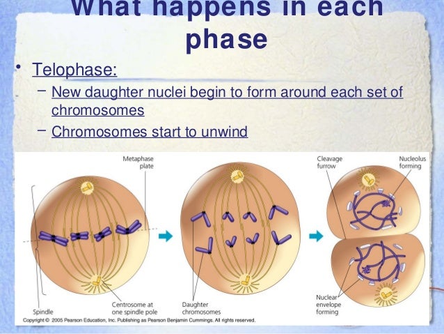
1) Prophase: chromatin into chromosomes, the nuclear envelope break down, chromosomes attach to spindle fibres by their centromeres 2) Metaphase: chromosomes line up along the metaphase plate (centre of the cell) 3) Anaphase: sister chromatids are pulled to opposite poles of the cell 4) Telophase: nuclear envelope ...
What happens in each step of mitosis?
Prophase – The chromosomes shorten and thicken. Metaphase – Chromosomes line up in the middle of the cell. Anaphase – Chromatids break apart at the centromere and move to opposite poles. Telophase – Two nuclei formed after nuclear envelopes reform around each group of chromosomes.
Is prometaphase same as late prophase?
No, the two phases are distinct. Late prophase displays thick and condensed chromosomes. Chromatids are visible. The centrosomes have moved to the...
What is the difference between prophase and prometaphase?
Prophase is the first stage of mitosis. The chromosome condenses and thickens. The sister chromatids are visible. The arms of the chromatids are in...
What happens during prometaphase?
During prometaphase, the nuclear membrane dissolves. The chromatids come out of the nuclear area. The chromatids develop kinetochores and kinetocho...
What is Prometaphase?
Mitosis is a process by which somatic cells produce two daughter cells. Somatic cells are the cells present in an organism. They have the normal number of chromosomes. They are distinct from reproductive cells which have only half the number of chromosomes.
What happens in Prometaphase?
In prometaphase, the nuclear membrane breaks down into numerous small vesicles. The spindle fibers are able to latch on the centromeres of the chromatids.
What is the important chromosomal event of prometaphase?
The important chromosomal event of prometaphase is the attachment of the chromosomes to the spindle and their movement towards the center of the spindle. Attachment of the chromosome to the spindle occurs at the kinetochore, which contains proteins for chromatid attachment. The breakdown of the nuclear envelope permits the kinetochores to attach to the spindle microtubules.
What is the prometaphase of the nuclear envelope?
Prometaphase begins when the nuclear envelope disassembles, exposing nuclear structures and its contents to the cytoplasm. The nuclear envelope is composed of two membrane barriers, the inner nuclear membrane (INM) and the outer nuclear membrane (ONM). The nuclear envelope is stabilized by nuclear lamina (polymerization of lamin proteins) underlying the INM. NEB is thought to involve early spindle microtubules directly piercing the nuclear envelope causing folds and invaginations that create mechanical tension in the nuclear lamina (Beaudouin et al., 2002; Georgatos et al., 1997 ). Lamins are subsequently phosphorylated by Cdk1/cyclin B1 triggering their depolymerization, causing the nuclear envelope to vesiculate ( Gallant and Nigg, 1992; Heald and McKeon, 1990; Peter et al., 1990 ). Concurrently, nuclear pore complexes (NPCs) are disassembled by phosphorylation of the core components, nucleoporins, resulting in complete dissolve of the membrane permeability barrier ( Terasaki et al., 2001 ).
How long does prophase arrest last?
In S. cerevisiae, where prophase I arrest lasts at least 3.5 h, this is achieved by regulated proteolysis of M-phase regulators by the meiosis-specific APC/C coactivator called Ama1 ( Okaz et al., 2012; Pesin and Orr-Weaver, 2008 ). In mammalian oocytes, no meiosis-specific activator of the APC/C has yet been identified, and it seems instead that mitotic Cdh1 takes the role of Ama1 in prophase I ( Pesin and Orr-Weaver, 2008; Reis et al., 2006 ). Cdh1 is a member of the Cdc20 family of APC/C regulators, and in mitosis it functions to target proteins for ubiquitination after the onset of anaphase. In oocytes, APC/C-Cdh1 activity is tightly regulated in order to maintain prophase arrest by controlling Cyclin B and Securin levels ( Marangos and Carroll, 2008; Marangos et al., 2007 ).
How do microtubules attach to chromosomes?
The attachment of microtubules to kinetochores can be reconstituted in vitro from mixtures of chromosomes, isolated centrosomes, and tubulin subunits. The plus ends of microtubules grow out from centrosomes and attach to the chromosomes. Surprisingly, chromosome-bound microtubules can either lengthen or shorten at the attached end without detaching from the chromosome. Similar experiments with kinetochores isolated from budding yeast cells showed that kinetochores can remain attached to a shortening microtubule plus end even against an applied force of 9 pN (piconewtons). Physiological levels of tension actually stabilize the attachments of kinetochores to microtubules in vitro, as in vivo. This tethering of kinetochores to disassembling microtubules is essential for chromosome movements during mitosis.
How do chromosomes move away from the spindle pole?
More recent studies revealed that chromosomes attached to only one spindle pole can move away from that pole if the unattached kinetochore associates with the kinetochore fiber of a chromosome already aligned at the spindle equator. In this case, the kinetochore of the mono-oriented chromosome glides toward the equator, where it is more likely to capture microtubules emanating from the opposite pole. This motion of one chromosome along the kinetochore fiber of another chromosome requires the kinesin-7 motor centromere protein E (CENP-E) (see Fig. 36.13) associated with the kinetochore of the moving chromosome. Recognition of a tubulin posttranslational modification leads CENP-E to move the chromosome toward the spindle equator, rather than out into the aster.
What is the mitotic spindle?
The mitotic spindle is a bipolar structure composed of microtubules and associated motor proteins. Microtubules initially emanate from the spindle poles/centrosomes toward the cell equator and with their plus-ends attach to a large protein assembly on the centrometric chromatin of a chromosome called the kinetochore ( Figure 3 ). This population of microtubules is termed kinetochore- or K-fibres. This attachment allows rapid movement of the bound chromosome such that the sister kinetochore attaches to a microtubule growing from the opposing centrosome. The result is a correctly bi-orientated chromosome at the metaphase plate with a stable microtubule–kinetochore attachment. Once all chromosomes have achieved this, the cell is considered to be in metaphase and chromosome segregation during anaphase can proceed. The mitotic spindle consists of two other populations of microtubules: (1) microtubules that do not attach to kinetochores, but also emanate toward the cell center and are called central spindle or non-kinetochore microtubules and (2) microtubules that emanate from the centrosomes circumferentially and anchor at the cell cortex are called astral microtubules, which function to maintain correct spindle orientation. Proteomics of purified mitotic spindles has identified approximately 800 spindle-associated proteins ( Sauer et al., 2005 ), indicating the breadth of signaling pathways required to establish and maintain this structure for correct chromosome attachment and alignment. Future investigation of these proteins in functional studies will aid in our understanding.
What are the components of the centromere?
Figure 3. The centromere–kinetochore association. The centromere is composed of borealin, survivin, Aurora B, inner centromere protein (INCENP), and mitotic centromere-associated kinesin (MCAK), which function to regulate stability of microtubule–kinetochore attachments. The kinetochore is a protein matrix attached to the centromere and is composed of several layers ( Musacchio and Salmon, 2007 ). The inner plate is in contact with the centromere (or centromeric DNA) and is marked by the specialized histone variant CENP-A (which substitutes histone H3 in this region) as well as additional auxiliary proteins. Adjacent to the inner plate is the outer plate, which consists primarily of proteins that assemble at the kinetochore upon NEB. Evolutionarily conserved proteins such as Spc105, MIS12, MSM21, and Hdc80 occupy this space to form attachment sites for bundles of microtubules known as K-fibres. In animal cells, kinetochores contain approximately 20 anchoring sites for the plus-ends of microtubules. The outer most region of the kinetochore is the fibrous corona and consists of proteins that are highly dynamic in their concentration. These proteins are either high in their concentration at the kinetochore before (molecular motors CENP-E and dynein as well as their target components ZW10 and ROD, and the spindle checkpoint proteins such as Mad1, Mad2, BubR1, and Cdc20) or after (EB1, APC, and proteins in the Ran pathway such as RanGap1 and RanBP2) K-fiber capture.
Learn about this topic in these articles
In prometaphase the nuclear envelope breaks down (in many but not all eukaryotes) and the chromosomes attach to the mitotic spindle. Both chromatids of each chromosome attach to the spindle at a specialized chromosomal region called the kinetochore. In metaphase the condensed chromosomes align in a…
cell division
In prometaphase the nuclear envelope breaks down (in many but not all eukaryotes) and the chromosomes attach to the mitotic spindle. Both chromatids of each chromosome attach to the spindle at a specialized chromosomal region called the kinetochore. In metaphase the condensed chromosomes align in a…
What is the major event marking a cell's entry to prometaphase?
The major event marking a cell's entry to prometaphase is the breakdown of the nuclear envelope into small vesicles. Kinetochores also become fully matured on the centromeres of the chromosomes. The disruption of the nuclear envelope allows for the mitotic spindles to gain access to the mature kinetochores. As the microtubles of the mitotic spindle enter the nuclear region, some attach to the kinetochores making them kinetochore microtubules. The remaining microtubules are called non-kinetochore microtubules. Sister chromatids are captured by microtubules stemming from centrosomes on opposite ends of the cell. Once they have captured chromosomes, the kinetochore mictrotubles begin to exert force on the chromosomes, moving them.
What are the kinetochores in prophase?
Late in prophase, kinetochores assemble on the centromeres. Specialized microtubules, called kinetochore microtubules later attach to these sites. Duplicated centrosomes, which are the organizing centers of microtubules, begin to separate towards opposite poles of the cell.
What are the microtubules that attach to the kinetochores called?
As the microtubles of the mitotic spindle enter the nuclear region, some attach to the kinetochores making them kinetochore microtubules. The remaining microtubules are called non-kinetochore microtubules. Sister chromatids are captured by microtubules stemming from centrosomes on opposite ends of the cell.
How are sister chromatids captured?
Sister chromatids are captured by microtubules stemming from centrosomes on opposite ends of the cell. Once they have captured chromosomes, the kinetochore mictrotubles begin to exert force on the chromosomes, moving them. Previous section Introduction to Mitosis Next section Problems.
What is the physical characteristic of cells beginning mitosis?
Another physical characteristic of cells beginning mitosis is the sprouting of microtubules from replicated centrosomes.
What are the requirements for entering the M phase?
As we discussed in cell cycle, before cells are allowed to enter M phase they must meet certain cellular requirements. Among these requirements are appropriate cell size and cellular environment. Following DNA replication in S phase, cells contain twice their normal number of chromosomes. Because cells that undergo mitosis are diploid, their number of chromosomes can be represented as 2 N, where N equals the number of distinct chromosomes in the cell. Cells about to enter M phase, which have passed through S phase and replicated their DNA, have 4 N chromosomes. Entry into M phase is allowed by the formation of the mitotic cyclin-Cdk complex known as M phase-promoting factor that occurs as a cell cycle regulatory mechanism in the G2 phase.
What is the first phase of mitosis?
The first phase of mitosis within M phase is called prophase. It follows G2, the final phase of interphase. A cell entering M phase manifests a number of physicsl signs. Among these are condensation, or thickening, of chromosomes. Chromosome condensation is visible through a microscope and is required for subsequent chromosome separation ...
What happens during prometaphase?
The short version of what happens during prometaphase is that the nuclear membrane breaks down .
What is the term for the early metaphase?
Prometaphase is often referred to as “late prophase.” (Though it’s also sometimes called “early metaphase” or referred to as a distinct phase entirely!) Regardless, some really important things occur during prometaphase that propel cell division along and that help explain what happens in metaphase.
What Is Mitosis?
Mitosis is a process that occurs during the cell cycle. The role of mitosis in the cell cycle is to replicate the genetic material in an existing cell—known as the “parent cell”—and distribute that genetic material to two new cells, known as “daughter cells.” In order to pass its genetic material to the two new daughter cells, a parent cell must undergo cell division, or mitosis. Mitosis results in two new nuclei—which contain DNA—that eventually become two identical cells during cytokinesis .
What is the line that divides the sister chromatids down the middle of the cell called?
This imaginary line dividing the cell down the middle is called the metaphase plate or equatorial plane .
How many phases does mitosis occur in?
In order to accomplish this goal, mitosis occurs in four discrete, consistently consecutive phases: 1) prophase, 2) metaphase, 3) anaphase, and 4) telophase . We have an overview of mitosis here, which is more of an intro to what mitosis is and how it works. If you're a little shaky on mitosis still, that's definitely where you should start.
What is interphase in biology?
We can think of interphase as a transitional phase. Interphase is when the parent cell prepares itself for mitosis. This phase isn’t considered part of mitosis, but understanding what happens during interphase can help the steps of mitosis make a little more sense.
What is the purpose of mitosis?
The main purpose of mitosis is to accomplish cell regeneration, cell replacement, and growth in living organisms. Mitosis is important because it ensures that all new cells that are generated in a given organism will have the same number of chromosomes and genetic information. In order to accomplish this goal, mitosis occurs in four discrete, consistently consecutive phases: 1) prophase, 2) metaphase, 3) anaphase, and 4) telophase .
